
National Model EMS
Clinical Guidelines
March 2022
VERSION 3.0
These guidelines will be maintained by the National Association of
State EMS Officials (NASEMSO) to facilitate the creation of state
and local EMS system clinical guidelines, protocols, or operating
procedures. System medical directors and other leaders are
invited to harvest content as will be useful. These guidelines are
either evidence-based or consensus-based and have been
formatted for use by field EMS professionals.
NASEMSO Medical Directors Council
www.nasemso.org

NASEMSO
National Model EMS Clinical Guidelines
Rev. March 2022
2
Version 3.0
Contents
INTRODUCTION ..................................................................................................................... 6
PURPOSE AND NOTES ........................................................................................................... 7
TARGET AUDIENCE .................................................................................................................. 8
WHAT IS NEW IN THE 2022 EDITION ........................................................................................... 8
ACKNOWLEDGEMENTS ............................................................................................................. 8
UNIVERSAL CARE ................................................................................................................... 9
UNIVERSAL CARE GUIDELINE ..................................................................................................... 9
FUNCTIONAL NEEDS .............................................................................................................. 19
PATIENT REFUSALS ................................................................................................................ 23
CARDIOVASCULAR .............................................................................................................. 26
ADULT AND PEDIATRIC SYNCOPE AND NEAR SYNCOPE .................................................................... 26
CHEST PAIN/ACUTE CORONARY SYNDROME (ACS)/ST-SEGMENT ELEVATION MYOCARDIAL INFARCTION (STEMI)
........................................................................................................................................ 31
BRADYCARDIA ..................................................................................................................... 35
IMPLANTABLE VENTRICULAR ASSIST DEVICES ............................................................................... 39
TACHYCARDIA WITH A PULSE ................................................................................................... 42
SUSPECTED STROKE/TRANSIENT ISCHEMIC ATTACK ........................................................................ 48
GENERAL MEDICAL .............................................................................................................. 51
ABDOMINAL PAIN ................................................................................................................ 51
ABUSE AND MALTREATMENT ................................................................................................... 55
AGITATED OR VIOLENT PATIENT/BEHAVIORAL EMERGENCY ............................................................. 59
ANAPHYLAXIS AND ALLERGIC REACTION ..................................................................................... 66
ALTERED MENTAL STATUS ...................................................................................................... 71
BACK PAIN ......................................................................................................................... 75
END-OF-LIFE CARE/HOSPICE CARE ............................................................................................ 78
HYPERGLYCEMIA .................................................................................................................. 81
HYPOGLYCEMIA ................................................................................................................... 84
NAUSEA-VOMITING .............................................................................................................. 89
PAIN MANAGEMENT ............................................................................................................. 93
SEIZURES .......................................................................................................................... 101
SHOCK ............................................................................................................................. 107
SICKLE CELL PAIN CRISIS ....................................................................................................... 114
RESUSCITATION ................................................................................................................ 117
CARDIAC ARREST (VF/VT/ASYSTOLE/PEA).............................................................................. 117
ADULT POST-ROSC (RETURN OF SPONTANEOUS CIRCULATION) CARE .............................................. 126
DETERMINATION OF DEATH/WITHHOLDING RESUSCITATIVE EFFORTS ............................................... 130
DO NOT RESUSCITATE STATUS/ADVANCE DIRECTIVES/HEALTHCARE POWER OF ATTORNEY (POA) STATUS 133
TERMINATION OF RESUSCITATIVE EFFORTS ................................................................................ 136

NASEMSO
National Model EMS Clinical Guidelines
Rev. March 2022
3
Version 3.0
RESUSCITATION IN TRAUMATIC CARDIAC ARREST ........................................................................ 141
PEDIATRIC-SPECIFIC GUIDELINES ....................................................................................... 144
BRIEF RESOLVED UNEXPLAINED EVENT (BRUE) & ACUTE EVENTS IN INFANTS .................................... 144
PEDIATRIC RESPIRATORY DISTRESS (BRONCHIOLITIS) .................................................................... 150
PEDIATRIC RESPIRATORY DISTRESS (CROUP) .............................................................................. 155
NEONATAL RESUSCITATION ................................................................................................... 159
OB/GYN ............................................................................................................................ 165
CHILDBIRTH....................................................................................................................... 165
ECLAMPSIA/PRE-ECLAMPSIA ................................................................................................. 171
OBSTETRICAL AND GYNECOLOGICAL CONDITIONS ........................................................................ 175
RESPIRATORY .................................................................................................................... 178
AIRWAY MANAGEMENT ....................................................................................................... 178
RESPIRATORY DISTRESS (INCLUDES BRONCHOSPASM, PULMONARY EDEMA) ...................................... 190
MECHANICAL VENTILATION (INVASIVE) .................................................................................... 198
TRACHEOSTOMY MANAGEMENT ............................................................................................. 203
TRAUMA ........................................................................................................................... 208
GENERAL TRAUMA MANAGEMENT .......................................................................................... 208
BLAST INJURIES .................................................................................................................. 215
BURNS ............................................................................................................................. 218
CRUSH INJURY/CRUSH SYNDROME .......................................................................................... 222
EXTREMITY TRAUMA/EXTERNAL HEMORRHAGE MANAGEMENT...................................................... 225
FACIAL/DENTAL TRAUMA ..................................................................................................... 230
HEAD INJURY..................................................................................................................... 233
HIGH THREAT CONSIDERATIONS/ACTIVE SHOOTER SCENARIO ........................................................ 238
SPINAL CARE ..................................................................................................................... 241
TRAUMA MASS CASUALTY INCIDENT ....................................................................................... 249
TOXINS AND ENVIRONMENTAL ......................................................................................... 252
POISONING/OVERDOSE UNIVERSAL CARE ................................................................................. 252
ACETYLCHOLINESTERASE INHIBITORS (CARBAMATES, NERVE AGENTS, ORGANOPHOSPHATES) EXPOSURE ... 260
RADIATION EXPOSURE ......................................................................................................... 271
TOPICAL CHEMICAL BURN ..................................................................................................... 275
STIMULANT POISONING/OVERDOSE ........................................................................................ 279
CYANIDE EXPOSURE ............................................................................................................ 283
BETA BLOCKER POISONING/OVERDOSE .................................................................................... 287
BITES AND ENVENOMATION .................................................................................................. 291
CALCIUM CHANNEL BLOCKER POISONING/OVERDOSE .................................................................. 295
OPIOID POISONING/OVERDOSE ............................................................................................. 303
AIRWAY RESPIRATORY IRRITANTS ........................................................................................... 308
RIOT CONTROL AGENTS ....................................................................................................... 317
HYPERTHERMIA/HEAT EXPOSURE ........................................................................................... 320
HYPOTHERMIA/COLD EXPOSURE ............................................................................................ 326
DROWNING ...................................................................................................................... 333

NASEMSO
National Model EMS Clinical Guidelines
Rev. March 2022
4
Version 3.0
DIVE (SCUBA) INJURY/ACCIDENTS......................................................................................... 337
ALTITUDE ILLNESS ............................................................................................................... 341
CONDUCTED ELECTRICAL WEAPON INJURY (I.E., TASER®) ............................................................ 345
ELECTRICAL INJURIES ........................................................................................................... 348
LIGHTNING/LIGHTNING STRIKE INJURY ..................................................................................... 352
APPENDICES ...................................................................................................................... 357
I. AUTHOR, REVIEWER AND STAFF INFORMATION ........................................................................ 357
II. UNIVERSAL DOCUMENTATION GUIDELINE .............................................................................. 363
III. MEDICATIONS ............................................................................................................... 377
IV. APPROVED ABBREVIATIONS .............................................................................................. 394
V. BURN AND BURN FLUID CHARTS ......................................................................................... 398
VI. NEUROLOGIC STATUS ASSESSMENT .................................................................................... 404
VII. ABNORMAL VITAL SIGNS ................................................................................................ 405
VIII. EVIDENCE-BASED GUIDELINES: GRADE METHODOLOGY ....................................................... 406
IX. 2022 NATIONAL GUIDELINE FOR THE FIELD TRIAGE OF INJURED PATIENTS .................................... 407

NASEMSO
National Model EMS Clinical Guidelines
Rev. March 2022
5
Version 3.0
This publication was developed with funding from the National Highway Traffic Safety Administration
(NHTSA), Office of Emergency Medical Services (Cooperative Agreement 693JJ92050001-0002) and the
Health Resources and Services Administration/Maternal and Child Health Bureau/EMS for Children
program. The opinions, findings, and conclusions expressed in this publication are those of the authors and
not necessarily those of the United States Government. The United States Government assumes no liability
for its content or use thereof. If trade or manufacturers’ names or products are mentioned, it is because
they are considered essential to the object of the publication and should not be construed as an
endorsement. The United States Government does not endorse products or manufacturers. For more
information, please visit EMS.gov and HRSA.gov.

NASEMSO
National Model EMS Clinical Guidelines
Rev. March 2022
6
Version 3.0
Introduction
We are honored to present the third edition of the National Association of State EMS Officials (NASEMSO)
National Model EMS Clinical Guidelines and want to thank the entire EMS community for contributing to its
evolution. The inaugural edition, released in September 2014, has been warmly welcomed by EMS
clinicians, agencies, medical directors, and healthcare organizations in our nation as well as abroad. The
creation of this document is a pinnacle event in the practice of EMS medicine as it fulfilled a
recommendation in The Future of Emergency Care: Emergency Medical Services at the Crossroads published
by the Institute of Medicine (now the National Academies of Sciences) in 2007. Specifically, this report
states “NHTSA, in partnership with professional organizations, should convene a panel of individuals with
multidisciplinary expertise to develop evidence-based model prehospital care protocols for the treatment,
triage, and transport of patients.”
The National Association of State EMS Officials (NASEMSO) recognizes the need for national EMS clinical
guidelines to help state EMS systems ensure a more standardized approach to the practice of patient care
now and, as experience dictates, the adoption of future practices. The value of EMS clinicians to the patient
has no boundaries as magnified by the historic 2019 novel coronavirus pandemic as well as other
interjurisdictional and global responses. Model EMS clinical guidelines promote uniformity in EMS medicine
which, in turn, fosters a more consistent skilled practice as EMS clinicians move across healthcare
systems. They also provide a standard to EMS medical directors upon which to base practice. Supported by
initial and subsequent grant funding from NHTSA’s Office of Emergency Medical Services (OEMS) and the
Health Resources and Services Administration (HRSA) Maternal and Child Health Bureau’s EMS for Children
program, NASEMSO continues to authorize its Medical Directors Council to partner with national
stakeholder organizations with expertise in EMS medical direction and subject matter experts to create a
unified set of patient care guidelines. For those aspects of clinical care where evidence-based guidelines
derived in accordance with the national evidence-based guideline model process were not available,
consensus-based clinical guidelines are developed utilizing current available research.
The NASEMSO Model EMS Clinical Guidelines are not mandatory, are not meant to be all-inclusive, nor are
they meant to determine local scope of practice. The focus of these guidelines is solely patient-centric. As
such, they are designed to provide a resource for EMS clinical practice, appropriate patient care, safety of
patients and clinicians, and outcomes regardless of the existing resources and capabilities within an EMS
system. This document provides a clinical standard that can be used as is or adapted for use on a state,
regional, local, or organizational level to enhance patient care and to set benchmark performance of EMS
practice. The Guidelines should be adapted to align with federal, state, regional, and jurisdictional laws and
regulations. NASEMSO’s ongoing support of this project underlines the critical evolution of the model EMS
clinical guidelines as new EMS research and evidence-based patient care measures emerge.
We are most grateful to be able to partner with a group of talented, committed individuals in this
worthwhile endeavor.
Carol Cunningham, M.D. Richard Kamin, M.D.
Co-Principal Investigator Co-Principal Investigator

NASEMSO
National Model EMS Clinical Guidelines
Rev. March 2022
7
Version 3.0
Purpose and Notes
These guidelines are intended to help state EMS systems ensure a more standardized approach to the
practice of patient care, and to encompass evidence-based guidelines (EBG) as they are developed.
The long-term goal is to develop a full range of evidence-based clinical guidelines for the practice of EMS
medicine. However, until there is a sufficient body of evidence to fully support this goal, there is a need for
this interim expert, consensus-based step.
The National Model EMS Clinical Guidelines can fill a significant gap in uniform clinical guidance for EMS
patient care, while also providing input to the evidence-based guideline (EBG) development process.
These guidelines will be maintained by the Medical Directors Council of the National Association of State
EMS Officials (NASEMSO) and will be reviewed and updated periodically. As EBG material is developed, it
will be substituted for the consensus-based guidelines now comprising the majority of the content of this
document. In the interim, additional consensus-based guidelines will also be added as the need is identified.
For guidelines to be considered for inclusion, they must be presented in the format followed by all
guidelines in the document.
Universal Care and Poisoning/Overdose Universal Care guidelines are included to reduce the need for
extensive reiteration of basic assessment and other considerations in every guideline.
The appendices contain material such as neurologic status assessment and burn assessment tools to which
many guidelines refer to increase consistency in internal standardization and to reduce duplication.
While some specific guidelines have been included for pediatric patients, considerations of patient age and
size (pediatric, geriatric, and bariatric) have been interwoven in the guidelines throughout the document.
Where IV access and drug routing are specified, it is intended to include IO access and drug routing when IV
access and drug routing is not possible.
Generic medication names are utilized throughout the guidelines. A complete list of these, along with
respective brand names, may be found in Appendix III. “Medications”.
Accurate and quality data collection is crucial to the advancement of EMS and a critical element of EMS
research. The National EMS Information System (NEMSIS) has the unique ability to unify EMS data on a
national scope to fulfill this need. Each guideline, therefore, is also listed by the closest NEMSIS Version 3
Label and Code corresponding to it, listed in parentheses below the guideline name.
Quality assurance (QA) and/or continued performance improvement (CPI) programs are an indispensable
element of medical direction as they facilitate the identification of gaps and potential avenues of their
resolution within an EMS system. The National EMS Quality Alliance (NEMSQA) Performance Measures is a
resource for these programs. This edition of the NASEMSO National Model EMS Clinical Guidelines
incorporates many of the NEMSQA performance measures into the key performance measures associated
with each clinical guideline.

NASEMSO
National Model EMS Clinical Guidelines
Rev. March 2022
8
Version 3.0
Target Audience
While this material is intended to be integrated into an EMS system’s operational guidance materials by its
medical director and other leaders, it is written with the intention that it will be consumed by field EMS
clinicians.
To the degree possible, it has been assembled in a format useful for guidance and quick reference so that
leaders may adopt it in whole or in part, harvesting and integrating as they deem appropriate to the format
of their guideline, protocol, or procedure materials.
Any set of guidelines must determine a balance between education and patient care. This document
purposefully focuses on the patient care aspect of EMS response. This does not preclude the individual
medical director from using these guidelines and including additional education as well as incorporation of
state, local, or jurisdictional operational procedures.
What is New in the 2022 Edition
All of the 2017 guidelines have been reviewed and updated, and additional guidelines and new evidence-
based guidelines have been added to this edition. While some of the new material has been added as
guidelines in the appropriate chapter, other topics have been incorporated into a previously existing
guideline. New guidelines have been added to the 2022 edition for the following clinical conditions or
scenarios:
•
Brief Resolved Unexplained Event (BRUE) & Acute Events in Infants
•
Resuscitation in Traumatic Cardiac Arrest
•
Tracheostomy Management
•
Trauma Mass Casualty Incident
In addition, with the permission and assistance of the American College of Surgeons – Committee on
Trauma, we have included the 2022 National Guideline for the Field Triage of Injured Patients as Appendix
IX.
Acknowledgements
The authors of this document are NASEMSO Medical Director Council members partnered with
representatives of seven EMS medical director stakeholder organizations. The stakeholder organizations are
the American Academy of Emergency Medicine (AAEM), the American Academy of Pediatrics (AAP), the
American College of Emergency Physicians (ACEP), the American College of Surgeons Committee on Trauma
(ACS-COT), the Air Medical Physician Association (AMPA), and the National Association of EMS Physicians
(NAEMSP).
In honor and gratitude, the authors of the inaugural NASEMSO National Model EMS Clinical Guidelines are
also included. Their invaluable contributions and expertise to build the foundation of this evolutionary
document will always be deeply respected and appreciated.

NASEMSO
National Model EMS Clinical Guidelines
________________________ Go To TOC
Universal Care Rev. March 2022
Universal Care Guideline 9
Version 3.0
Universal Care
Universal Care Guideline
Aliases
Patient assessment Patient history Physical assessment
Primary survey Secondary survey
Patient Care Goals
Facilitate appropriate initial assessment and management of any EMS patient and link to appropriate
specific guidelines as dictated by the findings within the Universal Care guideline
Patient Presentation
Inclusion Criteria
All patient encounters with and care delivery by EMS personnel
Exclusion Criteria
None
Patient Management
Assessment
1. Assess scene safety
a. Evaluate for hazards to EMS personnel, patient, bystanders
b. Safely remove patient from hazards prior to beginning medical care
c. Determine number of patients
d. Determine mechanism of injury or potential source of illness
e. Request additional resources if needed and weigh the benefits of waiting for additional
resources against rapid transport to definitive care
f. Consider declaration of mass casualty incident if needed
2. Use appropriate personal protective equipment (PPE)
a. Consider suspected or confirmed hazards on scene
b. Consider suspected or confirmed highly contagious infectious disease (e.g., contact [bodily
fluids], droplet, airborne)
3. Wear high-visibility, retro-reflective apparel when deemed appropriate (e.g., operations at night
or in darkness, on or near roadways)
4. Consider cervical spine stabilization and/or spinal care if traumatic injury suspected. [See Spine
Care Guideline]
5. Primary survey
(Airway, Breathing, Circulation (ABC) is cited below; although there are specific circumstances
where Circulation, Airway, Breathing (CAB) may be indicated, such as for cardiac arrest, or
Massive hemorrhage, Airway, Respirations, Circulation, Hypothermia and head injury (MARCH)
may be indicated for trauma or major arterial bleeding)

NASEMSO
National Model EMS Clinical Guidelines
________________________ Go To TOC
Universal Care Rev. March 2022
Universal Care Guideline 10
Version 3.0
a. Airway (assess for patency and open the airway as indicated) – go to Airway Management
Guideline
i. Patient is unable to maintain airway patency—open airway
1. Head tilt/chin lift
2. Jaw thrust
3. Suction
4. Consider use of the appropriate airway management adjuncts and devices: oral
airway, nasal airway, supraglottic airway device or endotracheal tube
5. For patients with laryngectomies or tracheostomies, remove all objects or clothing
that may obstruct the opening of these devices, maintain the flow of prescribed
oxygen, and reposition the head and/or neck
b. Breathing
i. Evaluate rate, breath sounds, accessory muscle use, retractions, patient positioning,
oxygen saturation
ii. Provide supplemental oxygen as appropriate to achieve the target of 94–98% oxygen
saturation (SPO
2
) based upon clinical presentation and assessment of ventilation (e.g.,
EtCO
2
)
iii. Apnea (not breathing) – go to Airway Management Guideline
c. Circulation
i. Control any major external bleeding [See General Trauma Management Guideline
and/or Extremity Trauma/External Hemorrhage Management Guideline]
ii. Assess pulse
1. If none – go to Resuscitation Section
2. Assess rate and quality of carotid and radial pulses
iii. Evaluate perfusion by assessing skin color and temperature
1. Evaluate capillary refill
d. Disability
i. Evaluate patient responsiveness: AVPU (Alert, Verbal, Painful, Unresponsive)
ii. Evaluate gross motor and sensory function in all extremities
iii. Check blood glucose in patients with altered mental status (AMS) or suspected stroke. If
blood glucose is less than 60 mg/dL – go to Hypoglycemia Guideline
iv. If acute stroke suspected – go to Suspected Stroke/Transient Ischemic Attack Guideline
e. Expose patient for exam as appropriate to complaint
i. Be considerate of patient modesty
ii. Keep patient warm
6. Assess for urgency of transport
7. Secondary survey
The performance of the secondary survey should not delay transport in critical patients. See
also secondary survey specific to individual complaints in other protocols. Secondary surveys
should be tailored to patient presentation and chief complaint. The following are suggested
considerations for secondary survey assessment:
a. Head
i. Pupils
ii. Ears
iii. Naso-oropharynx
iv. Skull and scalp

NASEMSO
National Model EMS Clinical Guidelines
________________________ Go To TOC
Universal Care Rev. March 2022
Universal Care Guideline 11
Version 3.0
b. Neck
i. Jugular venous distension
ii. Tracheal position
iii. Spinal tenderness
c. Chest
i. Retractions
ii. Breath sounds
iii. Chest wall tenderness, deformity, crepitus, and excursion
iv. Respiratory pattern, symmetry of chest movement with respiration
d. Abdomen/Back
i. Tenderness or bruising
ii. Abdominal distension, rebound, or guarding
iii. Spinal tenderness, crepitus, or step-offs
iv. Pelvic stability or tenderness
e. Extremities
i. Pulses
ii. Edema
iii. Deformity/crepitus
f. Neurologic
i. Mental status/orientation
ii. Motor/sensory
g. Evaluate for medical equipment (e.g., pacemaker/defibrillator, left ventricular assist device
(LVAD), insulin pump, dialysis fistula)
8. Obtain baseline vital signs (an initial full set of vital signs is required: pulse, blood pressure,
respiratory rate, neurologic status assessment and obtain pulse oximetry if indicated)
a. Neurologic status assessment [See Appendix VII. Neurologic Status Assessment] involves
establishing a baseline and then trending any change in patient neurologic status
i. Glasgow Coma Score (GCS) is frequently used, but there are often errors in applying and
calculating this score. With this in consideration, a more simple field approach may be
as valid as GCS. Either AVPU or only the motor component of the GCS may more
effectively serve in this capacity
ii. Sternal rub as a stimulus is discouraged
b. Patients with cardiac or respiratory complaints
i. Pulse oximetry
ii. 12-lead electrocardiogram (EKG) should be obtained promptly in patients with cardiac
or suspected cardiac complaints
iii. Continuous cardiac monitoring, if available
iv. Consider waveform capnography for patients with respiratory complaints (essential for
critical patients and those patients who require invasive airway management)
c. Patient with altered mental status
i. Check blood glucose. If low, go to Hypoglycemia Guideline
ii. Consider waveform capnography (essential for critical patients and those patients who
require invasive airway management) or digital capnometry
d. Stable patients should have at least two sets of pertinent vital signs. Ideally, one set should
be taken shortly before arrival at receiving facility
e. Critical patients should have pertinent vital signs frequently monitored

NASEMSO
National Model EMS Clinical Guidelines
________________________ Go To TOC
Universal Care Rev. March 2022
Universal Care Guideline 12
Version 3.0
9. Obtain OPQRST history:
a. Onset of symptoms
b. Provocation: location; any exacerbating or alleviating factors
c. Quality of pain
d. Radiation of pain
e. Severity of symptoms: pain scale
f. Time of onset and circumstances around onset
10. Obtain SAMPLE history:
a. Symptoms
b. Allergies: medication, environmental, and foods
c. Medications: prescription and over the counter; bring containers to ED if possible
d. Past medical history
i. Look for medical alert tags, portable medical records, advance directives
ii. Look for medical devices/implants (some common ones may be dialysis shunt, insulin
pump, pacemaker, central venous access port, gastric tubes, urinary catheter)
iii. For females of childbearing age, inquire of potential or recent pregnancy.
e. Last oral intake
f. Events leading up to the 911 call
In patients with syncope, seizure, altered mental status, or acute stroke, consider bringing
the witness to the hospital or obtain their contact phone number to provide to ED care
team
Treatment and Interventions
1. Administer oxygen as appropriate with a target of achieving 94–98% saturation and select the
appropriate method of oxygen delivery to mitigate or treat hypercarbia associated with
hypoventilation
2. Place appropriate monitoring equipment as dictated by assessment; these may include:
a. Continuous pulse oximetry
b. Cardiac rhythm monitoring
c. Waveform capnography or digital capnometry
d. Carbon monoxide assessment
3. Establish vascular access if indicated or in patients who are at risk for clinical deterioration.
a. If IO is to be used for a conscious patient, consider the use of 0.5 mg/kg of lidocaine 0.1
mg/mL with slow push through IO needle to a maximum of 40 mg to mitigate pain from IO
medication administration
4. Monitor pain scale if appropriate
5. Monitor agitation-sedation scale if appropriate
6. Reassess patient
Transfer of Care
1. The content and quality of information provided during the transfer of patient care to
another party is critical for seamless patient care and maintenance of patient safety
2. Ideally, a completed electronic or written medical record should be provided to the next
caregiver at the time of transfer of care

NASEMSO
National Model EMS Clinical Guidelines
________________________ Go To TOC
Universal Care Rev. March 2022
Universal Care Guideline 13
Version 3.0
3. If provision of the completed medical record is not possible at the time of transfer of care, a
verbal report and an abbreviated written run report should be provided to the next caregiver
4. The information provided during the transfer of care should include, but is not limited to,
a. Patient’s full name
b. Age
c. Chief complaint
d. History of present illness/Mechanism of injury
e. Past medical history
f. Medications
g. Allergies
h. Vital signs with documented times
i. Patient assessment and interventions along with the timing of any medication or
intervention and the patient’s response to such interventions
5. The verbal or abbreviated written run report provided at the time of transfer of care does not
take the place of or negate the requirement for the provision of a complete electronic or
written medical record of the care provided by EMS personnel
Patient Safety Considerations
1. Routine use of lights and sirens is not warranted
2. Even when lights and sirens are in use, always limit speeds to level that is safe for the
emergency vehicle being driven and road conditions on which it is being operated
3. Be aware of legal issues and patient rights as they pertain to and impact patient care (e.g.,
patients with functional needs or children with special healthcare needs)
4. Be aware of potential need to adjust management based on patient age and comorbidities,
including medication dosages
5. The maximum weight-based dose of medication administered to a pediatric patient should not
exceed the maximum adult dose except where specifically stated in a patient care guideline
6. Medical direction should be contacted when mandated or as needed
7. Consider air medical transport, if available, for patients with time-critical conditions where
ground transport time exceeds 30 minutes
Notes/Educational Pearls
Key Considerations
1. Pediatrics: use a weight-based assessment tool (length-based tape or other system) to estimate
patient weight and guide medication therapy and adjunct choice
a. Although the defined age varies by state, the pediatric population is generally defined by
those patients who weigh up to 40 kg or up to 14 years of age, whichever comes first
b. Consider using the pediatric assessment triangle (appearance, work of breathing,
circulation) when first approaching a child to help with assessment
2. Geriatrics: although the defined age varies by state, the geriatric population is generally defined
as those patients who are 65 years old or more
a. In these patients, as well as all adult patients, reduced medication dosages may apply to
patients with renal disease (i.e., on dialysis or a diagnosis of chronic renal insufficiency) or
hepatic disease (i.e., severe cirrhosis or end-stage liver disease)

NASEMSO
National Model EMS Clinical Guidelines
________________________ Go To TOC
Universal Care Rev. March 2022
Universal Care Guideline 14
Version 3.0
3. Co-morbidities: reduced medication dosages may apply to patients with renal disease (i.e., on
dialysis or a diagnosis of chronic renal insufficiency) or hepatic disease (i.e., severe cirrhosis or
end-stage liver disease)
4. Vital Signs:
a. Oxygen
i. Administer oxygen as appropriate with a target of achieving 94–98% saturation
ii. Supplemental oxygen administration is warranted to patients with oxygen saturations
below this level and titrated based upon clinical condition, clinical response, and
geographic location and altitude
iii. The method of oxygen delivery should minimize or treat hypercarbia associated with
hypoventilation (e.g., non-invasive positive airway pressure devices)
b. Normal vital signs (See Table 1. Normal Vital Signs)
i. Hypotension is considered a systolic blood pressure less than the lower limit on the
chart
ii. Tachycardia is considered a pulse above the upper limit on the chart
iii. Bradycardia is considered a pulse below the lower limit on the chart
iv. Tachypnea is considered a respiratory rate above the upper limit on the chart
v. Bradypnea is considered a respiratory rate below the lower limit on the chart
c. Hypertension. Although abnormal, may be an expected finding in many patients
i. Unless an intervention is specifically suggested based on the patient’s complaint or
presentation, the hypertension should be documented, but otherwise, no intervention
should be taken acutely to normalize the blood pressure
ii. The occurrence of symptoms (e.g., chest pain, dyspnea, vision change, headache, focal
weakness or change in sensation, altered mental status) in patients with hypertension
should be considered concerning, and care should be provided appropriate with the
patient’s complaint or presentation
5. Secondary Survey: if patient has critical primary survey problems, it may not be possible to
complete
6. Critical Patients: proactive patient management should occur simultaneously with assessment
a. Ideally, one clinician should be assigned to exclusively monitor and facilitate patient-
focused care
b. Other than lifesaving interventions that prevent deterioration en route, treatment and
Interventions should be initiated as soon as practical, but should not impede extrication or
delay transport to definitive care
7. Air Medical Transport: air transport of trauma patients should generally be reserved for higher
acuity trauma patients where there is a significant time saved over ground transport, where the
appropriate destination is not accessible by ground due to systemic or logistical issues, and for
patients who meet the American College of Surgeons Committee on Trauma (ACS-COT) 2022
National Guideline for the Field Triage of Injured Patients anatomic, physiologic, and situational
high-acuity triage criteria. In selected circumstances, air medical resources may be helpful for
non-trauma care (e.g., stroke, STEMI when geographically constrained)
8. Additional Protective Measures for the EMS Clinician: Due to suspected or confirmed hazards
and/or highly infectious contagious diseases, traditional patient treatment and care delivery
may be altered due to recommendations by federal, state, local or jurisdictional officials

NASEMSO
National Model EMS Clinical Guidelines
________________________ Go To TOC
Universal Care Rev. March 2022
Universal Care Guideline 15
Version 3.0
Pertinent Assessment Findings
Refer to individual guidelines
Quality Improvement
Associated NEMSIS Protocol(s) (eProtocol.01) (for additional information, go to www.nemsis.org)
•
9914075 – General - Universal Patient Care/Initial Patient Contact
Key Documentation Elements
• At least two sets of vital signs should be documented for every patient
• All patient interventions and response to care should be documented
• All major changes in clinical status including, but not limited to, vital signs and data from
monitoring equipment, should be documented
Performance Measures
• Abnormal vital signs should be addressed and reassessed
• Response to therapy provided should be documented including pain scale or agitation-sedation
scale (e.g., Richmond Agitation-Sedation Scale (RASS)) reassessment if appropriate
• Limit scene time for patients with time-critical illness or injury unless clinically indicated
• Appropriate utilization of air medical services
• Blood glucose level obtained when indicated
• Compliance with provision of critical information during patient transfer of care
• National EMS Quality Alliance (NEMSQA) Performance Measures (for additional information,
see www.nemsqa.org )

NASEMSO
National Model EMS Clinical Guidelines
________________________ Go To TOC
Universal Care Rev. March 2022
Universal Care Guideline 16
Version 3.0
Table 1. Normal Vital Signs
Age
Pulse-Awake
(beats/
minute)
Pulse-Sleeping
(beats/
minute)
Respiratory
Rate
(breaths/
minute)
Systolic
BP
(mmHg)
Preterm less than 1 kg
120–160
30–60
39–59
Preterm 1–3 kg
120–160
30–60
60–76
Newborn
100–205
85–160
30–60
67–84
Up to 1 year
100–190
90–160
30–60
72–104
1–2 years
100–190
90–160
24–40
86–106
2–3 years
98–140
60–120
24–40
86–106
3–4 years
80–140
60–100
24–40
89–112
4–5 years
80–140
60–100
22–34
89–112
5–6 years
75–140
58–90
22–34
89–112
6–10 years
75–140
58–90
18–30
97–115
10–12 years
75–118
58–90
18–30
102–120
12–13 years
60–100
58–90
15–20
110–131
13–15 years
60–100
50–90
15–20
110–131
15 years or older
60–100
50–90
15–20
110–131
Source: Extrapolated from the 2020 American Heart Association Pediatric Advanced Life Support’s
tables from the Nursing Care of the Critically Ill Child, and from Web Box 1: Existing reference ranges
for respiratory rate and heart rate in the appendix of the article by Fleming, et al, published in Lancet
Note: While many factors affect blood pressure (e.g., pain, activity, hydration), it is imperative to
rapidly recognize hypotension, especially in children. For children of the ages 1–10, hypotension is
present if the systolic blood pressure is less than 70 mmHg + (child’s age in years x 2) mmHg.

NASEMSO
National Model EMS Clinical Guidelines
________________________ Go To TOC
Universal Care Rev. March 2022
Universal Care Guideline 17
Version 3.0
Table 2. Glasgow Coma Scale
ADULT GLASGOW COMA SCALE
PEDIATRIC GLASGOW COMA SCALE
Eye Opening (4)
Eye Opening (4)
Spontaneous
4
Spontaneous
4
To Speech
3
To Speech
3
To Pain
2
To Pain
2
None
1
None
1
Best Motor Response (6)
Best Motor Response (6)
Obeys Commands
6
Spontaneous Movement
6
Localizes Pain
5
Withdraws to Touch
5
Withdraws from Pain
4
Withdraws from Pain
4
Abnormal Flexion
3
Abnormal Flexion
3
Abnormal Extension
2
Abnormal Extension
2
None
1
None
1
Verbal Response (5)
Verbal Response (5)
Oriented
5
Coos, Babbles
5
Confused
4
Irritable Cry
4
Inappropriate
3
Cries to Pain
3
Incomprehensible
2
Moans to Pain
2
None
1
None
1
Total
Total
Source: https://www.cdc.gov/masstrauma/resources/gcs.pdf
References
1. 2020 Pediatric Advanced Life Support Provider Manual, American Heart Association, 2020
2. Bass, R. R., Lawner, B., Lee, D. and Nable, J. V. 2015 Medical oversight of EMS systems, in
Emergency Medical Services: Clinical Practice and Systems Oversight, Second Edition (eds D. C.
Cone, J. H. Brice, T. R. Delbridge and J. B. Myers), John Wiley & Sons, Ltd, Chichester, UK
3. Bledsoe BE, Porter RS, Cherry RA. Paramedic Care: Principles & Practice, Volume 3, 4
th
Ed. Brady,
2012
4. Duckworth, Rom, EMS Trauma Care: ABCs vs. MARCH, Rescue Digest, September 1, 2017
5. Emergency Cardiovascular Care: For Healthcare Providers. American Heart Association, 2020.
6. Fleming, S, et al, Normal ranges of heart rate and respiratory rate in children from birth to 18
years: a systematic review of observational studies, Lancet, March 19, 2011,377(9770),1011–
1018
7. Gerecht, Ryan, et al, “Understanding when to Request a Helicopter for Your Patient”, Journal of
EMS, October 3, 2014. https://www.jems.com/operations/ambulances-vehicle-
ops/understanding-when-request-helicopter-yo/. Accessed March 11, 2022
8. Gill M, Steele R, Windemuth R, Green SM. A comparison of five simplified scales to the out-of-
hospital Glasgow Coma Scale for the prediction of traumatic brain injury outcomes. Acad Emerg
Med. 2006;13(9):968–73
9. Haziinski, MF, Children are Different, Nursing Care of the Critically Ill Child, 3rd ed, Mosby,
2013,1–18

NASEMSO
National Model EMS Clinical Guidelines
________________________ Go To TOC
Universal Care Rev. March 2022
Universal Care Guideline 18
Version 3.0
10. Kupas, D. Lights and Siren Use by Emergency Medical Services (EMS): Above All Do No Harm.
National Highway Traffic Safety Administration Contract DTNH22-14-F-00579. Published May
2017
11. National Association of State Emergency Medical Services Officials. State model rules for the
regulation of air medical services. September 2016
12. O’Driscoll BR, Howard LS, Davison AG. BTS guideline for emergency oxygen use in adult patients.
Thorax 2008;63:vi1-vi68
13. Thomas SH, Brown KM, Oliver ZJ, Spaite DW, Sahni R, Weik TS, et al. An evidence-based
guideline for the air medical transportation of trauma patients. Prehosp Emerg Care 2014;18
Suppl 1:35–44
14. U.S. Fire Administration. Traffic incident management systems, FA-330. March 2012.
https://www.usfa.fema.gov/downloads/pdf/publications/fa_330.pdf. Accessed March 11, 2022
Revision Date
March 24, 2022

NASEMSO
National Model EMS Clinical Guidelines
________________________ Go To TOC
Universal Care Rev. March 2022
Functional Needs 19
Version 3.0
Functional Needs
Aliases
Developmental delay Disabled Handicapped
Impaired Mental Illness Intellectual Disability
Special needs
Patient Care Goals
To meet and maintain the additional support required for patients with functional needs during the delivery
of prehospital care
Patient Presentation
Inclusion Criteria
Patients who are identified by the World Health Organization’s International Classification of
Functioning, Disability, and Health that have experienced a decrement in health resulting in some
degree of disability. According to the U.S. Department of Health and Human Services, this includes,
but is not limited to, individuals with physical, sensory, mental health, and cognitive and/or
intellectual disabilities affecting their ability to function independently without assistance
Exclusion Criteria
None noted
Patient Management
Assessment
1. Identify the functional need by means of information from the patient, the patient’s family,
bystanders, medic alert bracelets or documents, or the patient’s adjunct assistance devices
2. The physical examination should not be intentionally abbreviated, although the way the exam is
performed may need to be modified to accommodate the specific needs of the patient
Treatment and Interventions
Medical care should not intentionally be reduced or abbreviated during the triage, treatment, and
transport of patients with functional needs, although the way the care is provided may need to be
modified to accommodate the specific needs of the patient
Patient Safety Considerations
For patients with communication barriers (language or sensory), it may be desirable to obtain
secondary confirmation of pertinent data (e.g., allergies) from the patient’s family, interpreters, or
written or electronic medical records. The family members can be an excellent source of
information and the presence of a family member can have a calming influence on some of these
patients

NASEMSO
National Model EMS Clinical Guidelines
________________________ Go To TOC
Universal Care Rev. March 2022
Functional Needs 20
Version 3.0
Notes/Educational Pearls
Key Considerations
1. Communication Barriers
a. Language Barriers:
i. Expressive and/or receptive aphasia
ii. Nonverbal
iii. Fluency in a different language than that of the EMS professional
iv. Examples of tools to overcome language barriers include:
1. Transport of an individual who is fluent in the patient’s language along with the
patient to the hospital
2. Medical translation cards
3. Telephone-accessible services with live language interpreters
4. Methods through which the patient augments his/her communication skills (e.g.,
eye blinking, nodding) should be noted, utilized as able, and communicated to the
receiving facility
5. Electronic applications for translation
b. Sensory Barriers:
i. Visual impairment
ii. Auditory impairment
iii. Examples of tools to overcome sensory barriers include:
1. Braille communication card
2. Sign language
3. Lip reading
4. Hearing aids
5. Written communication
2. Physical Barriers:
a. Ambulatory impairment (e.g., limb amputation, bariatric)
b. Neuromuscular impairment
3. Cognitive Barriers:
a. Mental illness
b. Developmental challenge or delay
Pertinent Assessment Findings
1. Assistance Adjuncts. Examples of devices that facilitate the activities of daily living for the
patient with functional needs include, but are not limited to:
a. Extremity prostheses
b. Hearing aids
c. Magnifiers
d. Tracheostomy speaking valves
e. White or sensory canes
f. Wheelchairs or motorized scooters
2. Service Animals
As defined by the American Disabilities Act, “any guide dog, signal dog, or other animal
individually trained to do work or perform tasks for the benefit of an individual with a disability,
including, but not limited to guiding individuals with impaired vision, alerting individuals with

NASEMSO
National Model EMS Clinical Guidelines
________________________ Go To TOC
Universal Care Rev. March 2022
Functional Needs 21
Version 3.0
impaired hearing to intruders or sounds, providing minimal protection or rescue work, pulling a
wheelchair, or fetching dropped items”
a. Services animals are not classified as a pet and should, by law, always be permitted to
accompany the patient with the following exceptions:
i. A public entity may ask an individual with a disability to remove a service animal from
the premises if:
1. The animal is out of control and the animal's handler does not take effective action
to control it; or
2. The animal is not housebroken
b. Service animals are not required to wear a vest or a leash. It is illegal to make a request for
special identification or documentation from the service animal’s partner. EMS clinicians
may only ask the patient if the service animal is required because of a disability and the
form of assistance the animal has been trained to perform.
c. EMS clinicians are not responsible for the care of the service animal. If the patient is
incapacitated and cannot personally care for the service animal, a decision can be made
whether to transport the animal in this situation.
d. Animals that solely provide emotional support, comfort, or companionship do not qualify as
service animals
Quality Improvement
Associated NEMSIS Protocol(s) (eProtocol.01) (for additional information, go to www.nemsis.org)
•
9914063 – General - Individualized Patient Protocol
•
9914165 – Other
Key Documentation Elements
• Document all barriers in the NEMSIS element “eHistory.01 – Barriers to Patient Care” (NEMSIS
Required National Element)
• Document specific physical barriers in the appropriate exam elements (e.g., “blind” under Eye
Assessment; or paralysis, weakness, or speech problems under Neurological Assessment)
• Document any of the following, as appropriate in the narrative:
o Language barriers:
▪ The patient’s primary language of fluency
▪ The identification of the person assisting with the communication
▪ The methods through which the patient augments his/her communication skills
o Sensory barriers:
▪ The methods through which the patient augments his/her communication skills
▪ Written communication between the patient and the EMS professional is part of the
medical record, even if it is on a scrap sheet of paper, and it should be retained with the
same collation, storage, and confidentiality policies and procedures that are applicable
to the written or electronic patient care report
o Assistance adjuncts (devices that facilitate the activities of life for the patient)
Performance Measure
• Accuracy of key data elements (chief complaint, past medical history, medication, allergies)
• Utilization of the appropriate adjuncts to overcome communication barriers

NASEMSO
National Model EMS Clinical Guidelines
________________________ Go To TOC
Universal Care Rev. March 2022
Functional Needs 22
Version 3.0
• Documentation of the patient’s functional need and avenue exercised to support the patient
• Documentation of complete and accurate transfer of information regarding the functional need
to the receiving facility
• Barriers documented under “eHistory.01—Barriers to Patient Care”
References
1.
International classification of functioning, disability, and health. Presented at: 54
th
World Health
Assembly, WHA 54.21, Agenda Item 13.9; May 21, 2001
2.
U.S. Department of Health and Human Services, Office of the Assistant Secretary of
Preparedness and Response. FEMA’s Functional Needs Support Services Guidance. 2012.
http://www.phe.gov/Preparedness/planning/abc/Documents/fema-fnss.pdf. Accessed August
18, 2017
3.
US Department of Labor. Americans with Disabilities Act; 28 Code of Federal Regulations Part
35. July 23, 2010
4.
US Department of Labor. Americans with Disabilities Act; 42 U.S. Code, Chapter 126. 1990
5.
US Department of Labor. Americans with Disabilities Act; Amendments Act; 42 U.S. Code. 2008
Revision Date
March 11, 2022

NASEMSO
National Model EMS Clinical Guidelines
________________________ Go To TOC
Universal Care Rev. March 2022
Patient Refusals 23
Version 3.0
Patient Refusals
Aliases
Against medical advice Refusal of treatment Refusal of transport
Patient Care Goals/Patient Presentation (Overview)
If an individual (or the parent or legal guardian of the individual) refuses secondary care and/or ambulance
transport to a hospital after prehospital clinicians have been called to the scene, clinicians should determine
the patient’s capacity to make decisions. Competency is generally a legal status of a person’s ability to make
decisions. However, state laws vary in the definition of competency and its impact upon authority.
Therefore, one should consult with the respective state EMS office for clarification on legal definitions and
patient rights.
Patient Management
Assessment
1. Decision-Making Capacity
a. An individual who is alert, oriented, and can understand the circumstances surrounding
his/her illness or impairment, as well as the possible risks associated with refusing
treatment and/or transport, typically is considered to have decision-making capacity
b. The individual’s judgment must also not be significantly impaired by illness, injury, or
drugs/alcohol intoxication. Individuals who have attempted suicide, verbalized suicidal
intent, or had other factors that lead EMS clinicians to suspect suicidal intent, should not be
regarded as having decision-making capacity and may not decline transport to a medical
facility
Treatment and Interventions
1. Obtain a complete set of vital signs and complete an initial assessment, paying particular
attention to the individual’s neurologic and mental status
2. Determine the individual’s capacity to make a valid judgment concerning the extent of his/her
illness or injury; if the EMS clinician has doubts about whether the individual has the mental
capacity to refuse or if the patient lacks capacity, the EMS clinician should contact medical
direction
3. If patient has capacity, clearly explain to the individual and all responsible parties the possible
risks and overall concerns with regards to refusing care and that they may reengage the EMS
system if needed
4. Perform appropriate medical care with the consent of the individual
5. Complete the patient care report clearly documenting the initial assessment findings and the
discussions with all involved individuals regarding the possible consequences of refusing
additional prehospital care and/or transportation
Notes/Educational Pearls
Key Considerations
1. An adult or emancipated minor who has demonstrated possessing sufficient mental capacity for
making decisions has the right to determine the course of his/her medical care, including the
refusal of care

NASEMSO
National Model EMS Clinical Guidelines
________________________ Go To TOC
Universal Care Rev. March 2022
Patient Refusals 24
Version 3.0
2. Individuals must be advised of the risks and consequences resulting from refusal of medical care
to enable an informed decision regarding consent or refusal of treatment
3. An individual determined to lack decision-making capacity by EMS clinicians should not be
allowed to refuse care against medical advice or to be released at the scene. Mental illness,
drugs, alcohol intoxication, or physical/mental impairment may significantly impair an
individual’s decision-making capacity. Individuals who have attempted suicide, verbalized
suicidal intent, or had other factors that lead EMS clinicians to suspect suicidal intent, should
not be regarded as having demonstrated sufficient decision-making capacity
4. The determination of decision-making capacity may be challenged by communication barriers
or cultural differences
5. EMS clinicians should not put themselves in danger by attempting to treat and/or transport an
individual who refuses care. Law enforcement personnel should be requested if needed
6. Always act in the best interest of the patient. EMS clinicians, with the support of direct medical
oversight, must strike a balance between abandoning the patient and forcing care
7. Special Considerations – Minors
It is preferable for minors to have a parent or legal guardian who can provide consent for
treatment on behalf of the child
a. All states allow healthcare clinicians to provide emergency treatment when a parent is not
available to provide consent. This is known as the emergency exception rule or the doctrine
of implied consent. For minors, this doctrine means that the EMS clinician can presume
consent and proceed with appropriate treatment and transport if the following six
conditions are met:
i. The child is suffering from an emergent condition that places their life or health in
danger
ii. The child’s legal guardian is unavailable or unable to provide consent for treatment or
transport
iii. Treatment or transport cannot be safely delayed until consent can be obtained
iv. The EMS clinician administers only treatment for emergency conditions that pose an
immediate threat to the child
v. As a rule, when the EMS clinician’s authority to act is in doubt, EMS clinicians should
always do what they believe to be in the best interest of the minor
vi. If a minor is injured or ill and no parent contact is possible, the EMS clinician may
contact medical direction for additional instructions
Quality Improvement
Associated NEMSIS Protocol(s) (eProtocol.01) (for additional information, go to www.nemsis.org)
•
9914189 – General - Refusal of Care
Key Documentation Elements
• Document patient capacity with:
o All barriers to patient care in the NEMSIS element “eHistory.01—Barriers to Patient Care” (a
Required National Element of NEMSIS)
o Exam fields for “eExam.19—Mental Status” and “eExam.20—Neurological Assessment”
o Vitals for level of responsiveness and Glasgow Coma Scale
o Alcohol and drug use indicators

NASEMSO
National Model EMS Clinical Guidelines
________________________ Go To TOC
Universal Care Rev. March 2022
Patient Refusals 25
Version 3.0
o Blood glucose level (as appropriate to situation and patient history)
• Patient Age
• Minors who are not emancipated and adults with a legal guardian: guardian name, contact, and
relationship
• Any efforts made to contact guardians if contact could not be made
• What the patient’s plan is after refusal of care and/or transport
• Who will be with the patient after EMS departs
• Patient was advised that they can change their mind and EMS can be contacted again at any
time
• Patient was advised of possible risks to their health resulting from refusing care and/or
transport
• Patient voices understanding of risks. A quotation of the patient’s actual words, stating they
understand, is best
• Reason for patient refusing care. A quotation of the patient’s actual words, stating they
understand, is best
• Medical direction contact
• Any assessments and treatments performed
Performance Measures
• Patient decision-making capacity was determined and documented
• Medical direction was contacted as indicated by EMS agency protocol
• Guardians contacted or efforts to contact the guardians for minor patients who are not or
cannot be confirmed to be emancipated
References
1. Patient Autonomy and Destination Factors in Emergency Medical Services (EMS) and EMS-Affiliated
Mobile Integrated Healthcare/Community Paramedicine Programs. Acep.org.
https://www.acep.org/globalassets/new-pdfs/policy-statements/patient-autonomy-and-
destination-factors-in-ems.pdf Revised October 2015. Accessed March 11, 2022
Revision Date
March 11, 2022

NASEMSO
National Model EMS Clinical Guidelines
________________________ Go To TOC
Cardiovascular Rev. March 2022
Adult and Pediatric Syncope and Near Syncope 26
Version 3.0
Cardiovascular
Adult and Pediatric Syncope and Near Syncope
Aliases
Loss of consciousness
Patient Care Goals
1. Stabilize and resuscitate when necessary
2. Initiate monitoring and diagnostic procedures
3. Transfer for further evaluation
Patient Presentation
1. Syncope is heralded by both the loss of consciousness and the loss of postural tone and resolves
spontaneously without medical interventions. Syncope typically is abrupt in onset and resolves
equally quickly. EMS clinicians may find the patient awake and alert on initial evaluation
2. Near syncope is defined as the prodromal symptoms of syncope. The symptoms that can
precede syncope last for seconds to minutes with signs and symptoms that may include pallor,
sweating, lightheadedness, visual changes, or weakness. It may be described by the patient as
“nearly blacking out” or “nearly fainting”.
3. Rapid first aid during the onset may improve symptoms and prevent syncope
Inclusion Criteria
1. Abrupt loss of consciousness with loss of postural tone
2. Prodromal symptoms of syncope
Exclusion Criteria
Conditions other than the above, including:
1. Patients with alternate and obvious cause of loss of consciousness (e.g., trauma – See Head
Injury Guideline)
2. Patients with ongoing mental status changes or coma should be treated per the Altered Mental
Status Guideline
3. Patients with persistent new neurologic deficit [See Suspected Stroke/Transient Ischemic Attack
Guideline]
Patient Management
Assessment
1. Pertinent History
a. Review the patient’s past medical history including a history of:
i. Cardiovascular disease (e.g., cardiac disease/stroke, valvular disease, hypertrophic
cardiomyopathy, mitral valve prolapse)
ii. Seizure
iii. Recent trauma
iv. Active cancer diagnosis

NASEMSO
National Model EMS Clinical Guidelines
________________________ Go To TOC
Cardiovascular Rev. March 2022
Adult and Pediatric Syncope and Near Syncope 27
Version 3.0
v. Dysrhythmias including prior electrophysiology studies/pacemaker and/or implantable
cardioverter defibrillator (ICD)
vi. History of syncope
vii. History of thrombosis or emboli
b. History of Present Illness, including:
i. Conditions leading to the event: after transition from recumbent/sitting to standing;
occurring with strenuous exercise (notably in the young and seemingly healthy)
1. Syncope that occurs during exercise often indicates an ominous cardiac cause.
Patients should be evaluated in the emergency department
ii. Patient complaints before or after the event including prodromal symptoms
iii. History of symptoms described by others on scene, including seizures or shaking,
presence of pulse/breathing (if noted), duration of the event, events that lead to the
resolution of the event
c. Review of Systems:
i. Current medications (new medications, changes in doses)
ii. Fluid losses (nausea/vomiting/diarrhea) and fluid intake
iii. Last menstrual period/pregnant
iv. Occult blood loss (gastrointestinal (GI)/genitourinary (GU))
v. Palpitations
vi. Unilateral Leg swelling, history of recent travel, prolonged immobilization, malignancy
d. Pertinent Physical Exam including:
i. Attention to vital signs and evaluation for trauma
ii. Note overall patient appearance, diaphoresis, pallor
iii. Detailed neurologic exam (including stroke screening and mental status)
iv. Heart, lung, abdominal, and extremity exam
v. Additional Evaluation:
1. Cardiac monitoring
2. Oxygen saturation (SPO
2
)
3. Ongoing vital signs
4. 12-lead EKG
5. Blood glucose level (BGL)
Treatment and Interventions:
1. Should be directed at abnormalities discovered in the physical exam or on additional
examination and may include management of cardiac dysrhythmias, cardiac ischemia/infarct,
hemorrhage, shock, etc.
a. Manage airway as indicated
b. Oxygen as appropriate
c. Evaluate for hemorrhage and treat for shock if indicated
d. Establish IV access
e. Fluid bolus if appropriate
f. Cardiac monitor
g. 12-lead EKG
h. Monitor for and treat arrhythmias (if present, refer to appropriate guideline)

NASEMSO
National Model EMS Clinical Guidelines
________________________ Go To TOC
Cardiovascular Rev. March 2022
Adult and Pediatric Syncope and Near Syncope 28
Version 3.0
Patient Safety Considerations:
1. Patients suffering from syncope due to arrhythmia may experience recurrent arrhythmias and
should therefore be placed on a cardiac monitor
2. Geriatric patients suffering falls from standing may sustain significant injury and should be
diligently screened for trauma. [General Trauma Management Guideline]
Notes/Educational Pearls
Key Considerations
1. By being most proximate to the scene and to the patient’s presentation, EMS clinicians are
commonly in a unique position to identify the cause of syncope. Consideration of potential
causes, ongoing monitoring of vitals and cardiac rhythm and detailed exam and history are
essential pieces of information to pass on to hospital clinicians
2. For patients where a lower risk etiology is suspected, e.g., vasovagal syncope, decisions
regarding delayed or non-transport should be made in consultation with medical direction
3. High-risk causes of syncope include, but are not limited to, the following:
a. Cardiovascular
i. Myocardial infarction
ii. Aortic stenosis
iii. Hypertrophic cardiomyopathy (consider in young patient with unexplained syncope
during exertion)
iv. Pulmonary embolus
v. Aortic dissection
vi. Dysrhythmia
vii. Mitral valve prolapse is associated with higher risk for sudden death
b. Neurovascular
i. Intracranial hemorrhage
ii. Transient ischemic attack or stroke
iii. Vertebral basilar insufficiency
c. Hemorrhagic
i. Ruptured ectopic pregnancy
ii. GI bleed
iii. Aortic rupture
4. Consider high-risk 12-lead EKG features including, but not limited to:
a. Evidence of QT prolongation (generally over 500 msec)
b. Delta waves
c. Brugada syndrome (incomplete right bundle branch block (RBBB) pattern in V1/V2 with ST
segment elevation)
d. Hypertrophic obstructive cardiomyopathy
Pertinent Assessment Findings
1. 12-lead EKG findings
2. Evidence of alternate etiology, including seizure
3. Evidence of cardiac dysfunction (e.g., evidence of congestive heart failure (CHF), arrhythmia)
4. Evidence of hemorrhage
5. Evidence of neurologic compromise

NASEMSO
National Model EMS Clinical Guidelines
________________________ Go To TOC
Cardiovascular Rev. March 2022
Adult and Pediatric Syncope and Near Syncope 29
Version 3.0
6. Evidence of trauma
7. Initial and ongoing cardiac rhythm
Quality Improvement
Associated NEMSIS Protocol(s) (eProtocol.01) (for additional information, go to www.nemsis.org)
• 9914149 – Medical – Syncope
Key Documentation Elements
• Presenting cardiac rhythm
• Cardiac rhythm present when patient is symptomatic
• Any cardiac rhythm changes
• Blood pressure
• Pulse
• Blood glucose level (BGL)
• Symptoms immediately preceding event
• Patient status on EMS arrival: recovered or still symptomatic
Performance Measures
• Acquisition of 12-lead EKG
• Application of cardiac monitor
• National EMS Quality Alliance (NEMSQA) Performance Measures (for additional information,
see www.nemsqa.org )
o Stroke — 01: Suspected Stroke Receiving Prehospital Stroke Assessment
References
1. Anderson JB, Willis M, Lancaster H, Leonard K, Thomas C. The evaluation and management of
pediatric syncope. Pediatr Neurol. 2016; 55:6–13
https://doi.org/10.1016/j.pediatrneurol.2015.10.018
2. Benditt DG, Adkisson WO. Approach to the patient with syncope. Cardiol Clin. 2013;31(1):9–25
3. Dovgalyuk J, Holstege C, Mattu A, Brady WJ. The electrocardiogram in the patient with
syncope. Am J Emerg Med. 2007; 25:688–701
4. Fischer J, Choo CS. Pediatric syncope: cases from the emergency department. Emerg Med Clin
North Am. 2010;28(3):501–16
5. Herbert M, Spangler M, Swadron S, Mason J. Emergency Medicine Reviews and Perspectives
(EM:RAP). C3 Continuous Core Content Podcast. Syncope – Introduction. November 2016.
https://www.emrap.org/episode/c3syncope/syncope. Accessed March 11, 2022
6. Huff JS, Decker WW, Quinn JV, Perron AD, Napoli AM, Peeters S, et al; American College of
Emergency Physicians. Clinical policy: critical issues in the evaluation and management of adult
patients presenting to the emergency department with syncope. Ann Emerg
Med. 2007;49(4):431–44
7. Kessler C, Tristan JM, De Lorenzo R. The emergency department approach to syncope:
evidence-based guidelines and prediction rules. Emerg Med Clin North Am. 2010;28(3):248–500
8. Khoo C, Chakrabarti S, Arbour L, Krahn AD. Recognizing life-threatening causes of
syncope. Cardiol Clin. 2013;31(1):51–66

NASEMSO
National Model EMS Clinical Guidelines
________________________ Go To TOC
Cardiovascular Rev. March 2022
Adult and Pediatric Syncope and Near Syncope 30
Version 3.0
9. Orman R, Mattu A; Emergency Medicine Reviews and Perspectives (EMRAP). Spring Forward
into PE. Cardiology Corner – Syncope. March 2016.
https://www.emrap.org/episode/springforward/cardiology. Accessed March 11, 2022
10. Ouyang H, Quinn J. Diagnosis and management of syncope in the emergency
department. Emerg Med Clin North Am. 2010;28(3):471.485
Revision Date
March 11, 2022

NASEMSO
National Model EMS Clinical Guidelines
________________________ Go To TOC
Cardiovascular Rev. March 2022
Chest Pain/Acute Coronary Syndrome (ACS)/ST-segment Elevation Myocardial Infarction (STEMI) 31
Version 3.0
Chest Pain/Acute Coronary Syndrome (ACS)/ST-segment Elevation Myocardial
Infarction (STEMI)
Aliases
Heart attack Myocardial infarction (MI)
Patient Care Goals
1. Identify ST-elevation myocardial infarction (STEMI) quickly
2. Determine the time of symptom onset
3. Activate hospital-based STEMI system of care
4. Monitor vital signs and cardiac rhythm and be prepared to provide CPR and defibrillation if
needed
5. Administer appropriate medications
6. Transport to appropriate facility
Patient Presentation
Inclusion Criteria
1. Chest pain or discomfort in other areas of the body (e.g., arm, jaw, epigastrium) of suspected
cardiac origin, shortness of breath, associated or unexplained sweating, nausea, vomiting, or
dizziness. Atypical or unusual symptoms are more common in women, the elderly, and diabetic
patients. May also present with CHF, syncope, and/or shock
2. Chest pain associated sympathomimetic use (e.g., cocaine, methamphetamine)
3. Some patients will present with likely non-cardiac chest pain and otherwise have a low
likelihood of ACS (e.g., blunt trauma to the chest of a child). For these patients, defer the
administration of aspirin (ASA) and nitrates per the Pain Management Guideline
Exclusion Criteria
None noted
Patient Management
Assessment, Treatment, and Interventions
1. Signs and symptoms include chest pain, congestive heart failure (CHF), syncope, shock,
symptoms similar to a patient’s previous MI
2. Assess the patient’s cardiac rhythm and immediately address pulseless rhythms,
symptomatic tachycardia, or symptomatic bradycardia [See Cardiovascular
Section and Resuscitation Section]
3. If the patient is dyspneic, hypoxemic, or has obvious signs of heart failure, EMS clinicians
should administer oxygen as appropriate with a target of achieving 94–98% saturation
[Refer to Universal Care Guideline]
4. The 12-lead EKG is the primary diagnostic tool that identifies a STEMI; it is imperative that
EMS clinicians routinely acquire a 12-lead EKG within 10 minutes for all patients exhibiting
signs and symptoms of ACS

NASEMSO
National Model EMS Clinical Guidelines
________________________ Go To TOC
Cardiovascular Rev. March 2022
Chest Pain/Acute Coronary Syndrome (ACS)/ST-segment Elevation Myocardial Infarction (STEMI) 32
Version 3.0
a. The EKG may be transmitted for remote interpretation by a physician or screened for
STEMI by properly trained EMS clinicians or other healthcare providers with or without
the assistance of computer-interpretation
b. Advance notification should be provided to the receiving hospital for patients identified as
having a STEMI
c. Performance of serial EKGs is encouraged for symptomatic patients with EKGs initially non-
diagnostic for STEMI
d. All EKGs should be made available to treating personnel at the receiving hospital, whether
hand delivered as hard copy or transmitted from the field
5. Administer aspirin; chewable, non-enteric-coated aspirin preferred (162–325 mg)
6. Establish IV access
7. Nitroglycerin 0.4 mg sublingual (SL), can repeat q (quaque, every) 3–5 minutes if SBP greater
than 100 mmHg
a. The use of nitrates should be avoided in any patient who has used a phosphodiesterase
inhibitor within the past 48 hours
b. Examples include sildenafil (Viagra®, Revatio®), vardenafil (Levitra®, Staxyn®), tadalafil
(Cialis®, Adcirca®) which are used for erectile dysfunction and pulmonary hypertension.
Also avoid use in patients receiving intravenous epoprostenol (Flolan®)
or treporstenil (Remodulin®) which is used for pulmonary hypertension
c. Care should always be taken when giving nitroglycerin when the patient’s blood pressure
is marginal. If used in this setting, the clinician should weigh the risk and benefit of nitrate
administration over the administration of an opiate analgesic and be ready to respond to
hypotension with fluid bolus or pressor
8. The location of the infarct does not preclude the use of nitrates. Right-sided leads are of no
additional value if an inferior STEMI has been diagnosed and such findings (presumed RV
infarct) do not preclude the use of nitroglycerin: however, continually monitor the patient’s
hemodynamic status and be prepared to resuscitate if hypotension occurs
9. If the pain is unresponsive to nitrates, opiates are an acceptable alternative. Morphine should
be used with caution in unstable angina (UA)/non-STEMI due to an association with increased
mortality
10. Transport and destination decisions should be based on local resources and system of care
11. Early notification to receiving facility of any changes in patient condition or serial EKGs
Patient Safety Considerations
1. Observe for signs of clinical deterioration: dysrhythmias, chest pain, shortness of breath,
decreased level of consciousness/syncope, or other signs of shock/hypotension
2. Perform serial 12-lead EKGs (especially if clinical changes are noted)
3. Consider placing defibrillator pads on high-risk patients
4. Consider configuring monitor/defibrillator to allow automatic VT/VF alert
5. Consider configuring monitor/defibrillator to allow ST-segment trending if available
Notes/Educational Pearls
Key Considerations
Acute coronary syndrome may present with atypical pain, vague or only generalized complaints.

NASEMSO
National Model EMS Clinical Guidelines
________________________ Go To TOC
Cardiovascular Rev. March 2022
Chest Pain/Acute Coronary Syndrome (ACS)/ST-segment Elevation Myocardial Infarction (STEMI) 33
Version 3.0
Ischemic burden time is a risk for morbidity and mortality, EMS can help decrease first medical
contact to intervention time/reflow by efficient scripting/training of safely minimizing scene time
Pertinent Assessment Findings
A complete medication list should be obtained from each patient. It is especially important for the
treating physician and healthcare providers to be informed if the patient is taking beta-blockers,
calcium channel blockers, clonidine, digoxin, blood thinners (anticoagulants), and medications for
the treatment of erectile dysfunction or pulmonary hypertension
Quality Improvement
Associated NEMSIS Protocol(s) (eProtocol.01) (for additional information, go to www.nemsis.org)
• 9914117 – Medical - Cardiac Chest Pain
• 9914143 – Medical - ST-Elevation Myocardial Infarction (STEMI)
Key Documentation Elements
• The time of symptom onset
• The time of patient contact by EMS to the time of 12-lead EKG acquisition
• The time aspirin (ASA) administered, or reason why not given
• The time of STEMI notification
Performance Measures
• The time of patient contact by the first medical contact to the time of 12-lead EKG acquisition
within 10 minutes
• The time from first diagnostic 12-lead EKG to STEMI notification
• Confirmation patient received ASA (taken prior to EMS arrival, advised by dispatch, given by
EMS, or substantiated by other pertinent negatives)
• The time of a STEMI patient’s ultimate arrival to a receiving hospital
*The time of EMS notification to the time of activation of a cardiac catheterization laboratory
*The time of arrival at the percutaneous coronary intervention (PCI) center to the time of cardiac
catheterization (door-to-balloon time) or if patient not transported directly to PCI center, the time
of arrival at receiving hospital to thrombolytics
*The time of prehospital 12-lead EKG acquisition to the time of device deployment (formerly EKG-
to-balloon time)
*NOTE: These measures can only be evaluated if EMS documentation can be combined with
information provided by the receiving hospital
References
1. Bosson KN, Kaji AH, Niemann JT, et al The utility of prehospital EKG transmission in a large EMS
system. Prehosp Emerg Care. 2015;19(4):496–503
2. De Champlain F, Boothroyd LJ, Vadeboncoeur A, et al. Computerized interpretation of the
prehospital electrocardiogram: predictive value for ST-segment elevation myocardial infarction
and impact on on-scene time. CJEM. 2014;16(2):94–105
3. Meine TJ, Roe MT, Chen AY, et al. Association of intravenous morphine use and outcomes in
acute coronary syndromes: results from the CRUSADE quality improvement initiative. Am Heart
J. 2005;149(6):1043–9

NASEMSO
National Model EMS Clinical Guidelines
________________________ Go To TOC
Cardiovascular Rev. March 2022
Chest Pain/Acute Coronary Syndrome (ACS)/ST-segment Elevation Myocardial Infarction (STEMI) 34
Version 3.0
4. Mission: Lifeline EMS Recognition. American Heart Association. Heart.org.
https://www.heart.org/en/professional/quality-improvement/mission-lifeline/mission-lifeline-
ems-recognition. Accessed March 11, 2022
5. Nam J, Caners K, Bowen JM, O’Reilly D. Systematic review and meta-analysis of the benefits of
out-of-hospital 12-lead EKG and advance notification in ST-segment elevation myocardial
infarction patients. Ann Emerg Med. 2014;64(2):176–86
6. O’Connor RE, Abudulaziz AAS, Brady WJ, et al. Part 9: acute coronary syndromes. Circulation.
2015;132(18 Suppl 2):S483–500
7. Robichaud L, Ross D, Proulx M-H, et al. Prehospital Nitroglycerin Safety in Inferior ST Elevation
Myocardial Infarction. Prehospital Emergency Care. 2016;20(1):76–
81. doi:10.3109/10903127.2015.1037480
8. Squire BT, Tamaryo-Sarver JH, Rashi P, Koenig W, Niemann JT. Effect of prehospital cardiac
catheterization lab activation on door-to-balloon time, mortality, and false-positive
activation. Prehosp Emerg Care. 2014;18(1):1–8
9. Verbeek PR, Ryan D, Turner L, Craig AM. Serial prehospital 12-lead electrocardiograms increase
identification of ST-segment elevation myocardial infarction. Prehosp Emerg
Care. 2012;16(1):109–14
Revision Date
March 11, 2022

NASEMSO
National Model EMS Clinical Guidelines
________________________ Go To TOC
Cardiovascular Rev. March 2022
Bradycardia 35
Version 3.0
Bradycardia
Aliases
Heart block Junctional rhythm
Patient Care Goals
1. Maintain adequate perfusion
2. Treat underlying cause:
a. Hypoxia
b. Shock
c. Second- or third-degree atrioventricular (AV) block
d. Toxin exposure (beta-blocker, calcium channel blocker, organophosphates, digoxin)
e. Electrolyte disorder
f. Hypoglycemia
g. Increased intracranial pressure (ICP)
h. Other
Patient Presentation
Inclusion Criteria
1. Heart rate less than 60 beats per minute (BPM) with either symptoms (altered mental status
(AMS), chest pain (CP), congestive heart failure (CHF), seizure, syncope, shock, pallor,
diaphoresis) or evidence of hemodynamic instability
2. The major EKG rhythms classified as bradycardia include:
a. Sinus bradycardia
b. Second-degree AV block
i. Type I-Wenckebach/Mobitz I
ii. Type II-Mobitz II
c. Third-degree AV block, complete heart block
d. Ventricular escape rhythms
3. See additional inclusion criteria for pediatric patients
Exclusion Criteria
None noted
Patient Management
Assessment, Treatment, and Interventions
1. Adult Management
a. Manage airway as necessary
b. Administer oxygen as appropriate with a target of achieving 94–98% saturation
c. Initiate monitoring and perform 12-lead EKG
d. Establish IV access
e. Check blood glucose and treat hypoglycemia per the Hypoglycemia
Guideline and Hyperglycemia Guideline

NASEMSO
National Model EMS Clinical Guidelines
________________________ Go To TOC
Cardiovascular Rev. March 2022
Bradycardia 36
Version 3.0
f. Consider the following additional therapies if bradycardia and symptoms or hemodynamic
instability continue:
i.
Atropine 1 mg IV q 3–5 minutes (maximum total dose of 3 mg)
ii.
Vasopressor medications (in order of preference)
1. Epinephrine IV drip 0.02–0.2 mcg/kg/min titrated to a MAP greater than 65 mmHg
OR
2. Epinephrine by push dose (dilute boluses): for example, prepare 10 mcg/mL by
adding 1 mL of 0.1 mg/mL epinephrine to 9 mL of normal saline, then administer
10–20 mcg boluses (1–2 mL) q 2 minutes titrated MAP greater than 65 mmHg
OR
3. Norepinephrine 0.02–0.4 mcg/kg/minute IV titrated to a MAP greater than 65
mmHg
iii.
Transcutaneous Pacing – If pacing is performed, consider sedation or pain control
2. Pediatric Management
Treatment is only indicated for patients who are symptomatic (pale/cyanotic, diaphoretic,
altered mental status, hypoxic)
a. For infants and newborns, initiate chest compressions for heart rate less than 60 BPM and
signs of poor perfusion (altered mental status, hypoxia, hypotension, weak pulse, delayed
capillary refill, cyanosis)
b. Manage airway and assist ventilations as necessary with minimally interrupted chest
compressions using a compression-to-ventilation ratio 15:2 (30:2 if single clinician is
present)
c. Administer oxygen as appropriate with a target of achieving 94–98% saturation
d. Initiate monitoring and perform 12-lead EKG
e. Establish IV access
f. Check blood glucose and treat hypoglycemia per the Hypoglycemia Guideline
g. Consider the following additional therapies if bradycardia and symptoms or hemodynamic
instability continue:
i. Epinephrine by push dose (dilute boluses). For example, prepare 10 mcg/mL by adding
1 mL of 0.1 mg/mL epinephrine to 9 mL of normal saline, then administer 0.01 mg/kg
(0.1 mL/kg) maximum single dose 10 mcg (1 mL) q 3–5 minutes titrated to MAP greater
than 65 mmHg
ii. Also consider atropine 0.01–0.02 mg/kg IV with minimum dose of 0.1 mg if increased
vagal tone or cholinergic drug toxicity to maximum initial dose of 0.5 mg (maximum
total dose of 3 mg)
iii. Transcutaneous pacing: If pacing is performed, consider sedation or pain control
iv. Epinephrine may be used for bradycardia and poor perfusion unresponsive to
ventilation and oxygenation
1. It is reasonable to administer atropine for bradycardia caused by increased vagal
tone or cholinergic drug toxicity
Patient Safety Considerations
If pacing is performed, consider sedation or pain control

NASEMSO
National Model EMS Clinical Guidelines
________________________ Go To TOC
Cardiovascular Rev. March 2022
Bradycardia 37
Version 3.0
Notes/Educational Pearls
Key Considerations
1. Observe for signs of decreased end-organ perfusion: chest pain (CP), shortness of breath (SOB),
decreased level of consciousness, syncope, or other signs of shock/hypotension
2. Patients who have undergone cardiac transplant will not respond to atropine
3. Consider potential culprit medications including beta-blockers, calcium channel blockers,
sodium channel blockers/anti-depressants, digoxin, and clonidine
a. If medication overdose is considered, refer to appropriate guideline in the Toxins and
Environmental Section
4. The differential diagnosis includes the following: myocardial infarction (MI), hypoxia, pacemaker
failure, hypothermia, sinus bradycardia, athletes, head injury with increased intracranial
pressure (ICP), stroke, spinal cord lesion, sick sinus syndrome, AV blocks, overdose, cholinergic
nerve agents
5. Consider hyperkalemia in the patient with wide complex bradycardia
6. Bradycardia should be managed via the least invasive manner possible, escalating care as
needed
a. Third-degree heart block or the denervated heart (as in cardiac transplant) may not respond
to atropine and in these cases, proceed quickly to chronotropic agents (such as epinephrine
or dopamine) or transcutaneous pacing
b. Dopamine is not indicated for pediatric patients
c. In cases of impending hemodynamic collapse, proceed directly to transcutaneous pacing
d. For shock that is suspected to be from sepsis, norepinephrine is preferred over dopamine
due to its reduced risk of arrhythmias and its lower mortality rate
7. Be aware of acute coronary syndrome as a cause of bradycardia in adult patients
8. When dosing medications for pediatric patients, dose should be weight-based for non-obese
patients and based on ideal body weight for obese patients
9. Although dopamine is often recommended for the treatment of symptomatic bradycardia,
recent research suggests that patients in cardiogenic or septic shock treated with
norepinephrine have a lower mortality rate compared to those treated with dopamine
10. Caution: Norepinephrine can theoretically cause reflex bradycardia
Pertinent Assessment Findings
None noted
Quality Improvement
Associated NEMSIS Protocol(s) (eProtocol.01) (for additional information, go to www.nemsis.org)
• 9914115 – Medical - Bradycardia
Key Documentation Elements
• Cardiac rhythm/rate
• Time, dose, and response of medications given
• Pacing: Time started or stopped, rate, joules, capture, and response rate
• Patient weight
• Pediatric length-based tape color (for pediatrics who fit on tape)

NASEMSO
National Model EMS Clinical Guidelines
________________________ Go To TOC
Cardiovascular Rev. March 2022
Bradycardia 38
Version 3.0
• History of event supporting treatment of underlying causes
Performance Measures
• Blood sugar obtained
• Correct medication(s) and dose given for patient condition, age, and weight
• Correct application and use of cardiac pacing
• Use of sedation or pain management with cardiac pacing
• National EMS Quality Alliance (NEMSQA) Performance Measures (for additional information,
see www.nemsqa.org )
o Hypoglycemia—01: Treatment Administered for Hypoglycemia
o Pediatrics—03: Documentation of Estimated Weight in Kilograms
References
1. Berg KM, Soar J, Andersen LW, et al. Adult Advanced Life Support: 2020 International Consensus
on Cardiopulmonary Resuscitation and Emergency Cardiovascular Care Science With Treatment
Recommendations. Circulation. 2020;142(16_suppl_1): S92-S139.
doi:10.1161/CIR.0000000000000893
2. Brady W, Swart G, Mao R, Aufderheide TP. The efficacy of atropine in the treatment of
hemodynamically unstable bradycardia and atrioventricular block: prehospital and emergency
department considerations. Resuscitation. 1999;41(1):47–55
3. De Backer D, Biston P, Devriendt J, et al. Comparison of dopamine and norepinephrine in the
treatment of shock. N Engl J Med. 2010; 362:779–89
4. De Backer, Daniel et al. “Dopamine versus norepinephrine in the treatment of septic shock: a
meta-analysis*.” Critical care medicine vol. 40,3 (2012): 725-30.
doi:10.1097/CCM.0b013e31823778ee
5. Gottlieb M. Bolus dose of epinephrine for refractory post-arrest hypotension. CJEM. 2017;
10:1–5
6. Kleinman ME, Chameides L, Schexnayder SM, et al. Part 14: pediatric advanced life
support. Circulation. 2010;122(18 Suppl.3): S876-S908
7. Link MS, Berkow PJ, Kudenchuk HR, et. al. Part 7: adult advanced cardiovascular life
support. Circulation. 2015;132(18 Suppl 2): S444–64
8. Marik, Paul E., Dopamine increases mortality in pediatric septic shock. Current Best Evidence.
January 01, 2016; 168:253-256 doi.org/10.1016/j.jpeds.2015.10.073
9. Sherbino J, Verbeek PR, MacDonald RD, Sawadsky BV, McDonald AC, Morrison LJ. Prehospital
transcutaneous cardiac pacing for symptomatic bradycardia or bradyasystolic cardiac arrest: a
systematic review. Resuscitation. 2006;70(2):193–200
10. Weingart S. Push-dose pressors for immediate blood pressure control. Clin Exp Emerg Med.
2015;2(2):131–132
11. Xu, Xudong, Xu, Xianghua and Wu, Yueying. "Norepinephrine was superior in death risk
reducing and hemodynamics compared to dopamine in treatment of patients with septic shock"
Pteridines, 2021;32(1):5-10.
Revision Date
March 11, 2022

NASEMSO
National Model EMS Clinical Guidelines
________________________ Go To TOC
Cardiovascular Rev. March 2022
Implantable Ventricular Assist Devices 39
Version 3.0
Implantable Ventricular Assist Devices
Aliases
Biventricular assist device (BiVAD) Left ventricular assist device (LVAD)
Right ventricular assist device (RVAD) Ventricular assist device (VAD)
Patient Care Goals
1. Rapid identification of, and interventions for, cardiovascular compromise in patients with VADs
2. Rapid identification of, and interventions for, VAD-related malfunctions or complications
Patient Presentation
Inclusion Criteria
1. Adult patients that have had an implantable ventricular assist device (VAD), including a left
ventricular assist device (LVAD), right ventricular assist device (RVAD), or biventricular-assist
device (BiVAD) and have symptoms of cardiovascular compromise
2. Patients with VADs that are in cardiac arrest
3. Patients with VADs that are experiencing a medical or injury-related event not involving the
cardiovascular system or VAD malfunction
Exclusion Criteria
Adult patients who do not have a VAD in place
Patient Management
Assessment
1. Assess for possible pump malfunction
a. Assess for alarms
b. Auscultate for pump sound “hum”
c. Signs of hypoperfusion including pallor, diaphoresis, altered mental status
2. If the VAD pump has malfunctioned:
a. Utilize available resources to troubleshoot potential VAD malfunctions and to determine
appropriate corrective actions to restore normal VAD function:
i. Contact the patient’s VAD-trained companion, if available
ii. Contact the patient’s VAD coordinator, using the phone number on the device
iii. Check all the connections to system controller
iv. Change VAD batteries, and/or change system controller if indicated
v. Have patient stop all activity and assess for patient tolerance
vi. Follow appropriate cardiovascular condition-specific protocol(s) as indicated
Treatment and Interventions
1. Manage airway as indicated
2. Cardiac monitoring
3. IV access
4. Acquire 12-lead EKG

NASEMSO
National Model EMS Clinical Guidelines
________________________ Go To TOC
Cardiovascular Rev. March 2022
Implantable Ventricular Assist Devices 40
Version 3.0
5. If patient is experiencing VAD-related complications or cardiovascular problems, expedite
transport to the medical facility where VAD was placed if patient’s clinical condition and time
allows
6. If patient has a functioning VAD and is experiencing a non-cardiovascular-related problem,
transport to a facility that is appropriate for the patient’s main presenting problem without
manipulating the device
7. If patient has a functioning VAD and is hypoperfused:
a. Administer IV fluids (30 mL/kg isotonic fluid; maximum of 1 liter) over less than 15 minutes,
using a push-pull method of drawing up the fluid in a syringe and pushing it through the IV
b. May repeat up to 3 times based on patient’s condition and clinical impression for a total
cumulative dose not to exceed 3 L
8. If patient is in full cardiac arrest:
a. CPR should not be performed if there is any evidence the pump is still functioning. The
decision whether to perform CPR should be made based upon best clinical judgment in
consultation with the patient’s VAD-trained companion and the VAD coordinator (or direct
medical oversight if VAD coordinator unavailable)
b. CPR may be initiated only where:
i. You have confirmed the pump has stopped and troubleshooting efforts to restart it
have failed, and
ii. The patient is unresponsive and has no detectable signs of life
Notes/Educational Pearls
1. You do not need to disconnect the controller or batteries to:
a. Defibrillate or cardiovert
b. Acquire a 12-lead EKG
2. Automatic non-invasive cuff blood pressures may be difficult to obtain due to the narrow pulse
pressure created by the continuous flow pump
3. Flow though many VAD devices is not pulsatile, and patients may not have a palpable pulse or
accurate pulse oximetry
4. The blood pressure, if measurable, may not be an accurate measure of perfusion
5. Ventricular fibrillation, ventricular tachycardia, or asystole/PEA may be the patient’s “normal”
underlying rhythm. Evaluate clinical condition and provide care in consultation with VAD
coordinator
6. The patient’s travel bag should always accompany them with back-up controller and spare
batteries
7. If feasible, bring the patient’s power module, cable, and display module to the hospital
8. All patients should carry a spare pump controller with them
9. The most common cause for VAD alarms is low batteries or battery failures
10. Although automatic non-invasive blood pressure cuffs are often ineffective in measuring
systolic and diastolic pressure, if they do obtain a measurement, the MAP is usually accurate
11. Other VAD complications:
a. Infection
b. Stroke/Transient ischemic attack (TIA)
c. Bleeding
d. Arrhythmias
e. Cardiac tamponade

NASEMSO
National Model EMS Clinical Guidelines
________________________ Go To TOC
Cardiovascular Rev. March 2022
Implantable Ventricular Assist Devices 41
Version 3.0
f. Congestive heart failure (CHF)
g. Aortic insufficiency
Quality Improvement
Associated NEMSIS Protocol(s) (eProtocol.01) (for additional information, go to www.nemsis.org)
• 9914065 – General - Indwelling Medical Devices/Equipment
• 9914069 – General - Medical Device Malfunction
Key Documentation Elements
• Information gained from the VAD control box indicating any specific device malfunctions
• Interventions performed to restore a malfunctioning VAD to normal function
• Time of notification to and instructions from VAD-trained companion and/or VAD coordinator
Performance Measures
• Identify and mitigate any correctable VAD malfunctions
• Perform CPR for patients in cardiac arrest when indicated
References
1. Garg S, Ayers CR, Fitzsimmons C, et al. In-hospital cardiopulmonary arrests in patients with left
ventricular assist devices. J Card Fail. 2014;20(12):899–904
2. Mabvuure NT, Rodrigues JN. External cardiac compression during cardiopulmonary
resuscitation with left ventricular assist devices. Interact Cardiovasc Thorac Surg.
2014;19(2):286–9
3. Mechem M. Prehospital assessment and management of patients with ventricular-assist
devices. Prehosp Emerg Care. 2013;17(2):223–9
4. Shinar Z, Bellezzo J, Stahovich M, Cheskes S, Chillcott S, Dembitsky W. Chest compressions may
be safe in arresting patients with left ventricular assist devices (LVADs). Resuscitation.
2014;85(5):702–4
5. Sepsis: SIRS 4 Criteria, Severe Sepsis Criteria and Treatment Nursing Jobs Exam The Beginner's
Guide to Intensive Care: A Handbook for Junior Doctors and Allied Professionals. Nurses Notes.
2021. https://nursingjobsexam.com/sepsis-sirs-criteria-severe-sepsis-criteria/. Accessed March
11, 2022
Revision Date
March 11, 2022

NASEMSO
National Model EMS Clinical Guidelines
________________________ Go To TOC
Cardiovascular Rev. March 2022
Tachycardia with a Pulse 42
Version 3.0
Tachycardia with a Pulse
Aliases
Atrial fibrillation (A-FIB) Atrial flutter
Supraventricular tachycardia (SVT) Multifocal atrial tachycardia (MAT)
Torsades Ventricular tachycardia (VT)
Patient Care Goals
1. Maintain adequate oxygenation, ventilation, and perfusion
2. Control ventricular rate
3. Restore regular sinus rhythm in unstable patient
4. Search for underlying cause:
a. Medications (caffeine, diet pills, thyroid, decongestants)
b. Drugs (cocaine, amphetamines)
c. History of dysrhythmia
d. congestive heart failure (CHF)
Patient Presentation
Patients will manifest elevated heart rate for age and may or may not also present with associated signs or
symptoms such as palpitations, dyspnea, chest pain, syncope/near-syncope, hemodynamic compromise,
altered mental status, or other signs of end organ malperfusion
Inclusion Criteria
Heart rate greater than 100 BPM in adults or relative tachycardia in pediatric patients
Exclusion Criteria
Sinus tachycardia
Patient Management
Assessment, Treatments, and Interventions
1. Adult Management
a. Manage airway as necessary
b. Administer oxygen as appropriate with a target of achieving 94–98% saturation
c. Initiate monitoring and perform 12-lead EKG
d. Establish IV access
e. Check blood glucose and treat hypoglycemia per the Hypoglycemia Guideline
f. Consider the following additional therapies if tachycardia with signs and symptoms or
hemodynamic instability continues:
i. Regular Narrow Complex Tachycardia – Stable (SVT)
1. Perform vagal maneuvers
2. Adenosine 6 mg IV (proximal site) followed by 10 mL fluid bolus
a. If tachycardia continues, give adenosine 12 mg IV
b. A third dose of adenosine, 12 mg IV, can be given
3. Diltiazem 0.25 mg/kg slowly IV over 2 minutes

NASEMSO
National Model EMS Clinical Guidelines
________________________ Go To TOC
Cardiovascular Rev. March 2022
Tachycardia with a Pulse 43
Version 3.0
a. After 15 minutes, a second dose of diltiazem 0.35 mg/kg IV may be given if
needed
b. For patients older than 65 years old, recommend maximum initial dose of
diltiazem 10 mg IV and a maximum second dose of 20 mg
4. Metoprolol 5 mg IV given over 1–2 minutes. May repeat as needed q 5 minutes for
a total of 3 doses
5. Verapamil 2.5–5 mg IV given over 2 minutes. May repeat with verapamil 5–10 mg
after 15–30 minutes.
ii. Regular Narrow Complex Tachycardia – Unstable
1. Deliver a synchronized shock based on manufacturer’s recommendations
2. For responsive patients, consider sedation and analgesia
iii. Irregular Narrow Complex Tachycardia – Stable (atrial fibrillation (A-FIB), atrial flutter,
multifocal atrial tachycardia)
1. Diltiazem 0.25 mg/kg slowly IV over 2 minutes
a. After 15 minutes, a second dose of diltiazem 0.35 mg/kg IV may be given if
needed
b. For patients older than 65 years old, recommend maximum initial dose of
diltiazem 10 mg IV and a maximum second dose of 20 mg
2. Metoprolol 5 mg IV given over 1–2 minutes. May repeat as needed q 5 minutes for
a total of 3 doses
iv. Irregular Narrow Complex Tachycardia – Unstable
1. Deliver a synchronized shock based on manufacturer’s recommendation
2. For responsive patients, consider sedation
v. Regular Wide Complex Tachycardia – Stable (ventricular tachycardia, supraventricular
tachycardia, atrial fibrillation/flutter with aberrancy, accelerated idioventricular
rhythms, pre-excited tachycardias with accessory pathways)
1. Amiodarone 150 mg IV over 10 minutes
a. May repeat once as needed
2. Procainamide 20–50 mg/min until arrhythmia suppressed, hypotension ensues, QRS
duration increases greater than 50%, or maximum dose 17 mg/kg given
a. Maintenance infusion: 1–4 mg/min
b. Avoid if prolonged QT or CHF
3. Lidocaine 1–1.5 mg/kg IV
a. May be repeated at 5-minute intervals for a maximum dose of 3 mg/kg IV
4. Adenosine 6 mg IV (proximal site) followed by 10 mL fluid bolus
a. If monomorphic tachycardia continues, give adenosine 12 mg IV
vi. Regular Wide Complex Tachycardia – Unstable
1. Deliver a synchronized shock based on manufacturer’s recommendation
2. For responsive patients, consider sedation
vii. Irregular Wide Complex Tachycardia – Stable (A-FIB with aberrancy, pre-excited A-FIB
(i.e., A-FIB using an accessory pathway), multifocal atrial tachycardia (MAT) or
polymorphic VT/torsades de pointes
1. Procainamide 20–50 mg/min until arrhythmia suppressed, hypotension ensues, QRS
duration increases greater than 50%, or maximum dose 17 mg/kg given
a. Maintenance infusion: 1–4 mg/min
b. Avoid if prolonged QT or CHF

NASEMSO
National Model EMS Clinical Guidelines
________________________ Go To TOC
Cardiovascular Rev. March 2022
Tachycardia with a Pulse 44
Version 3.0
2. If torsades, give magnesium 1–2 g IV over 10 minutes
3. Amiodarone 150 mg IV over 10 minutes
a. May repeat once as needed
b. Administration of amiodarone, if needed, should follow procainamide in patients
with Wolff–Parkinson–White syndrome
viii. Irregular Wide Complex Tachycardia – Unstable
1. Deliver a synchronized shock based on manufacturer’s recommendation
2. For responsive patients, consider sedation
2. Pediatric Management
a. Manage airway as necessary
b. Administer oxygen as appropriate with a target of achieving 94–98% saturation
c. Initiate monitoring and perform 12-lead EKG
d. Establish IV access
e. Check blood glucose and treat hypoglycemia per the Hypoglycemia Guideline
f. Consider the following additional therapies if tachycardia and symptoms or hemodynamic
instability continue:
i. Regular Narrow Complex Tachycardia – Stable (SVT)
1. Perform vagal maneuvers
2. Adenosine 0.1 mg/kg (maximum of 6 mg)
a. If unsuccessful, may repeat with 0.2 mg/kg (maximum of 12 mg)
ii. Regular Narrow Complex Tachycardia – Unstable
1. Deliver a synchronized shock: 0.5–1 J/kg for the first dose
2. Repeat doses should be 2 J/kg
iii. Regular, Wide Complex Tachycardia — Stable
1. Consider adenosine 0.1 mg/kg (maximum of 6 mg) for SVT with aberrancy
2. Otherwise give amiodarone 5 mg/kg IV (maximum of 150 mg) over 10 minutes
iv. Regular, Wide Complex Tachycardia – Unstable
1. Synchronized cardioversion 0.5–1.0 J/kg
Notes/Educational Pearls
Key Considerations
1. Causes:
a. Hypovolemia
b. Hypoxia
c. Hydrogen (acidosis)
d. Myocardial infarction
e. Hypokalemia/Hyperkalemia
f. Hypoglycemia
g. Hypothermia
h. Toxins/Overdose
i. Tamponade
j. Tension pneumothorax
k. Thrombus – central or peripheral
l. Trauma
m. Hyperthyroidism

NASEMSO
National Model EMS Clinical Guidelines
________________________ Go To TOC
Cardiovascular Rev. March 2022
Tachycardia with a Pulse 45
Version 3.0
2. A-FIB rarely requires cardioversion in the field. As it is difficult to ascertain the onset of this
rhythm, the risk of stroke needs to be considered prior to cardioversion
3. A wide-complex irregular rhythm should be considered pre-excited A-FIB; extreme care must be
taken in these patients
a. Characteristic EKG findings include a short PR interval and, in some cases, a delta wave
b. Avoid AV nodal blocking agents such as adenosine, calcium channel blockers, digoxin, and
possibly beta-blockers in patients with pre-excitation A-FIB (e.g., Wolff-Parkinson-White
Syndrome, Lown-Ganong-Levine Syndrome) because these drugs may cause a paradoxical
increase in the ventricular response
c. Blocking the AV node in some of these patients may lead to impulses that are transmitted
exclusively down the accessory pathway, which can result in ventricular fibrillation
d. Amiodarone or procainamide may be used as an alternative
4. Amiodarone or procainamide can be used as a rate-controlling agent for patients who are
intolerant of or unresponsive to other agents, such as patients with CHF who may not otherwise
tolerate diltiazem or metoprolol
a. Caution should be exercised in those who are not receiving anticoagulation, as amiodarone
can promote cardioversion
5. Administer metoprolol to patients with SBP greater than 120 mmHg
a. Worsening CHF, chronic obstructive pulmonary disease (COPD), asthma, as well as
hypotension and bradycardia can occur with use of metoprolol
6. Biphasic waveforms have been proven to convert A-FIB at lower energies and higher rates of
success than monophasic waveforms
a. Strategies include dose escalation (70, 120, 150, 170 joules (J) for biphasic or 100, 200, 300,
360 J for monophasic) versus beginning with single high energy/highest success rate for
single shock delivered
7. Studies in infants and children have demonstrated the effectiveness of adenosine for the
treatment of hemodynamically stable or unstable SVT
8. Adenosine should be considered the preferred medication for stable SVT
a. Verapamil may be considered as alternative therapy in older children but should not be
routinely used in infants
b. Procainamide or amiodarone given by a slow IV infusion with careful hemodynamic
monitoring may be considered for refractory SVT
Pertinent Assessment Findings
None noted
Patient Safety Considerations
1. Only use one antidysrhythmic at a time
2. Patients who receive beta-blockers (e.g., metoprolol) with calcium channel blockers (e.g.,
diltiazem) are at increased risk for hypotension and bradycardia
3. If using cardioversion, consider sedation and pain control
4. With irregular wide complex tachycardia (A-FIB with aberrancy such as Wolff-Parkinson-White
and Lown-Ganong Levine), avoid use of AV nodal blocking agents (e.g., adenosine, calcium
channel blockers, beta-blockers)
5. Patients with Wolff–Parkinson–White should be given procainamide prior to amiodarone

NASEMSO
National Model EMS Clinical Guidelines
________________________ Go To TOC
Cardiovascular Rev. March 2022
Tachycardia with a Pulse 46
Version 3.0
Quality Improvement
Associated NEMSIS Protocol(s) (eProtocol.01) (for additional information, go to www.nemsis.org)
• 9914147 – Medical - Supraventricular Tachycardia (including A-FIB)
• 9914151 – Medical - Ventricular Tachycardia (with pulse)
• 9914199 – Medical-Tachycardia
Key Documentation Elements
• Initial rhythm and all rhythm changes
• Time, dose, and response to medications given
• Cardioversion times, synchronization, attempts, joules, and response
• Obtain monitor strips after each intervention
• Patient weight
• Pediatric length-based tape color (for pediatrics who fit on tape)
• History of event supporting treatment of underlying causes
Performance Measures
• Time to clinical improvement from patient contact
• Blood sugar obtained
• Correct medication(s) and dose given for patient condition, age, and weight
• Correct cardioversion joules delivered given patient weight and/or condition
• Use of sedation for responsive patient
• National EMS Quality Alliance (NEMSQA) Performance Measures (for additional information,
see www.nemsqa.org )
o Hypoglycemia—01: Treatment Administered for Hypoglycemia
o Pediatrics—03: Documentation of Estimated Weight in Kilograms
References
1. DeSouza IS, Martindale JL, Sinert R. Antidysrhythmic drug therapy for the termination of stable,
monomorphic ventricular tachycardia: a systematic review. Emerg Med J. 2015;32(2):161–7
2. Fengler BT, Brady WJ, Plautz CU. Atrial fibrillation in the Wolff-Parkinson-White Syndrome: EKG
recognition and treatment in the ED. Am J Emerg Med. 2007;25(5):576–83
3. Fuster V, Rydén LE, Cannom DS, et al. ACC/AHA/ESC 2006 guidelines for the management of
patients with atrial fibrillation – executive summary. Rev Port Cardiol. Apr;26(4):383–446
4. Link MS, Berkow LC, Kudenchuk HR, et al. Part 7: adult advanced cardiovascular life support.
Circulation. 2015;132(18 Suppl 2):S444–64
5. Long B, Koyfman A. Best clinical practice: emergency medicine management of stable
monomorphic ventricular tachycardia. J Emerg Med. Epub 2016 Oct 15. 2017;4(15):484–492.
doi:10.1016/j.jemermed.2016.09.010.
6. McNamara RL, Tamariz LJ, Segal JB, Bass EB. Management of atrial fibrillation: review of the
evidence for the role of pharmacologic therapy, electrical cardioversion, and echocardiography.
Ann Intern Med. 2003;139(12):1018–33

NASEMSO
National Model EMS Clinical Guidelines
________________________ Go To TOC
Cardiovascular Rev. March 2022
Tachycardia with a Pulse 47
Version 3.0
7. Ortiz M, Martin A, Arribas F, et al. Randomized comparison of intravenous procainamide vs.
intravenous amiodarone for the acute treatment of tolerated wide QRS tachycardia: the
PROCAMIO study. Eur Hear J. 2017;38(17):1329–35
8. Somberg JC, Bailin SJ, Haffajee CI, et al. Intravenous lidocaine versus intravenous amiodarone
(in a new aqueous formulation) for incessant ventricular tachycardia. Am J Cardiol.
2002;90(8):853–9
9. Wann LS, Curtis AB, January CT, et al. 2011 ACCF/AHA/HRS focused update on the management
of patients with atrial fibrillation (updating the 2006 guideline): a report of the American
College of Cardiology Foundation/American Heart Association task force on practice guidelines.
Circulation. 2011; 123:104–23
10. Zimetbaum P, Reynolds MR, Ho KK, et al. Impact of a practice guideline for patients with atrial
fibrillation on medical resource utilization and costs. Am J Cardiol, 2003;92(6):677–81
Revision Date
March 11, 2022

NASEMSO
National Model EMS Clinical Guidelines
________________________ Go To TOC
Cardiovascular Rev. March 2022
Suspected Stroke/Transient Ischemic Attack 48
Version 3.0
Suspected Stroke/Transient Ischemic Attack
Aliases
Cerebrovascular accident (CVA) Transient ischemic attack (TIA)
Patient Care Goals
1. Detect neurological deficits
2. Determine eligibility for transport to a stroke center
3. Identify patients who have potentially sustained a stroke involving a large vessel occlusion (LVO)
Patient Presentation
1. Neurologic deficit such as facial droop, localized weakness, gait disturbance, slurred speech,
altered mentation, sudden onset of dizziness/vertigo
2. Hemiparesis or hemiplegia
3. Dysconjugate gaze, forced or crossed gaze (if patient is unable to voluntarily respond to exam,
makes no discernible effort to respond, or is unresponsive)
4. Severe headache, neck pain/stiffness, difficulty seeing
Inclusion Criteria
Patient has signs and symptoms consistent with stroke or transient ischemic attack (TIA)
Exclusion Criteria
1. If glucose less than 60 mg/dL (deciliter), treat per the Hypoglycemia Guideline
2. If trauma and Glasgow Coma Score (GCS) less than or equal to 13, treat per the Head Injury
Guideline and General Trauma Management Guideline
Patient Management
Assessment
1. Use a validated prehospital stroke scale that may include, but is not limited to:
a. Facial smile/grimace – ask patient to smile
b. Arm drift – close eyes and hold out arms for count of 10 seconds
c. Speech – ask patient to say “You can’t teach an old dog new tricks”
2. Use a validated prehospital stroke severity scale that may include, but is not limited to:
a. Vision changes
b. Sensory neglect
c. Aphasia
3. Pertinent historical data includes:
a. History – “last known well” and source of that information
b. Neurologic status assessment [See Appendix VII. Neurologic Status Assessment]
c. Patient is taking warfarin or any anticoagulant medication
d. History of recent trauma
e. History of recent seizure
f. History of recent surgery
g. History of recent hemorrhage (e.g., GI bleed)

NASEMSO
National Model EMS Clinical Guidelines
________________________ Go To TOC
Cardiovascular Rev. March 2022
Suspected Stroke/Transient Ischemic Attack 49
Version 3.0
4. Evaluate for the presence of stroke mimics including:
a. Hypoglycemia
b. Seizure
c. Sepsis
d. Migraine
e. Intoxication
Treatment and Interventions
1. Determine “last known well” time
2. Administer oxygen as appropriate with a target of achieving 94–98% saturation
3. If seizure activity present, treat per Seizures Guideline
4. Check blood glucose level (BGL)
a. Treat only if glucose less than 60 mg/dL
5. Acquire 12-lead EKG, if possible
6. Early hospital notification per local stroke plan that should include any suspected large vessel
occlusion (LVO) stroke
Patient Safety Considerations
1. Prevent aspiration – elevate head of stretcher 15–30 degrees if systolic BP greater than 100
mmHg
a. Maintain head and neck in neutral alignment, without flexing the neck
2. Protect paralyzed limbs from injury
3. Avoid multiple IV attempts
Notes/Educational Pearls
Key Considerations
1. Transport and destination decisions should be based on local resources and stroke system of
care
a. Destination hospitals may include:
i. Stroke Ready
ii. Primary Stroke Center
iii. Thrombectomy-capable Stroke Center
iv. Comprehensive Stroke Center
2. Time of onset of stroke or last known well is critical data for patient treatment
a. Positive stroke scale with time of onset or last known well less than 4½ hours may be
eligible for thrombolytic agents
b. Positive stroke severity scale with time of onset or last known well less than 24 hours may
be eligible for mechanical thrombectomy
i. Consider transport to hospital capable of mechanical thrombectomy per local stroke
plan
3. Do not treat hypertension
4. Place on cardiac monitor
5. Pediatrics:
a. Treatment principles remain the same
b. Although rare, pediatric patients can have strokes

NASEMSO
National Model EMS Clinical Guidelines
________________________ Go To TOC
Cardiovascular Rev. March 2022
Suspected Stroke/Transient Ischemic Attack 50
Version 3.0
c. Stroke scales are not validated for pediatric patients
d. The EMS crew should call ahead to make sure that the hospital can manage the patient
Quality Improvement
Associated NEMSIS Protocol(s) (eProtocol.01) (for additional information, go to www.nemsis.org)
● 9914145 – Medical - Stroke/TIA
Key Documentation Elements
● “Last known well” must be specific
o If the patient was last known well prior to bedtime the night before, this is the time to be
documented (not time the patient woke up with symptoms present)
● Blood glucose results
● Specific validated stroke scale used and findings
● Time of notification to receiving hospital
Performance Measures
● Documentation of time “last known well”
● Use of validated stroke scale
● Blood glucose level obtained
● Minimize EMS scene time
● Hospital stroke team pre-arrival alert or activation occurred as early as possible after positive
stroke assessment finding
● National EMS Quality Alliance (NEMSQA) Performance Measures (for additional information,
see www.nemsqa.org)
o Stroke—01: Suspected Stroke Receiving Prehospital Stroke Assessment
NOTE: This measure can only be evaluated if EMS documentation can be combined with
information provided by the receiving hospital
References
1. Kleindorfer, D, Towfighi, A, et al. 2021 Guideline for the Prevention of Stroke in Patients With
Stroke and Transient Ischemic Attack: A Guideline From the American Heart
Association/American Stroke Association, Stroke 2021;52:e364–e467.
Revision Date
March 11, 2022

NASEMSO
National Model EMS Clinical Guidelines
________________________ Go To TOC
General Medical Rev. March 2022
Abdominal Pain 51
Version 3.0
General Medical
Abdominal Pain
Aliases
None noted
Patient Care Goals
1. Improve patient comfort
2. Identify life-threatening causes of abdominal pain
Patient Presentation
Inclusion Criteria
Abdominal pain or discomfort related to a non-traumatic cause
Exclusion Criteria
1. Abdominal pain due to trauma [See General Trauma Management Guideline]
2. Abdominal pain due to or related to pregnancy [See OB/GYN Section]
Patient Management
Assessment
1. Perform airway assessment and management per the Airway Management Guideline
2. Obtain vital signs including pulse, blood pressure, respiratory rate, neurologic status assessment
3. Obtain blood glucose if hyperglycemia is suspected per Hyperglycemia Guideline
4. Provide evaluation and management of pain per the Pain Management Guideline
5. Obtain vascular access as necessary to provide analgesia and/or fluid resuscitation
6. Assess for life-threatening causes of abdominal pain, which may include:
a. Signs and symptoms of ischemic, necrotic, or perforated bowel
i. Severe tenderness
ii. Abdominal pain with motion or palpation of the abdomen
iii. Fever
iv. Bloody stool
v. Nausea and vomiting
vi. Absence of passage of stool or gas
vii. Abdominal distention, with tympany to percussion
b. Signs and symptoms of dissecting or ruptured abdominal aortic aneurysm (AAA)
i. Unequal femoral or distal lower extremity pulses
ii. “Pulsatile” abdominal mass
iii. Associated back pain and/or chest pain
iv. Known history of abdominal aortic aneurysm
c. Signs and symptoms of ruptured ectopic pregnancy
i. Vaginal bleeding
ii. Recently diagnosed pregnancy
iii. Recent missed period/menstrual cycle in women of childbearing age

NASEMSO
National Model EMS Clinical Guidelines
________________________ Go To TOC
General Medical Rev. March 2022
Abdominal Pain 52
Version 3.0
d. Signs and symptoms of appendicitis
i. Focal right lower quadrant tenderness, possibly with rebound and guarding
ii. Right lower quadrant tenderness noted during palpation of the left lower
quadrant (positive Rovsing’s sign)
iii. Peri-umbilical or diffuse abdominal tenderness with palpation of the
abdomen/pelvis
iv. Fever
v. Nausea, vomiting
vi. Lack of appetite
e. Signs and symptoms of acute cholecystitis
i. Right upper quadrant or epigastric tenderness
ii. Fever
iii. Nausea and vomiting
iv. History of gallstones
f. Signs and symptoms of pyelonephritis
i. Fever
ii. Nausea, vomiting
iii. Urinary frequency/urgency
iv. Dysuria
v. Hematuria
vi. Back/flank pain
vii. Costovertebral angle tenderness to percussion
7. Assess for signs of shock
a. If shock is present, provide treatment per appropriate Shock Guideline
8. Assess for other non-life-threatening causes of abdominal pain
a. Signs and symptoms of kidney stone
i. Unilateral flank pain
ii. Nausea, vomiting
iii. Hematuria
Treatment and Interventions
1. Medication Administration:
a. Provide analgesia per the Pain Management Guideline
b. Administer antiemetics per the Nausea-Vomiting Guideline
c. Provide transport to an appropriate receiving facility. Consider specialty destination
centers for conditions such as suspected abdominal aortic aneurysm and aortic
dissection
d. Reassess vital signs and response to therapeutic interventions throughout transport
Patient Safety Considerations
Abdominal pain in older adults, patients with bleeding disorders, patients on anticoagulation
medications, children less than 2 years old and patients that are immunocompromised may be a
harbinger for severe illness.

NASEMSO
National Model EMS Clinical Guidelines
________________________ Go To TOC
General Medical Rev. March 2022
Abdominal Pain 53
Version 3.0
Notes/Educational Pearls
Key Considerations
1. Assess for life-threatening causes of abdominal pain
2. Provide appropriate treatment for pain, vomiting, and shock
3. Consider transport to a specialty surgical center if aortic aneurysm or aortic dissection is
suspected
Pertinent Assessment Findings
1. Rebound tenderness
2. Guarding
3. Abdominal distension
4. Abdominal tympany to percussion
5. Tenderness focal to a specific abdominal quadrant
6. Presence of “pulsatile” abdominal mass
7. Absence of or significant inequality of femoral or distal arterial pulses in lower extremities
8. Hyper or hypothermia
9. Rectal bleeding, hematemesis, vaginal bleeding
10. Jaundice
Quality Improvement
Associated NEMSIS Protocol(s) (eProtocol.01) (for additional information, go to www.nemsis.org)
• 9914109 – Medical - Abdominal Pain
Key Documentation Elements
• Assessment of abdomen to include findings on palpation/percussion including presence or
absence of masses and presence and nature of tenderness/pain
• Treatment and response to treatment
Performance Measures
• Assessment for life-threatening etiology
• Mitigation of pain per the Pain Management Guideline
References
1. Attard AR, Corlett MJ, Kidner NJ, Leslie AP, Fraser IA. Safety of early pain relief for acute
abdominal pain. BMJ. 1992;305(6853):554–6
2. Brewster GS, Herbert ME, Hoffman JR. Medical myth: analgesia should not be given to
patients with acute abdominal pain because it obscures the diagnosis. West J Med.
2000;172(3):209–10
3. LoVecchio F, Oster N, Sturmann K, Nelson LS, Flashner S, Finger R. The use of analgesics in
patients with acute abdominal pain. J Emerg Med 1997; 15:775–9
4. Manterola C, Astudillo P, Losada H, Pineda V, Sanhueza A, Vial M. Analgesia in patients with
acute abdominal pain. Cochrane Database of Syst Rev. 2011;1:CD005660
5. Pace S, Burke TF. Intravenous morphine for early pain relief in patients with acute
abdominal pain. Acad Emerg Med. 1996; 3:1086–92
6. Ranji SR, Goldman LE, Simel DL, Shojania KG. Do opiates affect the clinical evaluation of

NASEMSO
National Model EMS Clinical Guidelines
________________________ Go To TOC
General Medical Rev. March 2022
Abdominal Pain 54
Version 3.0
patients with acute abdominal pain? JAMA. 2006;296(14):1764–74
7. Vermuelen B, Morabia A, Unger PF, et al. Acute appendicitis: influence of early pain relief on
the accuracy of clinical and US findings in the decision to operate – a randomized trial.
Radiology. 1999; 210:639–43
Revision Date
March 11, 2022

NASEMSO
National Model EMS Clinical Guidelines
________________________ Go To TOC
General Medical Rev. March 2022
Abuse and Maltreatment 55
Version 3.0
Abuse and Maltreatment
Aliases
Maltreatment of vulnerable populations Non-accidental trauma
Definitions
1. Abuse/Maltreatment: Any act or series of acts of commission or omission by a caregiver or
person in a position of power over the patient that results in harm, potential for harm, or
threat of harm to a patient of any age group. EMS clinicians should have a heightened
awareness for vulnerable populations which include, but is not limited to, children, elderly,
and adults with mental or physical disabilities
2. Child Abuse/Maltreatment: Child maltreatment includes any act or series of acts of
commission or omission by a parent or other caregiver that results in harm, potential for
harm, or threat of harm to a child. An act of commission (child abuse) is the physical, sexual,
or emotional maltreatment or neglect of a child or children. An act of omission (child neglect)
includes, but is not limited to, failure to provide for the child’s needs (e.g., physical, emotional,
medical/dental, and educational neglect) and failure to supervise (e.g., inadequate supervision
or safety precautions, lack of appropriate car seat use, and exposure to violent or dangerous
environments)
3. Human Trafficking: when people are abducted or coerced into service (e.g., being forced into
servitude without compensation and/or prostitution). Signs may include, but are not limited
to, patient with branding/tattoos and environmental clues such as padlocks and/or doorknobs
removed on interior doors and intact windows that are boarded up
Patient Care Goals
1. Recognize any act or series of acts of commission or omission by a caregiver or person in a
position of power over the patient that results in harm, potential for harm, or threat of harm to
a patient
2. Take appropriate steps to protect the safety of the responders as well as bystanders
3. Remove the patient from immediate danger
4. Assess any patient injuries that may be the result of acute or chronic events
5. Attempt to preserve evidence whenever possible; however, the overriding concern should be
providing appropriate emergency care to the patient
6. Complete all mandatory reporting requirements per state guidelines
Patient Presentation
1. Clues to abuse or maltreatment can vary with age group of the patient and type of abuse
2. Not all abuse or maltreatment is physical
3. EMS role is to:
a. Document concerns
b. Assess potentially serious injuries
c. Disclose concerns to appropriate authorities
d. Initiate help to get the patient and any other vulnerable individuals at the scene into a safe
situation
e. Not to investigate or intervene beyond the steps above
f. Leave further intervention to law enforcement personnel

NASEMSO
National Model EMS Clinical Guidelines
________________________ Go To TOC
General Medical Rev. March 2022
Abuse and Maltreatment 56
Version 3.0
Inclusion/Exclusion Criteria
Absolute inclusion/exclusion criteria are not possible in this area. Rather, clues consistent with
different types of abuse/maltreatment should be sought:
1. Potential clues to abuse/maltreatment from caregivers or general environment:
a. Caregiver apathy about patient’s current situation
b. Caregiver overreaction to questions about situation
c. Inconsistent histories from caregivers or bystanders regarding what happened
d. Information provided by caregivers or patient that is not consistent with injury patterns
e. Injuries not appropriate for patient’s age or physical abilities (e.g., infants with injuries
usually associated with ambulatory children, elders who have limited mobility with
injury mechanisms inconsistent with their capabilities)
f. Caregiver not allowing adult patient to speak for themself, or who appears controlling –
pay special attention to patients who cannot communicate due to young age or language
and/or cultural barriers
g. Inadequate safety precautions or facilities where the patient lives and/or evidence of
security measures that appear to confine the patient inappropriately
2. Potential clues to abuse or maltreatment that can be obtained from the patient:
a. Multiple bruises in various stages of healing
b. Age-inappropriate behavior (e.g., adults who are submissive or fearful, children who act
in a sexually inappropriate way)
c. Pattern burns, bruises, or scars suggestive of specific weaponry used
d. Evidence of medical neglect for injuries or infections
e. Unexplained trauma to genitourinary systems or frequent infections to this system
f. Evidence of malnourishment and/or serious dental problems
3. Have a high index of suspicion for abuse in children presenting with a Brief Resolved
Unexplained Event (BRUE) [See Brief Resolved Unexplained Event (BRUE) & Acute
Events in Infants Guideline]
Patient Management
Assessment
1. Primary survey and identify any potentially life-threatening issues
2. Document thorough secondary survey to identify clues of for potential abuse/maltreatment:
a. Multiple bruises in various stages of healing. A complete skin exam can help identify
suggestive findings that would otherwise be missed
b. Age-inappropriate behavior (e.g., adults who are submissive or fearful, children who act
in a sexually inappropriate way)
c. Pattern burns, bruises, or scars suggestive of specific weaponry used
d. Evidence of medical neglect for injuries or infections
e. Unexplained trauma to genitourinary systems or frequent infections to this system
f. Evidence of malnourishment and/or serious dental problems
3. Assess physical issues and avoid extensive investigation of the specifics of abuse or
maltreatment, but document any statements made spontaneously by patient
a. Avoid asking directed questions of a child

NASEMSO
National Model EMS Clinical Guidelines
________________________ Go To TOC
General Medical Rev. March 2022
Abuse and Maltreatment 57
Version 3.0
Treatment and Interventions
1. Address life-threatening issues
2. Remove the patient to a safe place even if no medical indication for transport
3. Report concerns about potential abuse/maltreatment to law enforcement immediately, in
accordance with state law, including:
a. Caregivers impeding your ability to assess/transport patient
b. Caregivers refusing care for the patient
4. For patients transported, report concerns to hospital and/or law enforcement personnel
(including Child Protective Services agencies where appropriate) per mandatory reporting laws
Patient Safety Considerations
1. If no medical emergency exists, the next priority is safe patient disposition/removal from the
potentially abusive situation
2. Do not confront suspected perpetrators of abuse/maltreatment. This can create an unsafe
situation for EMS and for the patient
3. In situations of parental or religious objections to life-saving medical care when EMS
suspects abuse, law enforcement should be notified for assistance
Notes/Educational Pearls
Key Considerations
1. All states have specific mandatory reporting laws that dictate which specific crimes such as
suspected abuse or maltreatment must be reported and to whom they must be reported. It is
important to be familiar with the specific laws in your state including specifically who must
make disclosures, what the thresholds are for disclosures, and to whom the disclosures must
be made
2. Clues to abuse or maltreatment can vary depending on the age group of the patient and on
the nature of the abuse. Remember that not all abuse or maltreatment involves physical
harm. EMS clinicians are often unique in being the only members of the medical team to
observe the home environment or injury scene. It is important to realize that the job of EMS is
to document their concerns, assess the patient for potentially serious injuries, make sure that
their concerns are disclosed to the appropriate legal authorities, and work towards getting the
patient into a safe situation. EMS personnel should not take it upon themselves to investigate,
interview, or intervene above and beyond those concepts and should leave further
intervention to the appropriate law enforcement personnel
3. Abuse and maltreatment can happen to patients of all ages
4. Patients may be unwilling or unable to disclose abuse or maltreatment, so the responsibility
falls on EMS personnel to assess the situation, document appropriately, and take appropriate
action to secure a safe place for the patient
5. Document findings by describing what you see and not ascribing possible causes (e.g., “0.5-
inch round burn to back” as opposed to “burn consistent with cigarette burn”)
Pertinent Assessment Findings
As noted above

NASEMSO
National Model EMS Clinical Guidelines
________________________ Go To TOC
General Medical Rev. March 2022
Abuse and Maltreatment 58
Version 3.0
Quality Improvement
Associated NEMSIS Protocol(s) (eProtocol.01) (for additional information, go to www.nemsis.org)
• 9914187 – General - Neglect or Abuse Suspected
Key Documentation Elements
Meticulous documentation of any statements by the patient and any physical findings on the
patient or the surroundings are critical in abuse or maltreatment cases
Performance Measures
None noted
References
1. Blue Campaign. DHS.gov. https://www.dhs.gov/blue-campaign. Accessed March 11,
2022
2. Child Abuse and Neglect Prevention. CDC.gov.
https://www.cdc.gov/violenceprevention/childabuseandneglect/index.html. Accessed
March 11, 2022
3. Christian, Committee on Child Abuse and Neglect. The Evaluation of Suspected Child Physical
Abuse. Pediatrics. 2015;135(5): e1337-e1354
4. COMMITTEE ON BIOETHICS. Conflicts between religious or spiritual beliefs and pediatric
care: informed refusal, exemptions, and public funding. Pediatrics 2013; 132:962.
5. Elder Abuse. CDC.gov. https://www.cdc.gov/violenceprevention/elderabuse/index.html.
Accessed March 11, 2022
Revision Date
March 11, 2022

NASEMSO
National Model EMS Clinical Guidelines
________________________ Go To TOC
General Medical Rev. March 2022
Agitated or Violent Patient/Behavioral Emergency 59
Version 3.0
Agitated or Violent Patient/Behavioral Emergency
Aliases
Acute psychosis Patient restraint
Patient Care Goals
1. Provision of emergency medical care to the agitated, violent, or uncooperative patient
2. Maximizing and maintaining safety for the patient, EMS personnel, and others
Patient Presentation
Inclusion Criteria
Patients of all ages who are exhibiting agitated or violent behavior, are a danger to self or others
and in the sole assessment of the EMS clinician require physical and/or pharmacologic restraint to
mitigate injury to self or others
Exclusion Criteria
1. Patients exhibiting agitated or violent behavior due to medical conditions including, but not
limited to:
a. Head injury
b. Metabolic disorders (e.g., hypoglycemia, hypoxia)
Patient Management
Assessment
1. Note medications/substances on scene that may contribute to the agitation, or may be relevant
to the treatment of a contributing medical condition
2. Maintain and support airway
3. Note respiratory rate and effort – If possible, monitor pulse oximetry and/or capnography
4. Assess circulatory status:
a. Blood pressure (if possible)
b. Pulse rate
c. Capillary refill
5. Assess mental status
a. Check blood glucose (if possible)
6. Obtain temperature (if possible)
7. Assess for evidence of traumatic injuries
8. Use a validated risk assessment tool such as RASS (Richmond Agitation Sedation Score), AMSS
(Altered Mental Status Score), or BARS (Behavioral Activity Rating Scale) to risk stratify violent
patients to help guide interventions
Treatment and Interventions
1. Establish patient rapport
a. Attempt verbal reassurance and calm patient prior to use of pharmacologic and/or physical
management devices
b. Engage family members/loved ones to encourage patient cooperation if their presence
does not exacerbate the patient’s agitation

NASEMSO
National Model EMS Clinical Guidelines
________________________ Go To TOC
General Medical Rev. March 2022
Agitated or Violent Patient/Behavioral Emergency 60
Version 3.0
c. Continued verbal reassurance and calming of patient following use of chemical/physical
management devices
2. Pharmacologic management
a. Notes:
i. Selection of medications for pharmacologic management should be based upon the
patient’s clinical condition, current medications, and allergies in addition to EMS
resources and medical direction
ii. The medications are annotated to indicate when they are preferred for patients that
are particularly high-risk for violence as assessed by a validated scale – note that the
dosing can be adjusted to achieve different levels of sedation
iii. The numbering of medications below is not intended to indicate a hierarchy/preference
of administration
b. Benzodiazepines
i. Diazepam
1. Adults:
a. 5 mg IV; 2–5 minute onset of action
OR
b. 10 mg IM; 15–30 minute onset of action
2. Pediatrics:
a. 0.05–0.1 mg/kg IV (maximum dose is 5 mg)
OR
b. 0.1–0.2 mg/kg IM (maximum dose is 10 mg)
ii. Lorazepam
1. Adults:
a. 2 mg IV; 2–5 minute onset of action
OR
b. 4 mg IM; 15–30 minute onset of action
2. Pediatrics:
a. 0.05 mg/kg IV (maximum dose is 2 mg)
OR
b. 0.05 mg/kg IM (maximum dose is 2 mg)
iii. Midazolam
1. Adults:
a. 5 mg IV; 3–5 minute onset of action
OR
b. 5 mg IM; 10–15 minute onset of action
OR
c. 5 mg IN; 3–5 minute onset of action
2. Pediatrics:
a. 0.05–0.1 mg/kg IV (maximum dose 5 mg)
OR
b. 0.1–0.15 mg/kg IM (maximum dose is 5 mg)
OR
c. 0.3 mg/kg IN (maximum dose is 5 mg)
c. Antipsychotics
i. Droperidol (option for high violence risk)
1. Adults:

NASEMSO
National Model EMS Clinical Guidelines
________________________ Go To TOC
General Medical Rev. March 2022
Agitated or Violent Patient/Behavioral Emergency 61
Version 3.0
a. 2.5 mg IV; 10-minute onset of action
OR
b. 5–10 mg IM; 20-minute onset of action
2. Pediatrics: Not routinely recommended
ii. Haloperidol (Limited data available, optimal dose not established)
1. Adults:
a. 5 mg IV; 5–10 minute onset of action
OR
b. 5–10 mg IM; 10–20 minute onset of action
2. Pediatrics: Age 6–12 years old: 1–3 mg IM (maximum dose 0.15 mg/kg)
iii. Olanzapine
(Note: Concurrent use of IM/IV benzodiazepines and olanzapine IM is not recommended
as fatalities have been reported)
1. Adults:
a. 10 mg IM; 15–30 minute onset of action
b. 10 mg ODT PO or SL
2. Pediatrics:
a. Age 6–11 years old: 5 mg IM (limited data available for pediatric use)
b. Age 12–18 years old: 10 mg IM
c. Age 6–18 years old: 5 mg ODT PO or SL
iv. Ziprasidone
1. Adults: 10 mg IM; 10-minute onset of action
2. Pediatrics:
a. Age 6–11 years old: 5 mg IM (limited data available for pediatric use)
b. Age 12–18 years old: 10 mg IM
d. Dissociative Agents (provide sedation and anesthesia)
i. Ketamine (option for high violence risk)
1. Adults:
a. 2 mg/kg IV; 1 minute onset of action
OR
b. 4 mg/kg IM; 3–5 minute onset of action
2. Pediatrics:
a. 1 mg/kg IV
OR
b. 3 mg/kg IM
e. Antihistamines
i. Diphenhydramine
1. Adults: 25–50 mg IM/IV/PO
2. Pediatrics: 1 mg/kg IM/IV/PO (maximum dose of 25 mg)
2. Physical Management Devices
a. Body
i. Stretcher straps should be applied as the standard procedure for all patients during
transport
ii. Physical management devices, including stretcher straps, should never restrict the
patient’s chest wall motion
iii. If necessary, sheets may be used as improvised supplemental stretcher straps. Other
forms of improvised physical management devices should be discouraged

NASEMSO
National Model EMS Clinical Guidelines
________________________ Go To TOC
General Medical Rev. March 2022
Agitated or Violent Patient/Behavioral Emergency 62
Version 3.0
iv. Supplemental straps or sheets may be necessary to prevent flexion/extension of torso,
hips, legs by being placed around the lower lumbar region, below the buttocks, and
over the thighs, knees, and legs
b. Extremities
i. Soft or leather devices should not require a key to release them
ii. Secure all four extremities to maximize safety for patient, staff, and others
iii. Secure all extremities to the stationary frame of the stretcher
iv. Multiple knots should not be used to secure a device
Patient Safety Considerations
The management of violent patients requires a constant reevaluation of the risk/benefit balance for
the patient and bystanders to provide the safest care for all involved. These are complex and high-
risk encounters. There is no “one size fits all” solution for addressing these patients
1. Don PPE
2. Do not attempt to enter or control a scene where physical violence or weapons are present
3. Dispatch law enforcement immediately to secure and maintain scene safety
4. Urgent de-escalation of patient agitation is imperative in the interest of patient safety as well as
for EMS personnel and others on scene
5. Uncontrolled or poorly controlled patient agitation and physical violence can place the patient
at risk for sudden cardiopulmonary arrest due to the following etiologies:
a. Delirium with agitated behavior: A postmortem diagnosis of exclusion for sudden death
thought to result from metabolic acidosis (most likely from lactate) stemming from physical
agitation or physical control measures and potentially exacerbated by stimulant drugs (e.g.,
cocaine) or alcohol withdrawal
b. Positional asphyxia: Sudden death from restriction of chest wall movement and/or
obstruction of the airway secondary to restricted head or neck positioning resulting in
hypercarbia and/or hypoxia
6. Apply a cardiac monitor as soon as possible, particularly when pharmacologic management
medications have been administered
7. All patients who have received pharmacologic management medications must be monitored
closely for the development of hypoventilation and oversedation
a. Utilize capnography if available
8. Patients who have received antipsychotic medication for pharmacologic management must be
monitored closely for the potential development of:
a. Dystonic reactions (this can easily be treated with diphenhydramine/benzodiazepines)
b. Mydriasis (dilated pupils)
c. Ataxia
d. Cessation of perspiration
e. Dry mucous membranes
f. Cardiac arrhythmias (particularly QT prolongation)
9. Patients who require physical management should also receive pharmacological treatment for
agitation to prevent consequences of delirium with agitated behavior
10. Placement of stretcher in sitting position prevents aspiration and reduces the patient’s physical
strength by placing the abdominal muscles in the flexed position
11. Patients who are more physically uncooperative should be physically secured with one arm
above the head and the other arm below the waist, and both lower extremities individually
secured

NASEMSO
National Model EMS Clinical Guidelines
________________________ Go To TOC
General Medical Rev. March 2022
Agitated or Violent Patient/Behavioral Emergency 63
Version 3.0
12. The following techniques should be expressly prohibited for use by EMS clinicians:
a. Secure or transport in a prone position with or without hands and feet behind the back
(hobbling or “hog-tying”)
b. "Sandwiching” patients between backboards
c. Techniques that constrict the neck or compromise the airway
13. Concurrent use of IM/IV benzodiazepines and olanzapine IM is not recommended as fatalities
have been reported
Notes/Educational Pearls
Key considerations
1. Direct medical direction should be contacted at any time for advice, especially when patient’s
level of agitation is such that transport may place all parties at risk
2. Transport by air is not advised
3. Stretchers with adequate foam padding, particularly around the head, facilitates patient’s
ability to self-position the head and neck to maintain airway patency
4. For patients with key-locking devices, applied by another agency, consider the following
options:
a. Remove device and replace it with a device that does not require a key
b. Administer pharmacologic management medication then remove and replace device with
another non-key-locking device after patient has become more cooperative
c. Transport patient accompanied in patient compartment by person who has device key
d. Transport patient in the vehicle of person who has the device key if medical condition of
patient is deemed stable, direct medical direction so authorizes, and law allows
Pertinent Assessment Findings
1. Continuous monitoring of:
a. Airway patency
b. Respiratory status with pulse oximetry and/or capnography
c. Circulatory status with frequent blood pressure measurements
d. Mental status and trends in level of patient cooperation
e. Cardiac status, especially if the patient has received pharmacologic management
medication
f. Extremity perfusion with capillary refill in patients in physical management device
Quality Improvement
Associated NEMSIS Protocol(s) (eProtocol.01) (for additional information, go to www.nemsis.org)
•
9914053 – General - Behavioral/Patient Restraint
Key Documentation Elements
• Etiology of agitated or violent behavior if known
• Patient’s medications, other medications or substances found on scene
• Patient’s medical history or other historic factors reported by patient, family, or bystanders
• Physical evidence or history of trauma
• Adequate oxygenation by pulse oximetry
• Blood glucose measurement

NASEMSO
National Model EMS Clinical Guidelines
________________________ Go To TOC
General Medical Rev. March 2022
Agitated or Violent Patient/Behavioral Emergency 64
Version 3.0
• Measures taken to establish patient rapport
• Dose, route, and number of doses of pharmacologic management medications administered
• Clinical response to pharmacologic management medications
• Number and physical sites of placement of physical management devices
• Duration of placement of physical management devices
• Repeated assessment of airway patency
• Repeated assessment of respiratory rate, effort, pulse oximetry/capnography
• Repeated assessment of circulatory status with blood pressure, capillary refill, cardiac
monitoring
• Repeated assessment of mental status and trends in the level of patient cooperation
• Repeated assessment of capillary refill in patient with extremity securing devices
• Communications with EMS medical direction
• Initiation and duration of engagement with law enforcement
Performance Measures
• Incidence of injuries to patient, EMS personnel, or others on scene
• Incidence of injuries to patient, EMS personnel, or others during transport
• Medical or physical complications (including sudden death) in patients
• Advance informational communication of EMS protocols for the management of agitated and
violent patients to others within the emergency care system and law enforcement
• Initiation and engagement with EMS medical direction
• Initiation and duration of engagement with law enforcement
• National EMS Quality Alliance (NEMSQA) Performance Measures (for additional information,
see www.nemsqa.org)
o Pediatrics—03: Documentation of Estimated Weight in Kilograms
References
1. Adimando AJ, Poncin YB, Baum CR. Pharmacological management of the agitated pediatric
patient. Pediatr Emerg Care. 2010;26(11):856–60
2. Calver L, Drinkwater V, Gupta R, Page CB, Isbister GK. Droperidol v. haloperidol for sedation of
aggressive behaviour in acute mental health: randomized controlled trial. Br J Psychiatry.
2015;206(3):223-228.
3. Calver L, Page CB, Downes MA, et al. The Safety and Effectiveness of Droperidol for Sedation of
Acute Behavioral Disturbance in the Emergency Department. Ann Emerg Med. 2015;66(3):230-
238.e1.
4. Calver L, Isbister GK. High dose droperidol and QT prolongation: analysis of continuous 12‐lead
recordings. British Journal of Clinical Pharmacology. 2014;77(5):880-886
5. Drayna PC, Estrada C, Wang W, Saville BR, Arnold DH. Ketamine sedation is not associated with
clinically meaningful elevation of intraocular pressure. Am J Emerg Med. 2012;30(7):1215–8.
6. Ely EW, Truman B, Shintani A, et al. Monitoring sedation status over time in ICU patients:
reliability and validity of the Richmond Agitation-Sedation Scale (RASS). JAMA.
2003;289(22):2983–91.
7. Gerson R, Malas N, Feuer V, Silver GH, Prasad R, Mroczkowski MM. Best Practices for Evaluation
and Treatment of Agitated Children and Adolescents (BETA) in the Emergency Department:
Consensus Statement of the American Association for Emergency Psychiatry [published
correction appears in West J Emerg Med. 2019 May;20(3):537] [published correction appears in

NASEMSO
National Model EMS Clinical Guidelines
________________________ Go To TOC
General Medical Rev. March 2022
Agitated or Violent Patient/Behavioral Emergency 65
Version 3.0
West J Emerg Med. 2019 Jul;20(4):688-689]. West J Emerg Med. 2019;20(2):409-418.
doi:10.5811/westjem.2019.1.41344
8. Halstead SM, Deakyne SJ, Bajaj L, Enzenauer R, Roosevelt GE. The effect of ketamine on
intraocular pressure in pediatric patients during procedural sedation. Acad Emerg Med.
2012;19(10):1145–50.
9. Ho JD, Smith SW, Nystrom PC, et al. Successful management of excited delirium syndrome with
prehospital ketamine: two case examples. Prehosp Emerg Care, 2013;17(2): 274–9.
10. Isbister GK, Calver LA, Page CB, Stokes B, Bryant JL, Downes MA. Randomized controlled trial of
intramuscular droperidol versus midazolam for violence and acute behavioral disturbance: the
DORM study. Ann Emerg Med. 2010;56(4):392-401 e1.
11. Kupas DF, Wydro GC. Patient restraint in emergency medical services systems. Prehosp Emerg
Care. 2002;6(3):340–5.
12. Sonnier L, Barzman D. Pharmacologic management of acutely agitated pediatric patients.
Paediatr Drugs. 2011 1;13(1):1–10.
13. Swift RH, Harrigan EP, Cappelleri JC, Kramer D, Chandler LP. Validation of the behavioural
activity rating scale (BARS): a novel measure of activity in agitated patients. J Psychiatr Res.
2002;36(2):87–95.
14. Tsze DS, Steele DW, Machan JT, Akhlaghi F, Linakis JG. Intranasal ketamine for procedural
sedation in pediatric laceration repair: a preliminary report. Pediatr Emerg Care.
2012;28(8):767–70
15. White Paper Report on Excited Delirium Syndrome. ACEP Excited Delirium Task Force, American
College of Emergency Physicians; September 10, 2009.
Revision Date
March 11, 2022

NASEMSO
National Model EMS Clinical Guidelines
________________________ Go To TOC
General Medical Rev. March 2022
Anaphylaxis and Allergic Reaction 66
Version 3.0
Anaphylaxis and Allergic Reaction
(Adapted from an evidence-based guideline created using the National Prehospital Evidence-Based
Prehospital Guideline Model Process)
Aliases
Anaphylactic Shock
Patient Care Goals
1. Provide timely therapy for potentially life-threatening reactions to known or suspected
allergens to prevent cardiorespiratory collapse and shock
2. Provide symptomatic relief for symptoms due to known or suspected allergens
Patient Presentation
Inclusion Criteria
Patients of all ages with suspected allergic reaction and/or anaphylaxis
Exclusion Criteria
None noted
Patient Management
Assessment
1. Evaluate for patent airway and presence of oropharyngeal edema
2. Auscultate for wheezing and assess level of respiratory effort
3. Assess for adequacy of perfusion
4. Assess for presence of signs and symptoms of anaphylaxis
a. Anaphylaxis – More severe and is characterized by an acute onset involving:
i. The skin (urticaria) and/or mucosa with either respiratory compromise or decreased BP
or signs of end-organ dysfunction
OR
ii. Hypotension for that patient after exposure to a known allergen
1. Adults: Systolic BP less than 90
2. Pediatrics: See Appendix VIII. Abnormal Vital Signs
OR
iii. Two or more of the following occurring rapidly after exposure to a likely allergen:
1. Skin and/or mucosal involvement (urticaria, itchy, swollen tongue/lips)
a. Skin involvement may be ABSENT in up to 40% of cases of anaphylaxis
2. Respiratory compromise (dyspnea, wheezing, stridor, hypoxemia)
3. Persistent gastrointestinal symptoms (vomiting, abdominal pain, diarrhea)
4. Hypotension or associated symptoms (syncope, hypotonia, chest tightness,
incontinence)
b. Non-anaphylactic Allergic Reaction
i. Signs involving only one organ system (e.g., localized angioedema that does not
compromise the airway, or not associated with vomiting; hives alone)

NASEMSO
National Model EMS Clinical Guidelines
________________________ Go To TOC
General Medical Rev. March 2022
Anaphylaxis and Allergic Reaction 67
Version 3.0
Treatment and Interventions
1. If signs of allergic reaction without signs of anaphylaxis, go to Step 8
2. Epinephrine administration is the primary treatment for anaphylaxis. If signs of anaphylaxis,
administer epinephrine 1 mg/mL at the following dose and route:
a. Adult (25 kg or more) 0.3 mg IM in the anterolateral thigh
b. Pediatric (less than 25 kg) 0.15 mg in the anterolateral thigh
c. Epinephrine 1 mg/mL may be administered from a vial or via auto-injector, if available
3. If respiratory distress with wheezing is present, consider administering
a. Albuterol 2.5–5 mg nebulized
AND/OR
b. Epinephrine 1 mg/mL, 5 mL nebulized
4. If stridor is present, consider administering epinephrine 1 mg/mL, 5 mL nebulized
5. If signs of anaphylaxis and hypoperfusion persist following the first dose of epinephrine,
additional IM epinephrine can be repeated q5–15 minutes at above noted doses
6. For signs of hypoperfusion, also administer 20 mL/kg isotonic fluid (normal saline or lactated
Ringer’s) rapidly (over 15 minutes) via IV or IO, and repeat as needed for ongoing
hypoperfusion
7. Consider an epinephrine IV drip (0.5 mcg/kg/minute) when cardiovascular collapse
(hypotension with altered mental status, pallor, diaphoresis and/or delayed capillary refill) is
present despite repeated IM doses of epinephrine in conjunction with at least 60 mL/kg isotonic
fluid boluses
8. For urticaria or pruritus, administer a diphenhydramine 1 mg/kg, up to maximum dose of 50
mg IM, IV, or PO)
a. The IV route is preferred for the patient in severe symptoms
b. As a supplement to diphenhydramine given for urticaria, any H2-blocking antihistamine
(e.g., famotidine, cimetidine) can be given IV or PO in conjunction with diphenhydramine
9. Transport as soon as possible, and perform ongoing assessment as indicated. Cardiac monitoring
is not required, but should be considered for those with known heart problems or who received
multiple doses of epinephrine
Patient Safety Considerations
1. Time to epinephrine delivery
2. Concentration of epinephrine in relation to route
3. Weight-based dosing of medications
Notes/Educational Pearls
Key Considerations
1. When anaphylaxis is suspected, EMS personnel should always consider epinephrine as first-
line treatment
2. Allergic reactions and anaphylaxis are serious and potentially life-threatening medical
emergencies. It is the body’s adverse reaction to a foreign protein (e.g., food, medicine, pollen,
insect sting or any ingested, inhaled, or injected substance). A localized allergic reaction (e.g.,
urticaria or angioedema that does not compromise the airway) may be treated with
antihistamine therapy. Cardiovascular collapse may occur abruptly, without the prior
development of skin or respiratory symptoms. Constant monitoring of the patient’s airway and
breathing is essential

NASEMSO
National Model EMS Clinical Guidelines
________________________ Go To TOC
General Medical Rev. March 2022
Anaphylaxis and Allergic Reaction 68
Version 3.0
3. Contrary to common belief that all cases of anaphylaxis present with cutaneous manifestations,
such as urticaria or mucocutaneous swelling, a significant portion of anaphylactic episodes may
not involve these signs and symptoms on initial presentation. Moreover, most fatal reactions to
food-induced anaphylaxis in children were not associated with cutaneous manifestations
4. A thorough assessment and a high index of suspicion are required for all potential allergic
reaction patients – consider:
a. History of Present Illness
i. Onset and location
ii. Insect sting or bite
iii. Food allergy/exposure
iv. New clothing, soap, detergent
v. Past history of reactions
vi. Medication history
b. Signs and Symptoms
i. Itching or urticaria
ii. Coughing, wheezing, or respiratory distress
iii. Chest tightness or throat constriction
iv. Hypotension or shock
v. Persistent gastrointestinal symptoms (nausea, vomiting, and diarrhea)
vi. Altered mental status (AMS)
c. Other Considerations
i. Angioedema (drug-induced)
ii. Aspiration/airway obstruction
iii. Vasovagal event
iv. Asthma or chronic obstructive pulmonary disease (COPD)
v. Heart failure
5. Gastrointestinal symptoms occur most commonly in food-induced anaphylaxis, but can occur
with other causes
a. Oral pruritus is often the first symptom observed in patients experiencing food-induced
anaphylaxis
b. Abdominal cramping is also common, but nausea, vomiting, and diarrhea are frequently
observed as well
6. Patients with asthma are at high-risk for a severe allergic reaction
7. There is no proven benefit to using steroids in the management of allergic reactions and/or
anaphylaxis
8. There is controversy among experts with very low-quality evidence to guide management for
the use of empiric IM epinephrine after exposure to a known allergen in asymptomatic patients
with a history of prior anaphylaxis
Pertinent Assessment Findings
1. Presence or absence of angioedema
2. Presence or absence of respiratory compromise
3. Presence or absence of circulatory compromise
4. Localized or generalized urticaria
5. Response to therapy

NASEMSO
National Model EMS Clinical Guidelines
________________________ Go To TOC
General Medical Rev. March 2022
Anaphylaxis and Allergic Reaction 69
Version 3.0
Quality Improvement
Associated NEMSIS Protocol(s) (eProtocol.01) (for additional information, go to www.nemsis.org)
•
9914111 – Medical - Allergic Reaction/Anaphylaxis
Key Documentation Elements
• Medications given
• Dose and concentration of epinephrine given
• Route of epinephrine administration
• Time of epinephrine administration
• Signs and symptoms of the patient
Performance Measures
• Percentage of patients with anaphylaxis that receive epinephrine for anaphylaxis:
o Via the IM route (vs. other routes)
o Via the IM route in the anterolateral thigh (vs. other locations)
• Percentage of patients with anaphylaxis who receive:
o Epinephrine within 10 minutes of arrival
o The appropriate weight-based dose of epinephrine
• Percentage of patients that require airway management in the prehospital setting (and/or the
emergency department)
• National EMS Quality Alliance (NEMSQA) Performance Measures (for additional information,
see www.nemsqa.org )
o Pediatrics—03: Documentation of Estimated Weight in Kilograms
References
1. Banerji, A, Rudders SA, Corel B, Garth AP, Clark S, Camargo, CA Jr. Predictors of hospital
admission for food-related allergic reactions that present to the emergency department. Ann
Allergy Asthma Immunol. 2011;106(1):42–8
2. Breuer C, Wachall B, Gerbeth K, Abdel-Tawab M, Fuhr U. Pharmacokinetics, and
pharmacodynamics of moist inhalation epinephrine using a mobile inhaler. Eur J Clin Pharmacol.
2013;69(6):303–10
3. Capps JA, Sharma V, Arkwright, PD. Prevalence, outcome, and pre–hospital management of
anaphylaxis by first aiders and paramedical ambulance staff in Manchester, UK. Resuscitation.
2010;81(6):653–7
4. Dahlof C, Mellstrand T, Svedmyr N. Systemic absorption of adrenaline after aerosol, eye–drop
and subcutaneous administration to healthy volunteers. Allergy. 1987;42(3):215–21
5. Hauswald M. Can paramedics safely decide which patients do not need ambulance transport or
emergency department care? Prehosp Emerg Care. 2002;6(4):383–6
6. Heilborn H, Hjemdahl P, Daleskog M, Adamsson U. Comparison of subcutaneous injection and
high-dose inhalation of epinephrine – implications for self-treatment to prevent anaphylaxis. J
Allergy Clin Immunol. 1986;78(6):1174–9
7. Hompes S, Köhli A, Nemat K, et al. Provoking allergens and treatment of anaphylaxis in children
and adolescents – data from the anaphylaxis registry of German-speaking countries. Pediatr
Allergy Immunol, 2011;22(6):568–74
8. Huang F, Chawla K, Jarvinen KM, Nowak-Wegrzyn A. Anaphylaxis in a New York City pediatric
emergency department: Triggers, treatments, and outcomes. J Allergy Clin Immunol.
2012;129(1):162–168.e1–3

NASEMSO
National Model EMS Clinical Guidelines
________________________ Go To TOC
General Medical Rev. March 2022
Anaphylaxis and Allergic Reaction 70
Version 3.0
9. Iribarren C, Tolstykh IV, Miller MK, Eisner, MD. Asthma and the prospective risk of anaphylactic
shock and other allergy diagnoses in a large integrated health care delivery system. Ann Allergy
Asthma Immunol. 2010;104(5):371–7
10. Kanwar M, Irvin CB, Frank JJ, Weber K, Rosman H. Confusion about epinephrine dosing leading
to iatrogenic overdose: a life-threatening problem with a potential solution. Ann Emerg Med.
2010;55(4):341–4
11. Lieberman P, Nicklas RA, Randolph C, et al. Anaphylaxis-a practice parameter update 2015. Ann
Allergy Asthma Immunol. 2015;115(5):341–84
12. Pointer JE, Levitt MA, Young JC, Promes SB, Messana BJ, Ader ME. Can paramedics using
guidelines accurately triage patients? Ann Emerg Med. 2011;38(3):268–77
13. Rea TD, Edwards C, Murray JA, Cloyd DJ, Eisenberg, MS. Epinephrine use by emergency medical
technicians for presumed anaphylaxis. Prehosp Emerg Care. 2004;8(4):405–10
14. Runge JW, Martinez JC, Caravati EM, Williamson SG, Hartsell, SC. Histamine antagonists in the
treatment of acute allergic reactions. Ann Emerg Med. 1992;21(3):237–42
15. Sampson HA. Anaphylaxis and emergency treatment. Pediatrics. 2003;111(6 Pt 3):1601–8
16. Sampson HA, Munoz–Furlong A, Campbell RL, et al. Second symposium on the definition and
management of anaphylaxis: summary report – Second National Institute of Allergy and
Infectious Disease/Food Allergy and Anaphylaxis Network Symposium. J Allergy Clin Immunol.
2004;117(2):391–7
17. Sheikh A, Shehata YA, Brown SG, Simons FE. Adrenaline (epinephrine) for the treatment of
anaphylaxis with and without shock. Cochrane Database Syst Rev. (4) 2008 CD006312
18. Sheikh A, Simons FE, Barbour V, Worth A. Adrenaline auto-injectors for the treatment of
anaphylaxis with and without cardiovascular collapse in the community. Cochrane Database
Syst Rev. 2012 Aug 15;(8):CD008935
19. Sheikh A, ten Broek V, Brown SG, Simons FE. H1-antihistamines for the treatment of anaphylaxis
with and without shock. Cochrane Database Syst Rev. 2007 Jan 24;(1):CD006160
20. Silvestri S, Rothrock SG, Kennedy D, Ladde J, Bryant M, Pagane J. Can paramedics accurately
identify patients who do not require emergency department care? Prehosp Emerg Care.
2002;6(4):387–90
21. Simons FE, Chan ES, Gu X, Simons KJ. Epinephrine for the out-of-hospital (first aid) treatment of
anaphylaxis in infants: is the ampule/syringe/needle method practical? J Allergy Clin Immunol.
2001;108(6):1040–4
22. Simons FE, Gu X, Johnston, LM, Simons KJ. Can epinephrine inhalations be substituted for
epinephrine injection in children at risk for systemic anaphylaxis? Pediatrics. 2000;106(5):1040–
4
23. Simons FE, Roberts JR, Gu X, Simons KJ. Epinephrine absorption in children with a history of
anaphylaxis. J Allergy Clin Immunol. 1998;101(1 Pt 1):33–7
24. Taillac PP, Brown L, Lubogo N, Nichols J, Shah MI. An evidence-based guideline for pediatric
prehospital allergic reaction management using GRADE methodology. Manuscript in
preparation
25. Watson NT, Weiss EL, Harter PM. Famotidine in the treatment of acute urticaria. Clin Exp
Dermatol. 2000;25(3):186–9
26. Yavuz ST, Sahiner UM, Buyuktiryaki B, et al. Clinical features of children with venom allergy and
risk factors for severe systemic reactions. Int Arch Allergy Immunol. 2013;160(3):313–21
Revision Date
March 11, 2022

NASEMSO
National Model EMS Clinical Guidelines
________________________ Go To TOC
General Medical Rev. March 2022
Altered Mental Status 71
Version 3.0
Altered Mental Status
Aliases
Altered level of consciousness Confusion
Patient Care Goals
1. Identify treatable causes
2. Perform appropriate assessment and diagnostics (e.g., oxygen saturation, glucose check,
monitor)
3. Protect patient from complications of altered mental status (e.g., respiratory failure, shock,
cardiopulmonary arrest)
Patient Presentation
Inclusion Criteria
Impaired decision-making capacity
Exclusion Criteria
Traumatic brain injury
Patient Management
Assessment
Look for treatable causes of altered mental status (AMS):
1. Airway: Make sure airway remains patent; reposition patient as needed
2. Breathing: Look for respiratory depression; check SPO
2
, EtCO
2
, and CO detector readings
3. Circulation: Look for signs of poor perfusion
4. Glasgow Coma Score and/or AVPU
5. Pupils
6. Head and neck: Evaluate for signs of trauma
7. Neck: Rigidity or pain with range of motion
8. Stroke assessment tool including focal neurologic findings
9. Blood glucose level
10. EKG or cardiac monitor: arrhythmia limiting perfusion
11. Breath odor: Possible unusual odors include alcohol, acidosis, ammonia
12. Chest/Abdominal: Intra-thoracic hardware, assist devices, abdominal pain or distention, signs of
trauma
13. Extremities/skin: Track marks, hydration, edema, dialysis shunt, temperature to touch (or if
able, use a thermometer), signs of trauma
14. Signs of infection: Fever, cough, skin changes, dysuria
15. Environment: Survey for pills, paraphernalia, substance use, medication patches, medical
devices, ambient temperature, social indicators of neglect, carbon monoxide exposures,
multiple casualties with same complaint
Treatment and Interventions
1. Oxygen [Refer to Universal Care Guideline]
2. Glucose [Refer to Hypoglycemia Guideline or Hyperglycemia Guideline]

NASEMSO
National Model EMS Clinical Guidelines
________________________ Go To TOC
General Medical Rev. March 2022
Altered Mental Status 72
Version 3.0
3. Naloxone [Refer to Opioid Poisoning/Overdose Guideline]
4. Restraint: physical and chemical [See Agitated or Violent Patient/Behavioral Emergency
Guideline]
5. Anti-dysrhythmic medication [See Cardiovascular Section for specific dysrhythmia guidelines]
6. Active cooling or warming [See Hypothermia/Cold Exposure Guideline or Hyperthermia/Heat
Exposure Guideline]
7. IV fluids [See fluid administration doses in Shock Guideline and Hypoglycemia Guideline or
Hyperglycemia Guideline]
8. Vasopressors [See Shock Guideline]
Patient Safety Considerations
1. With depressed mental status, initial focus is on airway protection, oxygenation, ventilation,
and perfusion
2. The violent patient may need pharmacologic and/or physical management to insure proper
assessment and treatment
3. Hypoglycemic and hypoxic patients can be irritable and violent [See Agitated or Violent
Patient/Behavioral Emergency Guideline]
Notes/Educational Pearls
Key Considerations
1. History from bystanders and caregivers
2. Age of the patient
3. Development age and baseline functional status
4. Consider the following differential using the mnemonic AEIOU-TIPS:
A – Alcohol, Abuse, Atypical migraine
E – Epilepsy, Electrolytes
I – Insulin (hypoglycemia)
O – Oxygen, Overdose
U – Uremia (kidney failure)
T – Trauma, Tumor
I – Infection
P – Psych, Poisoning
S – Seizure, Subarachnoid hemorrhage, Sepsis
5. Environment where patient found
6. Recent complaints (e.g., headache, chest pain, difficulty breathing, vomiting, fever)
7. Medical alert tags and accessory medical devices
8. Evaluate for reduced PO intake and/or vomiting and/or diarrhea or dehydration as a cause of
AMS in the pediatric and geriatric populations
9. Evidence of ingestion or topical placement (e.g., pill bottles/medications, patches, detergent
pods)
10. Medications a child may have access to including but not limited to (includes patches, drops,
pills, injectables):
a. Analgesics
b. Antidepressants
c. Antihypertensives/Cardiac medications
d. Oral hypoglycemic

NASEMSO
National Model EMS Clinical Guidelines
________________________ Go To TOC
General Medical Rev. March 2022
Altered Mental Status 73
Version 3.0
e. Opioids
f. Benzodiazepines
g. Antiepileptics
h. Prenatal vitamins
11. Substance use in the home (e.g., tobacco, marijuana, cocaine, amphetamines, PCP, alcohol)
12. Use of herbal or holistic medications
Pertinent Assessment Findings
1. Track marks
2. Breath odor
3. Skin temperature
4. Rash and/or petechiae
5. Evidence of trauma
6. Focal neurologic changes
7. Location
Quality Improvement
Associated NEMSIS Protocol(s) (eProtocol.01) (for additional information, go to www.nemsis.org)
•
9914113 – Medical - Altered Mental Status
Key Documentation Elements
• Glasgow Coma Score (GCS) or AVPU description
• Baseline developmental status and change from baseline
• Temperature was taken when able
• Patient and medic safety were considered
• Pupil and neck exam were done
• Evaluation of perfusion and skin exam were performed
• IV fluids given for poor perfusion
Performance Measure
• Hypoglycemia considered and treated appropriately
o Blood glucose level obtained
• National EMS Quality Alliance (NEMSQA) Performance Measures (for additional information,
see www.nemsqa.org )
o Hypoglycemia—01: Treatment Administered for Hypoglycemia
• Sepsis considered as a possible cause of hypotension
• Hypotension appropriately treated
• Naloxone is used as therapeutic intervention, not a diagnostic tool
• CO detector is used when available
References
1. Frisch A, Miller T, Haag A, Martin-Gill C, Guyette FX, Suffoletto BP. Diagnostic accuracy of a rapid
checklist to identify delirium in older patients transported by EMS. Prehosp Emerg Care, 2013
Apr-Jun; 17(2): 230–4

NASEMSO
National Model EMS Clinical Guidelines
________________________ Go To TOC
General Medical Rev. March 2022
Altered Mental Status 74
Version 3.0
2. Kumar A, Roberts D et al. Duration of hypotension before initiation of effective antimicrobial
therapy is the critical determinant of survival in human septic shock. Crit Care Med, 2006 Jun;
34(6): 1,589–96
3. Leong LB, Jian KH, Vasu A, Seow E. Prospective study of patients with altered mental status:
clinical features and outcome. Int J Emerg Med, 2008 Sep; 1(3): 179–82
4. Sanello A, Gausche-Hill M, Mulkerin W, Sporer KA, Brown JF, Koenig KL, Rudnick EM, Salvucci
AA, Gilbert GH. Altered mental status: Current evidenced-based guidelines for prehospital care.
West J Emerg Med 2018;19(3):527–541
5. Thomas AA, Mazor S. Unintentional marijuana exposure presenting as altered mental status in
the pediatric emergency department: A case series. J Emerg Med. 2017 Dec;53(6): e119–e123.
Pediatric Education for Prehospital Professionals, Fourth Edition; https://www.peppsite.com.
Accessed March 11, 2022
Revision Date
March 11, 2022

NASEMSO
National Model EMS Clinical Guidelines
________________________ Go To TOC
General Medical Rev. March 2022
Back Pain 75
Version 3.0
Back Pain
Aliases
None noted
Patient Care Goals
1. Improve patient discomfort
2. Identify life-threatening causes of back pain
Patient Presentation
Inclusion Criteria
Back pain or discomfort related to a non-traumatic cause
Exclusion Criteria
1. Back pain from spinal trauma [See Trauma Section]
2. Back pain due to sickle cell pain crisis [See Sickle Cell Pain Crisis Guideline]
3. Back pain from suspected labor [See OB/GYN Section]
Patient Management
Assessment
1. Perform airway assessment and management, per the Airway Management Guideline
2. Obtain vital signs including pulse, blood pressure, respiratory rate, neurologic status
assessment, pulse oximetry, temperature
3. Provide evaluation and management of pain, per the Pain Management Guideline
4. Obtain vascular access as necessary to provide analgesia and/or fluid resuscitation
5. Assess for life-threatening causes of back pain, which may include:
a. Spinal cord compression (e.g., from spinal epidural abscess, malignancy, spinal epidural
hematoma for patients on anticoagulants)
i. Urinary and/or bowel incontinence
ii. Inability to walk due to weakness
iii. New neurologic deficits in extremities
iv. Loss of sensation in saddle distribution
b. Aortic dissection or ruptured abdominal aortic aneurysm
i. Unequal femoral or distal lower extremity pulses
ii. “Pulsatile” abdominal mass
iii. Associated abdominal pain and/or chest pain
iv. Known history of abdominal aortic aneurysm or dissection
c. Pyelonephritis
i. Fever
ii. Nausea, vomiting
iii. Urinary frequency/urgency
iv. Dysuria
v. Hematuria
vi. Abdominal pain
vii. Costovertebral angle tenderness to percussion

NASEMSO
National Model EMS Clinical Guidelines
________________________ Go To TOC
General Medical Rev. March 2022
Back Pain 76
Version 3.0
6. Assess for signs of shock. If shock is present, provide treatment per appropriate Shock
Guideline
7. Assess for other non-life-threatening causes of back pain
a. Kidney stone
i. Unilateral flank pain
ii. Nausea, vomiting
iii. Possible hematuria
iv. History of kidney stones
Treatment and Interventions
1. Medication Administration
a. Provide analgesia, per Pain Management Guideline
b. Administer antiemetics, per Nausea-Vomiting Guideline
c. Provide transport to an appropriate receiving facility. Consider specialty destination
centers for conditions such as suspected aortic emergency
d. Reassess vital signs and response to therapeutic interventions throughout transport
Patient Safety Considerations
None noted
Notes/Educational Pearls
Key Considerations
1. Assess for life-threatening causes of back pain
2. Provide appropriate treatment for pain, vomiting, and shock
3. Consider transport to appropriate specialty center if aortic emergency suspected
4. Back and abdominal pain can often coexist with similar disease processes
5. Identify patients on anticoagulants since they are higher risk for spinal epidural hematoma or
retroperitoneal hemorrhage which can present as back pain
6. Identify patients with intravenous drug abuse (IVDA) history and/or impaired immune system
since they are higher risk for spinal epidural abscess
7. Identify patients with a history of cancer or with one suspicious for cancer – spinal
metastases can cause spinal cord compression
8. Identify older adults or patients with prolonged use of corticosteroids at risk for
vertebral body compression fracture
Pertinent Assessment Findings
1. Midline back tenderness
2. Back erythema or swelling
3. Motor and/or sensory loss in arms or legs
4. Loss of perianal sensation
5. Absence of or significant inequality of femoral or distal arterial pulses in lower extremities
6. Hyper or hypothermia
7. Rectal bleeding or hematemesis

NASEMSO
National Model EMS Clinical Guidelines
________________________ Go To TOC
General Medical Rev. March 2022
Back Pain 77
Version 3.0
Quality Improvement
Associated NEMSIS Protocol(s) (eProtocol.01) (for additional information, go to www.nemsis.org)
• 9914051 – General - Back Pain
Key Documentation Elements
• Assessment of back and abdomen to include findings on palpation/percussion including
presence or absence of masses and presence and nature of tenderness/pain
• Assesses initial and changes in neurologic status
• Assesses initial and changes in perfusion/pulses
Performance Measures
• Assessment for life-threatening etiology
• Mitigation of pain, per the Pain Management Guideline
References
None note
d
Revision Date
March 11, 2022

NASEMSO
National Model EMS Clinical Guidelines
________________________ Go To TOC
General Medical Rev. March 2022
End-of-Life Care/Hospice Care 78
Version 3.0
End-of-Life Care/Hospice Care
Aliases
None noted
Patient Care Goals
1. When providing care for a patient near end-of-life:
a. Provide relief from pain and other distressing symptoms
b. Affirm dying as a normal process
c. Integrate psychological and spiritual aspects of patient care
d. Offer a support system to help the family cope during the patient’s illness and in their
own bereavement
Patient Presentation
Inclusion Criteria
Patients enrolled in hospice or end-of-life care, or who have advance care directives, experiencing
complaints related to the illness for which the patient is receiving those services
Exclusion Criteria
Complaints unrelated to the illness for which the patient is receiving those services
Patient Management
Assessment, Treatment, and Interventions
1. Perform general patient management
2. Engage with the patient’s hospice or end-of-life care team or their primary care
physician if possible. If not a viable option, contact medical direction
3. If the patient can communicate and has the capacity to make decisions regarding
treatment and transport, consult directly with the patient before treatment and/or
transport
4. If the patient lacks the capacity to make decisions regarding treatment and/or transport,
identify any advanced care planning in place for information relating to advanced care
planning and consent for treatment
a. Advance directives
b. Medical/Physician Order for Life-Sustaining Treatment (MOLST/POLST) or similar directing
forms
c. Guardian, power of attorney, or other accepted healthcare proxy
5. If the patient requires pain relief [See Pain Management Guideline]
a. Opioid medications are frequently the most appropriate choices for pain management
b. Multimodal analgesia may be required for pain relief
c. Do not withhold opioids for fear of respiratory depression as patient comfort is the primary
goal for hospice and end-of-life care
6. If the patient is experiencing severe respiratory distress, consider:
a. Oxygen and bedside/handheld fan
b. Noninvasive ventilation (BiPAP/CPAP) if aligned with patient care goals
c. Opioids are the drug of choice for dyspnea for hospice and end-of-life care. Morphine 1–5

NASEMSO
National Model EMS Clinical Guidelines
________________________ Go To TOC
General Medical Rev. March 2022
End-of-Life Care/Hospice Care 79
Version 3.0
mg IV, IM or SQ initially and repeat as needed. If symptoms are unrelieved, follow written
hospice orders or contact medical direction for additional doses to administer
d. Anxiolytic if needed for anxiety, lorazepam 1 mg SL (pediatric 0.1 mg/kg) If not avail,
consider the administration of diazepam or midazolam.
7. If the patient has nausea [See Nausea-Vomiting Guideline]
8. If the patient has excessive secretions or aspiration, provide suctioning
9. If the patient is anxious or has delirium, in addition to nonpharmacologic interventions such as
creating a quiet environment, frequent reassurance, touch and verbal orientation, consider:
a. Benzodiazepines (diazepam, lorazepam, midazolam)
OR
b. Haloperidol 5 mg PO/IM/IV (pediatric: 0.5-1 mg)
OR
c. Ziprasidone 20 mg IM (pediatric 5 years old or older 0.2 mg/kg IM
10. If the patient appears dehydrated
a. Encourage PO fluid intake if patient can swallow
b. If available, offer ice chips and swabs soaked in ice water
c. Consider administration of normal saline at 10–20 mL/kg IV
11. In collaboration with hospice or end-of-life care clinician, coordinate with guardian, power of
attorney, or other accepted healthcare proxy if non-transport is considered
Patient Safety Considerations
1. Careful and thorough assessments should be performed to identify complaints not related
to the illness for which the patient is receiving hospice or end-of-life care
2. Care should be delivered with the utmost patience and compassion
Notes/Educational Pearls
Key Considerations
1. Social interactions with family may affect end-of-life care
2. Scene safety should be considered when deciding on management
Pertinent Assessment Findings
1. Vital signs
2. Pain score
3. Neurologic exam
4. Lung sounds
Quality Improvement
Associated NEMSIS Protocol(s) (eProtocol.01) (for additional information, go to www.nemsis.org)
• 9914169 – Cardiac Arrest - Do Not Resuscitate
• 9914171 – Cardiac Arrest - Special Resuscitation Orders
• 9914177 – General - Exception Protocol
Key Documentation Elements
• Interaction with hospice or end-of-life care clinician
• Confirmation of advanced directive or other advanced care documentation

NASEMSO
National Model EMS Clinical Guidelines
________________________ Go To TOC
General Medical Rev. March 2022
End-of-Life Care/Hospice Care 80
Version 3.0
• Pain score if applicable
Performance Measures
• If patient in pain, pain score change
• If patient is nauseated, symptom relief
• If patient is dehydrated, symptom relief or vital sign change
References
1. Coyne PJ, Viswanathan R, Smith TJ. Nebulized fentanyl citrate improves patients' perception of
breathing, respiratory rate, and oxygen saturation in dyspnea. J Pain Symptom Manage.
2002;23(2):157–60
2. Farahmand S, Shiralizadeh S, Talebian MT, et al. Nebulized fentanyl vs intravenous morphine for
ED patients with acute limb pain: a randomized clinical trial. Am J Emerg Med. 2014;32(9):1011–
5
3. Portenoy RK, Mehta Z, Ahmed E. Cancer pain management with opioids: Optimizing analgesia.
Post TW, ed. UpToDate. Waltham, MA: UpToDate Inc. https://www.uptodate.com/contents/
cancer-pain-management-with-opioids-optimizing-analgesia.. Accessed March 11, 2022
4. Prehospital Evidence Based Practice Program (PEP) [Internet]. Halifax, Nova Scotia: Dalhousie
University - Division of Emergency Medical Services https://emspep.cdha.nshealth.ca.Accessed
March 11, 2022
5. Shirk MB, Donahue KR, Shirvai J. Unlabeled uses of nebulized medications. Am J Health Syst
Pharm. 2006;63(18):1704–16
Revision Date
March 11, 2022

NASEMSO
National Model EMS Clinical Guidelines
________________________ Go To TOC
General Medical Rev. March 2022
Hyperglycemia 81
Version 3.0
Hyperglycemia
Aliases
Diabetes Diabetic ketoacidosis (DKA)
Hyperosmolar hyperglycemic state (HHS)
Patient Care Goals
1. Limit morbidity from hyperglycemia by:
a. Appropriate use of glucose monitoring
b. Appropriate hydration for hyperglycemia
Patient Presentation
Inclusion Criteria
1. Adult or pediatric patient with altered level of consciousness [See Altered Mental Status
Guideline]
2. Adult or pediatric patient with stroke symptoms (e.g., hemiparesis, dysarthria) [See
Suspected Stroke/Transient Ischemic Attack Guideline]
3. Adult or pediatric patient with seizure [See Seizures Guideline]
4. Adult or pediatric patient with symptoms of hyperglycemia (e.g., polyuria, polydipsia,
weakness, dizziness, abdominal pain, tachypnea)
5. Adult or pediatric patient with history of diabetes and other medical symptoms
Exclusion Criteria
Patient in cardiac arrest
Patient Management
Assessment
1. Monitoring:
a. Check blood glucose level
2. Secondary survey pertinent to altered blood glucose level:
a. Constitutional: assess for tachycardia, hypotension, and tachypnea
b. Eyes: assess for sunken eyes from dehydration
c. Nose/mouth/ears: assess for dry mucous membranes or tongue bite from seizure
d. Abdominal pain including nausea and vomiting especially in children
e. Neurologic:
i. Assess Glasgow Coma Score (GCS) and mental status
ii. Assess for focal neurologic deficit: motor and sensory
3. Evaluate for possible concomitant sepsis and septic shock [See Shock Guideline]
4. Obtain 12-lead EKG to assess for findings consistent with hyperkalemia or acute
coronary syndrome
Treatment and Interventions
1. If altered level of consciousness, stroke, or sepsis/septic shock, treat per Altered Mental
Status Guideline, Suspected Stroke/Transient Ischemic Attack Guideline, or Shock
Guideline accordingly
2. If glucose greater than 250 mg/dL with symptoms of dehydration, vomiting, abdominal pain,
or altered level of consciousness:

NASEMSO
National Model EMS Clinical Guidelines
________________________ Go To TOC
General Medical Rev. March 2022
Hyperglycemia 82
Version 3.0
a. Provide volume expansion with normal saline bolus
i. Adult: Normal saline 20 mL/kg at rate of 1000 mL/hr; if symptoms of hypovolemic
shock, follow Shock Guideline.
ii. Pediatric: Normal saline 10 mL/kg bolus IV, reassess, and repeat up to 40 mL/kg
total; if symptoms of hypovolemic shock, follow Shock Guideline.
3. If findings of hyperkalemia are present, administer IV fluids and consider administration of:
a. Calcium chloride: 1 gm IV/IO over 5 minutes, ensure IV patency and do not exceed 1
mL per minute
OR
b. Calcium gluconate: 2 gm IV/IO over 5 minutes, with constant cardiac monitoring
4. If findings of hyperkalemia, consider administration of sodium bicarbonate 1 mEq/kg (max
dose of 50 mEq) IV bolus over 5 minutes and consider albuterol 5 mg via nebulizer (can be
repeated if no response is seen) to the two places in the document where the administration
of albuterol is suggested for the treatment of hyperkalemia
5. Reassess patient
a. Reassess vital signs (pulse, blood pressure, respiratory rate, neurologic status assessment),
mental status, and signs of dehydration
b. If mental status changes, reassess blood glucose level and provide appropriate
treatment if hypoglycemia has developed
6. Disposition
a. Transport to closest appropriate receiving facility
Patient Safety Considerations
1. Overly aggressive administration of fluid in hyperglycemic patients may cause cerebral
edema or dangerous hyponatremia. Cerebral edema is a leading cause of death in
children with DKA but is very rare in adults
a. Closely monitor for signs of altered mental status, increased intracranial pressure, and
immediately discontinue IV fluids and elevate head of bed if signs of increased ICP
develop
b. Reassess and manage airway as needed
2. Asymptomatic hyperglycemia poses no risk to the patient while inappropriately aggressive
interventions to manage blood sugar may harm patients
Notes/Educational Pearls
Key Considerations
1. New onset DKA in pediatric patients commonly presents with nausea, vomiting,
abdominal pain, and/or urinary frequency
2. Consider causes for hyperglycemia by thinking about the 3 I’s:
a. Insulin: This refers to any medication changes for insulin or oral medications including
poor compliance or malfunctioning insulin pump
b. Ischemia: This refers to hyperglycemia sometimes being an indication of physiologic
stress in a patient and can be a clue to myocardial ischemia in particular
c. Infection: Underlying infection can cause derangements in glucose control
Pertinent Assessment Findings
1. Concomitant trauma
2. Abdominal pain, “fruity breath,” and rapid-deep respirations (Kussmaul respirations) may

NASEMSO
National Model EMS Clinical Guidelines
________________________ Go To TOC
General Medical Rev. March 2022
Hyperglycemia 83
Version 3.0
be associated with DKA
Quality Improvement
Associated NEMSIS Protocol(s) (eProtocol.01) (for additional information, go to www.nemsis.org)
• 9914121 – Medical - Hyperglycemia
Key Documentation Elements
• Document reassessment of vital signs and mental status after administration of IV fluids
• Document glucose level (if in scope of practice) when indicated
Performance Measures
• When in scope of practice, point of care blood glucose checked for all patients with
symptoms of altered level of consciousness, seizure, stroke, or hyperglycemia
• When hyperglycemia documented, appropriate volume replacement given while avoiding
overzealous repletion before insulin therapy at receiving center
• 12-lead EKG obtained
• National EMS Quality Alliance (NEMSQA) Performance Measures (for additional information,
see www.nemsqa.org )
o Pediatrics—03: Documentation of Estimated Weight in Kilograms
References
1. Corwell B, Knight B, Olivieri L, Willis GC. Current diagnosis and treatment of hyperglycemic
emergencies. Emerg Med Clin North Am. 2014;32(2):437–52
2. Desachy A, Vuagnat AC, Ghazali AD, et al. Accuracy of bedside glucometry in critically ill
patients: influence of clinical characteristics and perfusion index. Mayo Clin Proc.
2008;83(4):400–5
3. Funk DL, Chan L, Lutz N, Verdile VP. Comparison of capillary and venous glucose
measurements in healthy volunteers. Prehosp Emerg Care. 2001;5(3):275–7
4. Holstein A, Kuhne D, Elsing HG, et al. Practicality and accuracy of prehospital rapid venous
blood glucose determination. Am J Emerg Med. 2000;18(6):690–4
5. Holstein A, Plaschke A, Vogel MY, Egberts EH. Prehospital management of diabetic
emergencies – a population–based intervention study. Acta Anaesthesiol Scand.
2003;47(5):610–5
6. Jones JL, Ray VG, Gough JE, Garrison HG, Whitley TW. Determination of prehospital blood
glucose: a prospective, controlled study. J Emerg Med. 1992;10(6):679–82
7. Kitabchi AE, Umpierrez GE, Miles JM, et al. Hyperglycemic crises in adult patients with
diabetes. Diabetes Care. 2009;32(7):1335–43
8. Kulkarni A, Saxena M, Price G., et al. Analysis of blood glucose measurements using capillary
and arterial blood samples in intensive care patients. Intensive Care Med. 2005; 31:142
9. Kumar G, Sng BL, Kumar S. Correlation of capillary and venous glucometry with laboratory
determination. Prehosp Emerg Care. 2004;8(4):378–83
10. Roberts K, Smith A. Outcome of diabetic patients treated in the prehospital arena after a
hypoglycemic episode, and an exploration of treat and release protocols: a review of the
literature. Emerg J Med. 2003;20(3):274–6
Revision Date
March 11, 2022

NASEMSO
National Model EMS Clinical Guidelines
________________________ Go To TOC
General Medical Rev. March 2022
Hypoglycemia 84
Version 3.0
Hypoglycemia
Aliases
None noted
Patient Care Goals
1. Limit morbidity from hypoglycemia by:
a. Describing appropriate use of glucose monitoring
b. Treating symptomatic hypoglycemia
Patient Presentation
Inclusion Criteria
1. Patients with blood glucose less than 60 mg/dL with symptoms of hypoglycemia
2. Patients with altered level of consciousness [See Altered Mental Status Guideline]
3. Patients with stroke symptoms (e.g., hemiparesis, dysarthria) [See Suspected
Stroke/Transient Ischemic Attack Guideline]
4. Patients with seizure [See Seizures Guideline]
5. Patients with history of diabetes and other medical symptoms
6. Patients with suspected alcohol ingestion
7. Patients with metabolic disorders (glycogen storage disease, fatty oxidation or organic acid
disorders, maple syrup urine disease)
8. Patients who appear to be intoxicated
Exclusion Criteria
Patient in cardiac arrest
Patient with normal mental status in absence of inclusion criteria listed above
Patient Management
Assessment
1. Monitoring:
a. Check blood glucose level
2. Secondary survey pertinent to altered blood glucose level:
a. Evaluate for presence of an automated external insulin delivery device (insulin pump)
b. Constitutional: assess for tachycardia and hypotension
c. Eyes: assess for sunken eyes from dehydration
d. Nose/mouth/ears: assess for dry mucous membranes or tongue bite from seizure
e. Neurologic:
i. Assess GCS and mental status
ii. Assess for focal neurologic deficit: motor and sensory
Treatment and Interventions
1. If altered level of consciousness or stroke, treat per Altered Mental Status Guideline or
Suspected Stroke/Transient Ischemic Attack Guideline accordingly
2. If blood glucose is 60 mg/dL or less administer one of the following:
a. Conscious patient with a patent airway:

NASEMSO
National Model EMS Clinical Guidelines
________________________ Go To TOC
General Medical Rev. March 2022
Hypoglycemia 85
Version 3.0
i. Glucose, oral (in form of glucose tablets, glucose gel, tube of cake icing, etc.)
1. Adult Dosing: 25 g
2. Pediatric Dosing: 0.5–1 g/kg
b. Unconscious patient, or patients who are unable to protect their own airway:
i. Dextrose IV – administer in incremental doses until mental status improves or
maximum field dosing is reached (if available, D10% is preferred)
1. Maximum field adult dosing: 25 g of 10–50% dextrose IV
a. 50 mL of 50% dextrose
b. 100 mL of 25% dextrose
c. 250 mL of 10% dextrose
2. Maximum field pediatric dosing: 0.5–1 g/kg of 10–25% dextrose IV
a. 2–4 mL/kg of 25% dextrose for those greater than 8 years old
b. 5–10 mL/kg of 10% dextrose (newborns 2 mL/kg)
ii. Glucagon IM/IN – an option for patients for whom IV access cannot be established
1. Adult dosing: 1 mg IM/IN (or prefilled 3 mg dry powder IN or prefilled IM
autoinjector)
2. Pediatric dosing:
a. 1 mg IM/IN if ≥ 20 kg (or ≥ 5 years old (or prefilled 4 mg dry powder IN for
patients greater than 4 years old or prefilled IM autoinjector)
b. 0.5 mg IM/IN if less than 20 kg (or less than 5 years old)
iii. Remove or disable insulin pump if above treatments cannot be completed
a. For patients with an insulin pump who are hypoglycemic with associated altered mental
status (GCS less than 15):
i. Stop the pump, disconnect, or remove at insertion site if patient cannot ingest oral
glucose or ALS is not available
ii. Leave the pump connected and running if able to ingest oral glucose or receive ALS
interventions
2. Reassess patient
a. Reassess vital signs (pulse, blood pressure, respiratory rate, neurologic status assessment)
and mental status
b. Repeat check of blood glucose level if previous hypoglycemia and mental status has not
returned to normal
i. It is not necessary to repeat check of blood glucose level blood sugar if mental status
has returned to normal
c. If maximal field dosage of dextrose solution does not achieve euglycemia and
normalization of mental status:
i. Initiate transport to closest appropriate receiving facility for further treatment of
refractory hypoglycemia
ii. Evaluate for alternative causes of altered mental status
iii. Continue treatment of hypoglycemia using dextrose solutions as noted above
3. Disposition
a. If hypoglycemia with continued symptoms, transport to closest appropriate receiving
facility
b. Hypoglycemic patients who have had a seizure should be transported to the hospital
regardless of their mental status and response to therapy
c. If symptoms of hypoglycemia resolve after treatment, release without transport should
only be considered if all the following are true:

NASEMSO
National Model EMS Clinical Guidelines
________________________ Go To TOC
General Medical Rev. March 2022
Hypoglycemia 86
Version 3.0
i. Repeat glucose is greater than 80 mg/dL
ii. Patient takes insulin or metformin to control diabetes and does not take long-acting
oral sulphunylurea agents (e.g., glipizide, glyburide, or others)
iii. Patient returns to normal mental status, with no focal neurologic
signs/symptoms after receiving glucose/dextrose
iv. Patient can promptly obtain and will eat a carbohydrate meal
v. Patient or legal guardian refuses transport and EMS clinicians agree transport not
indicated
vi. A reliable adult will be staying with patient
vii. No major co-morbid symptoms exist, like chest pain, shortness of breath,
seizures, intoxication
viii. A clear cause of the hypoglycemia is identified (e.g., missed meal)
Patient Safety Considerations
1. Dextrose 10% can be safely used in all ages of patient. Dextrose 10% works as effectively and
quickly as other concentrations
2. Dextrose 50% can cause local tissue damage if it extravasates from vein and may cause
hyperglycemia. Dextrose 50% carries risk for little clinical gain. EMS systems may consider
carrying no more than 25% concentration of dextrose for treating hypoglycemia in adults
3. For children less than 8 years old, dextrose concentration of no more than 25% should be used
4. For neonates and infants less than 1 month of age, dextrose concentration of no more than 10–
12.5% should be used
5. Sulfonylureas (e.g., glyburide, glipizide) have long half-lives ranging from 12–60 hrs. Patients
with corrected hypoglycemia who are taking these agents are at particular risk for recurrent
symptoms and frequently require hospital admission
Notes/Educational Pearls
A formula for calculating a 0.5 g/kg dose of IV dextrose:
(____% concentration of glucose) x (_____mL/kg) = 50
For example:
Desired
Fluid type
mL of fluid Dose
0.5 g/kg
25% dextrose
2mL/kg
10% dextrose
5mL/kg
1 g/kg
25% dextrose
4mL/kg
10% dextrose
10mL/kg
Key Considerations
1. Using 10% dextrose is as effective and safer than other stronger concentrations
2. Consider contribution of oral diabetic medications to hypoglycemia
3. If possible, have family/patient turn off insulin pump
4. Consider potential for intentional overdose of hypoglycemic agents
5. Avoid overshoot hyperglycemia when correcting hypoglycemia. Administer dextrose-
containing IV fluids in small doses until either mental status improves or a maximum field
dose is achieved

NASEMSO
National Model EMS Clinical Guidelines
________________________ Go To TOC
General Medical Rev. March 2022
Hypoglycemia 87
Version 3.0
Pertinent Assessment Findings
1. Concomitant trauma
2. Diaphoresis or hypothermia may be associated with hypoglycemia
Quality Improvement
Associated NEMSIS Protocol(s) (eProtocol.01) (for additional information, go to www.nemsis.org)
• 9914125 – Medical - Hypoglycemia/Diabetic Emergency
Key Documentation Elements
• Document reassessment of vital signs and mental status after administration of
glucose/dextrose/glucagon
• Document point of care glucose level (if in scope of practice) when indicated
Performance Measures
• When in scope of practice, blood glucose is checked for all patients with symptoms of
altered level of consciousness, seizure, stroke, or hypoglycemia
• If patient released at scene, criteria documented for safe release
• National EMS Quality Alliance (NEMSQA) Performance Measures (for additional information,
see www.nemsqa.org)
o Hypoglycemia—01: Treatment Administered for Hypoglycemia
o Pediatrics—03: Documentation of Estimated Weight in Kilograms
References
1. A review of the efficiency of 10% dextrose as an alternative to high concentration glucose in
the treatment of out-of-hospital hypoglycemia. J Emerg Prim Health Care. 2009;7(3):990341
2. Desachy A, Vuagnat AC, Ghazali AD, et al. Accuracy of bedside glucometry in critically ill
patients: influence of clinical characteristics and perfusion index. Mayo Clin Proc.
2008;83(4):400–5
3. Funk DL, Chan L, Lutz N, Verdile VP. Comparison of capillary and venous glucose
measurements in healthy volunteers. Prehosp Emerg Care. 2001;5(3):275–7
4. Hern HG, Kiefer M, Louie D, Barger J, Alter HJ. D10 in the treatment of prehospital
hypoglycemia: a 24-month observational cohort study. Prehosp Emerg Care. 2017;21(1):63–7
5. Holstein A, Kuhne D, Elsing HG, et al. Practicality and accuracy of prehospital rapid venous
blood glucose determination. Am J Emerg Med. 2000;18(6):690–4
6. Holstein A, Plaschke A, Vogel MY, Egberts EH. Prehospital management of diabetic
emergencies – a population-based intervention study. Acta Anaesthesiol Scand.
2003;47(5):610–5
7. Jones JL, Ray VG, Gough JE, Garrison HG, Whitley TW. Determination of prehospital blood
glucose: a prospective, controlled study. J Emerg Med. 1992;10(6):679–82
8. Kulkarni A, Saxena M, Price G, O'Leary MJ, Jacques T, Myburgh JA. Analysis of blood glucose
measurements using capillary and arterial blood samples in intensive care patients. Intensive
Care Med. 2005;31(1):142–5
9. Kumar G, Sng BL, Kumar S. Correlation of capillary and venous glucometry with laboratory
determination. Prehosp Emerg Care. 2004;8(4):378–83
10. Moore C, Woollard M. Dextrose 10% or 50% in the treatment of hypoglycaemia out of
hospital? a randomized controlled trial. Emerg Med J. 2005; 22:512–5

NASEMSO
National Model EMS Clinical Guidelines
________________________ Go To TOC
General Medical Rev. March 2022
Hypoglycemia 88
Version 3.0
11. Roberts K, Smith A. Outcome of diabetic patients treated in the prehospital arena after a
hypoglycemic episode, and an exploration of treat and release protocols: a review of the
literature. Emerg J Med. 2003;20(3):274–6
12. Vilke GM, Castillo EM, Ray LU, Murrin PA, Chan TC. Evaluation of pediatric glucose
monitoring and hypoglycemic therapy in the field. Pediatr Emerg Care. 2005;21(1):1–5
Revision Date
March 11, 2022

NASEMSO
National Model EMS Clinical Guidelines
________________________ Go To TOC
General Medical Rev. March 2022
Nausea-Vomiting 89
Version 3.0
Nausea-Vomiting
Aliases
Emesis Gastroenteritis
Patient Care Goals
Identify hypoglycemia or hyperglycemia
Prevent dehydration
Patient Presentation
Inclusion Criteria
Currently nauseated and/or vomiting
Exclusion Criteria
None noted
Patient Management
Assessment
1. Routine patient care (e.g., vital signs)
2. History and physical examination focused on potential causes of nausea and vomiting (e.g.,
gastrointestinal, cardiovascular, obstetric, gynecologic, hypoglycemia, hyperglycemia,
neurologic, oncologic, psychogenic, or toxidrome) as well as medications that may prolong
the QT interval
3. Obtain glucose level
Treatment and Interventions
1. Antiemetic medication administration
a. Isopropyl alcohol: Allow patient to inhale vapor from isopropyl alcohol wipe 3 times q
(quaque, every) 15 minutes as tolerated
b. Ondansetron (contraindicated for suspected or known diagnosis of prolonged QT
syndrome)
i. Adult:
1. 4 mg IV/PO/SL
OR
2. 4 or 8 mg SL of the ODT formulation
ii. Pediatric (6 months – 14 years old):
1. 0.15 mg/kg IV/PO (maximum dose of 4 mg)
OR
2. 2 mg SL for ages 1–5 years old; age 6 and older use 4 mg of the ODT formulation
c. Metoclopramide
i. Adult: 10 mg IV/IM
ii. Pediatric (greater than 2 years old only and greater than 12 kg):
1. 0.1 mg/kg IM
OR

NASEMSO
National Model EMS Clinical Guidelines
________________________ Go To TOC
General Medical Rev. March 2022
Nausea-Vomiting 90
Version 3.0
2. 0.1 mg/kg IV (maximum 10 mg)
a. May repeat x 1 in 20–30 minutes if no relief
d. Prochlorperazine
i. Adult: 5 mg IV/IM
ii. Pediatric (over 2 years old only and greater than 12 kg):
1. 0.1 mg/kg slow IV
OR
2. 0.1 mg/kg deep IM (maximum 10 mg)
e. Droperidol
i. Adult: 1.25 mg IV/IM (contraindicated for suspected or known diagnosis of prolonged
QT syndrome)
f. Diphenhydramine
i. Adult: 12.5–25 mg IV/IM/PO
ii. Pediatric (over 2 years old only and greater than 12 kg): 0.1 mg/kg IV (maximum 25 mg)
Patient Safety Considerations
1. Ondansetron should not be administered to patients who have a prolonged QT interval as it can
cause torsades.
2. For very young pediatric patients, ondansetron can be sedating
3. Dystonic and extrapyramidal symptoms are possible side effects of antiemetics – If
encountered, consider diphenhydramine:
a. Adult: 25–50 mg IV/IM/PO
b. Pediatric: 1 mg/kg IV/IM/PO (maximum dose 50 mg)
4. Medications that prolong the QT interval may alter treatment options.
Notes/Educational Pearls
Key Considerations
1. Ondansetron is preferred in children for the treatment of nausea and vomiting
2. Metoclopramide has fewer adverse effects than prochlorperazine in children
3. Prochlorperazine and metoclopramide (phenothiazines) have an increased risk of dystonic
reactions
a. Some phenothiazines also have an increased risk of respiratory depression when used
with other medications that cause respiratory depression, and some phenothiazines can
cause neuroleptic malignant syndrome
b. Prochlorperazine carries a black box warning for use in elderly patients with dementia-
related psychosis.
4. IV form of ondansetron may be given PO in same dose
5. Nausea and vomiting are symptoms of illness – in addition to treating the patient’s nausea and
vomiting a thorough history and physical are key to identifying what may be a disease in need
of emergent treatment (e.g., bowel obstruction, myocardial infarction, pregnancy)
6. While ondansetron has not been adequately studied in pregnancy to determine safety,
women should be counseled regarding the available data. In the first trimester of
pregnancy, the administration of metoclopramide 5–10 mg IV with diphenhydramine 25
mg IV is recommended over the administration of ondansetron

NASEMSO
National Model EMS Clinical Guidelines
________________________ Go To TOC
General Medical Rev. March 2022
Nausea-Vomiting 91
Version 3.0
Pertinent Assessment Findings
1. Vital signs (pulse, blood pressure, respiratory rate, neurologic status assessment)
2. Risk factors for heart disease/EKG if applicable
3. Pregnancy status
4. Abdominal exam
5. Blood glucose levels
Quality Improvement
Associated NEMSIS Protocol(s) (eProtocol.01) (for additional information, go to www.nemsis.org)
• 9914131 – Medical - Nausea/Vomiting
Key Documentation Elements
• Patient age
• Patient weight and/or length-based weight measure for pediatric patients
• Medications given, including time, clinician level, dose, dose units, route, response, and
complications
• Vital signs before and after medication administration
• History and physical regarding etiology of nausea/vomiting
• EKG performed and interpretation documented if cardiac risk factors are present
Performance Measures
• In patients with nausea and vomiting, appropriate medication(s) was/were administered
(including proper dosage) and the patient's response to treatment is documented
• Any event where complications occurred, such as a dystonic reaction, should have event
and appropriate responsive interventions performed and documented
• EMS® NEMSQA Measure (for additional information, see www.nemsqa.org)
o Pediatrics—03: Documentation of Estimated Weight in Kilograms
References
1. ACOG Practice Bulletin No. 189: Nausea and Vomiting of Pregnancy. Committee on Practice
Bulletins-Obstetrics. Obstet Gynecol. 2018;131(1): e15
2. Beadle KL, Helbling AR, Love SL, April MD, Hunter CJ. Isopropyl alcohol nasal inhalation for
nausea in the emergency department: a randomized controlled trial. Ann Emerg Med.
2016;68(1):1–9
3. Colletti J. Brown KM, Sharieff GQ, Barata IA, Ishimine P; ACEP Pediatric Emergency Medicine
Committee. The management of children with gastroenteritis and dehydration in the
emergency department. J Emerg Med. 2010;38(5):686–98
4. Kenneday D. Ondansetron and pregnancy: understanding the data. Obstet Med.
2016;9(1):28–33
5. Nausea and Vomiting of Pregnancy. The American College of Obstetricians and
Gynecologists; September 2015. Practice Bulletin Number 153
6. Niño-Serna LF, Acosta-Reyes J, Veroniki A, et al. Antiemetics in Children with Acute
Gastroenteritis: A Meta-analysis. Pediatrics. 2020;145(4): e20193260
7. Patanwala A, Amini R, Hays DP, Rosen P. Antiemetic therapy for nausea and vomiting in the
emergency department. J Emerg Med. 2010;39(3):330–6
8. Salvucci AA, Squire B, Burdick M, Luoto M, Brazzel D, Vaezazizi R. Ondansetron is safe and

NASEMSO
National Model EMS Clinical Guidelines
________________________ Go To TOC
General Medical Rev. March 2022
Nausea-Vomiting 92
Version 3.0
effective for prehospital treatment of nausea and vomiting by paramedics. Prehosp Emerg
Care. 2011;15(1):34–8
9. Warden CR, Moreno R, Daya M. Prospective evaluation of ondansetron for undifferentiated
nausea and vomiting in the prehospital setting. Prehosp Emerg Care. 2008;12(1):87–91
Revision Date
March 11, 2022

NASEMSO
National Model EMS Clinical Guidelines
________________________ Go To TOC
General Medical Rev. March 2022
Pain Management
93
Version 3.0
Pain Management
Aliases
Analgesia Pain control
Patient Care Goals
1. Compassionately manage all patients with pain
2. Minimize adverse events in the treatment of pain
Patient Presentation
Inclusion Criteria
Patients who are experiencing pain regardless of transport interval
Exclusion Criteria
Pregnancy with active labor
Patient Management
Assessment, Treatment, and Interventions
1. Choice of medication class, route of administration, dosing and frequency are based on pain
severity and the need for escalation from oral to parenteral routes
2. The dosing guidelines apply to patients of all ages except where noted
3. Determine patient’s pain score assessment using standard pain scale
a. Less than 4 years old or those with cognitive impairment unable to self-report:
i. Observational Scales
1. Faces, Legs, Arms, Cry, Consolability (FLACC)
2. Children’s Hospital of Eastern Ontario Pain Scale (CHEOPS)
b. 4–12 years old:
i. Self-report scale
1. Wong Baker Faces
2. Faces Pain Scale (FPS)
3. Faces Pain Scale Revised (FPS-R)
c. Greater than 12 years old:
i. Self-report scale
1. Numeric Rating Scale (NRS)
4. Non-pharmacologic pain management options include
a. Placement of the patient in a position of comfort
b. Application of ice packs and/or splints for pain secondary to trauma
c. Verbal reassurance to control anxiety
5. Minor pain or as an adjunct for moderate/severe pain consider the following non-opioid
analgesic options:
a. Acetaminophen 15 mg/kg PO or IV (maximum dose 1 g)
b. Nonsteroidal anti-inflammatories
i. Ibuprofen 10 mg/kg PO for patients greater than 6 months of age (maximum dose
800 mg) OR

NASEMSO
National Model EMS Clinical Guidelines
________________________ Go To TOC
General Medical Rev. March 2022
Pain Management
94
Version 3.0
ii. Ketorolac
1. Adult: 30 mg IM or 15 mg IV
2. Pediatric age 2–16 years old: 0.5 mg/kg (maximum dose of 30 mg IM or 15 mg IV)
c. Nitrous Oxide
6. For Moderate to Severe pain, analgesics include:
a. Morphine sulfate:
i. 0.1 mg/kg IM, IV or IO (maximum initial dose is 10 mg)
b. Fentanyl:
i. 1 mcg/kg IN, IM, IV or IO (maximum initial dose of 100 mcg)
c. Hydromorphone:
i. 0.015 mg/kg IM, IV, or IO (maximum initial dose 2 mg; maximum cumulative
dose of 4 mg)
d. Ketamine:
i. 0.25 mg/kg IM, IV or IO (maximum initial dose 25 mg; maximum cumulative dose 100
mg)
7. Use of non-invasive capnography is an earlier predictor of hypoventilation than pulse
oximetry if opioid medications are administered
8. Consider administration of oral, sublingual, or IV antiemetics to prevent nausea [See
Nausea/Vomiting Guideline]
9. If indicated based on pain assessment, and vital signs allow, repeat pain medication
administration (excluding acetaminophen and nonsteroidal anti-inflammatory
medicines) after 5 minutes of the previous dose
10. Transport in position of comfort and reassess as indicated
Patient Safety Considerations
1. All patients should have drug allergies identified prior to administration of pain medication
2. Administer opioids with caution to patients with Glasgow Coma Score (GCS) less than 15,
hypotension, identified medication allergy, hypoxia (SPO
2
less than 90%) after maximal
supplemental oxygen therapy, or signs of hypoventilation
3. Opioids are contraindicated for patients who have taken monoamine oxidase inhibitors
(MAOI) during the previous 14 days
4. Avoid non-steroidal anti-inflammatory medications such as ibuprofen and ketorolac in patients
with NSAID allergy, aspirin-sensitive asthma, renal insufficiency, pregnancy, or known peptic
ulcer disease
5. Ketorolac should not be used in patients with hypotension (due to renal toxicity)
6. Use of splinting techniques and application of ice should be done to reduce the total
amount of medication used to keep the patient comfortable
Notes/Educational Pearls
Key Considerations
1. Intranasal routes of opioid analgesia are preferred as the initial dosing route in
pediatrics where IV access may be problematic; consider in other patient
populations when an IV in not otherwise indicated
2. Onset of action is dependent on the pharmacokinetics of the drug class as well
as route of administration; oral analgesics are effective for pain control but

NASEMSO
National Model EMS Clinical Guidelines
________________________ Go To TOC
General Medical Rev. March 2022
Pain Management
95
Version 3.0
have a slower onset of action so plan accordingly
3. Pain severity scores should be recorded before and after analgesic medication
administration and upon arrival at destination
4. Patients with acute abdominal pain should receive analgesic interventions – Use of
analgesics for acute abdominal pain does not mask clinical findings or delay diagnosis
5. Opiates may cause a rise in intracranial pressure
Pertinent Assessment Findings
1. Mental status (Glasgow Coma Score (GCS) and pain level)
2. Respiratory system (tidal volume, chest rigidity)
3. Gastrointestinal (assess for tenderness, rebound, guarding, and nausea)
Quality Improvement
Associated NEMSIS Protocol(s) (eProtocol.01) (for additional information, go to www.nemsis.org)
• 9914071 – General - Pain Control
Key Documentation Elements
• Documentation of patient vital signs (pulse, blood pressure, respiratory rate, neurologic status
assessment) with pulse oximetry
• Acquisition of patient’s allergies prior to administration of medication
• Documentation of initial patient pain scale assessment
• Documentation of medication administration with correct dose
• Documentation of patient reassessment with repeat vital signs and patient pain scale
assessment
Performance Measures
• The clinical efficacy of prehospital analgesia in terms of adequacy of dosing parameters
• National EMS Quality Alliance (NEMSQA) Performance Measures (for additional information,
see www.nemsqa.org)
o Pediatrics—03: Documentation of Estimated Weight in Kilograms
o Trauma—01: Pain Assessment of Injured Patients

NASEMSO
National Model EMS Clinical Guidelines
________________________ Go To TOC
General Medical Rev. March 2022
Pain Management
96
Version 3.0
Table 1. Adult Nonverbal Pain Scale University of Rochester Medical Center
Source: Odhner M, Wegman D, Freeland N, Ingersoll G. Evaluation of a newly developed
non-verbal pain scale (NVPS) for assessment of pain in sedated critically ill patients.

NASEMSO
National Model EMS Clinical Guidelines
________________________ Go To TOC
General Medical Rev. March 2022
Pain Management
97
Version 3.0
Table 2. Universal Pain Assessment Tool
Verbal
Descriptor
Scale
No Pain
Mild
Pain
Moderate
Pain
Severe Pain
Very
Severe Pain
Excruciating
Pain
Descriptive
Scale
Alert
Smiling
No
Humor
Serious,
Flat
Furrowed
Brow
Pursed Lips
Breath
Holding
Wrinkled Nose
Raised Upper
Lip
Rapid
Breathing
Slow Blink
Open Mouth
Eyes Closed
Moaning
Crying
Activity
Tolerance
Scale
No Pain
Can be
Ignored
Interferes
with Tasks
Interferes with
Concentration
Interferes with
Basic Needs
Bed Rest
Required
Spanish
Nada de
Dolor
Un
Poquito
de Dolor
Un Dolor Leve
Dolor Fuerte
Dolor
Desmasiado
Fuerte
Un Dolor
Insoportable
Source: Hybrid of scales by authors. Wong-Baker FACES® Pain Scale Rating license grants this use.
Reproduction of the Wong-Baker FACES® material requires licensing at www.wongbakerfaces.org.
Wong- Baker
FACES®

NASEMSO
National Model EMS Clinical Guidelines
________________________ Go To TOC
General Medical Rev. March 2022
Pain Management
98
Version 3.0
Pediatric-Appropriate Pain Assessment Tools
Table 3. Faces, Legs, Activity, Cry, Consolability (FLACC) Behavioral Scale
Categories
Appropriate age for use (per guideline): less than 4 years
Scoring
Face
No particular
expression or smile
Occasional grimace or
frown, withdrawn,
disinterested
Frequent to constant
frown, clenched jaw,
quivering chin
Legs
Normal position or
relaxed
Uneasy, restless, tense
Kicking,
or legs drawn up
Activity
Lying quietly, normal
position, moves easily
Squirming, shifting
back and forth, tense
Arched, rigid, or jerking
Cry
No cry (awake or
asleep)
Moans or whimpers,
occasional complaint
Crying steadily, screams
or sobs, frequent
complaints
Consolability
Content, relaxed
Reassured by
occasional touching,
hugging,
or being talked to,
distractible
Difficult to console or
comfort
Each of the five categories (F) Face; (L) Legs; (A) Activity; (C) Cry; (C) Consolability is scored from 0–2, which
results in a total score between zero and ten.
Source: © 2002, The Regents of the University of Michigan. All Rights Reserved.
Instructions:
• Patients who are awake: Observe for at least 1–2 minutes. Observe legs and body uncovered.
Reposition patient or observe activity, assess body for tenseness and tone. Initiate consoling
interventions if needed
• Patients who are asleep: Observe for at least 2 minutes or longer. Observe body and legs uncovered. If
possible, reposition the patient. Touch the body and assess for tenseness and tone.
Face
• Score 0 point if patient has a relaxed face, eye contact and interest in surroundings
• Score 1 point if patient has a worried look to face, with eyebrows lowered, eyes partially closed, cheeks
raised, mouth pursed
• Score 2 points if patient has deep furrows in the forehead, with closed eyes, open mouth and deep
lines around nose/lips
Legs
• Score 0 points if patient has usual tone and motion to limbs (legs and arms)
• Score 1 point if patient has increase tone, rigidity, tense, intermittent flexion/extension of limbs
• Score 2 points if patient has hyper tonicity, legs pulled tight, exaggerated flexion/extension of limbs,
tremors
Activity
• Score 0 points if patient moves easily and freely, normal activity/restrictions
• Score 1 point if patient shifts positions, hesitant to move, guarding, tense torso, pressure on body part
• Score 2 points if patient is in fixed position, rocking, side-to-side head movement, rubbing body part
Cry

NASEMSO
National Model EMS Clinical Guidelines
________________________ Go To TOC
General Medical Rev. March 2022
Pain Management 99
Version 3.0
• Score 0 points if patient has no cry/moan awake or asleep
• Score 1 point if patient has occasional moans, cries, whimpers, sighs
• Score 2 points if patient has frequent/continuous moans, cries, grunts
Consolability
• Score 0 points if patient is calm and does not require consoling
• Score 1 point if patient responds to comfort by touch or talk in ½ – 1 minute
• Score 2 points if patient require constant consoling or is unconsoled after an extended time
Whenever feasible, behavioral measurement of pain should be used in conjunction with self-report. When self-
report is not possible, interpretation of pain behaviors and decision-making regarding treatment of pain
requires careful consideration of the context in which the pain behaviors were observed.
Each category is scored on a 0–2 scale, which results in a total score of 0–10
Assessment of Behavioral Score:
0 = Relaxed and comfortable
1–3 = Mild discomfort
4–6 = Moderate pain
7–10 = Severe discomfort/pain
© 2002, The Regents of the University of Michigan. All Rights Reserved.
Source: The FLACC: A behavioral scale for scoring postoperative pain in young children, by S Merkel and others,
1997, Pediatr Nurse 23(3), p. 293–297.
Graphic 1. Faces Pain Scale – Revised (FPS-R)
In the following instructions, say "hurt" or "pain", whichever seems right for a particular child. "These faces show
how much something can hurt. This face [point to face on far left] shows no pain. The faces show more and more
pain [point to each from left to right] up to this one [point to face on far right] — it shows very much pain. Point to
the face that shows how much you hurt [right now]."
Score the chosen face 0, 2, 4, 6, 8, or 10, counting left to right, so “0” = “no pain” and “10” = “very much pain”. Do not
use words like “happy” or “sad.” This scale is intended to measure how children feel inside, not how their face looks.
Source: Permission for Use. Copyright of the FPS-R is held by the International Association for
the Study of Pain (IASP) ©2001. This material may be photocopied for non-commercial clinical, educational and
research use. For reproduction of the FPS-R in a journal, book or web page, or for any commercial use of the scale,
request permission from IASP online at https://www.iasp-pain.org/publications/copyright-permissions/.
References
1. Attard AR, Corlett MJ, Kidner NJ, Leslie AP, Fraser IA. Safety of early pain relief for acute
abdominal pain. BMJ. 1992;305(6853):554–6
2. Bieri D, Reeve R, Champion GD, Addico at L, Ziegler J. The Faces Pain Scale for the self-
assessment of the severity of pain experienced by children: Development, initial validation

NASEMSO
National Model EMS Clinical Guidelines
________________________ Go To TOC
General Medical Rev. March 2022
Pain Management 100
Version 3.0
and preliminary investigation for ratio scale properties. Pain 1990;41:139–150
3. Brewster GS, Herbert ME. Hoffman JR. Medical myth: analgesia should not be given to
patients with acute abdominal pain because it obscures the diagnosis. West J Med.
2000;172(3):209–10
4. De Nadal M, Munar F, Poca MA, Sahuquillo J, Garnacho A, Rosselló J. Cerebral hemodynamic
effects of morphine and fentanyl in patients with severe head injury: absence of correlation to
cerebral autoregulation. Anesthesia. 2000; 92:1–11
5. Hicks CL, von Baeyer CL, Spafford P, van Korlaar I, Goodenough B. The Faces Pain Scale –
Revised: Toward a common metric in pediatric pain measurement. Pain. 2001; 93:173–83
6. Jennings PA, Cameron P, Bernard S. Ketamine as an analgesic in the pre-hospital setting: a
systematic review. Acta Anaesthsiol Scand. 2011;55(6):638–43
7. Lindbeck, George et al. (Evidence-Based Guidelines for Prehospital Pain Management:
Literature and Methods (nasemso.org). Accessed March 11, 2022
8. LoVecchio F, Oster N, Sturmann K, Nelson LS, Flashner S, Finger R. The use of analgesics in
patients with acute abdominal pain. J Emerg Med. 1997;15(6):775–9
9. Manterola C, Astudillo P, Losada H, Pineda V, Sanhueza A, Vial M. Analgesia in patients with
acute abdominal pain. Cochrane Database Syst Rev. 2007 Jul 18;(3) CD005660
10. Merkel S, e al. The FLACC: A behavioral scale for scoring postoperative pain in young
children., Pediatr Nurse. 1997;23(3):293–7
11. Pace S, Burke TF. Intravenous morphine for early pain relief in patients with acute
abdominal pain. Acad Emerg Med. 1996;3(12):1086–92
12. Prehospital use of Ketamine in Battlefield Analgesia 2012–13. Falls Church, VA: Defense
Health Agency; March 8, 2012. Correspondence to Assistant Secretary of Defense (Health
Affairs)
13. Porter K. Ketamine in prehospital care. Emerg Med J 2004; 21:351–4
14. Powell, Jonathan R., et al. Evidence-Based Guidelines for Prehospital Pain Management:
Literature and Methods. Prehospital Emergency Care.
15. Ranji SR, Goldman LE, Simel DL, Shojania KG. Do opiates affect the clinical evaluation of
patients with acute abdominal pain? JAMA. 2006;296(14):1764–74
16. Svenson JE, Abernathy MK. Ketamine for prehospital use: new look at an old drug. Am J
Emerg Med. 2007; 25:977–80
17. Vermuelen B, Morabia A, Unger PF, et al. Acute appendicitis: influence of early pain relief on
the accuracy of clinical and US findings in the decision to operate – a randomized trial.
Radiology. 1999;210(3):639–43
18. Wiel E, Zitouni D, Assez N, et al. Continuous infusion of ketamine for out-of-hospital isolated
orthopedic injuries secondary to trauma: a randomized controlled trial. Prehosp Emerg Care.
2015;19(1);10–16
19. Wood PR. Ketamine: prehospital and in-hospital use. Trauma. 2003;5(2):137–40
Revision Date
March 11, 2022

NASEMSO
National Model EMS Clinical Guidelines
________________________ Go To TOC
General Medical Rev. March 2022
Seizures 101
Version 3.0
Seizures
(Adapted from an evidence-based guideline created using the National Prehospital Evidence-Based Guideline
Model Process)
Aliases
Convulsions Eclampsia Febrile seizure
Status epilepticus
Patient Care Goals
1. Prompt cessation of seizures in the prehospital setting
2. Minimizing adverse events in the treatment of seizures in the prehospital setting
3. Minimizing seizure recurrence during transport
Patient Presentation
Seizures due to trauma, pregnancy, hyperthermia, or toxic exposure should be managed according to those
condition-specific guidelines
Inclusion Criteria
Seizure activity upon arrival of prehospital personnel or new/recurrent seizure activity lasting
greater than 5 minutes
Exclusion Criteria
None noted
Patient Management
Assessment
1. History
a. Duration of current seizure
b. Prior history of seizures, diabetes, or hypoglycemia
c. Typical appearance of seizures
d. Baseline seizure frequency and duration
e. Focality of onset, direction of eye deviation
f. Concurrent symptoms of apnea, cyanosis, vomiting, bowel/bladder incontinence, or fever
g. Bystander administration of medications to stop the seizure
h. Current medications, including anticonvulsants
i. Recent dose changes or non-compliance with anticonvulsants
j. History of trauma, pregnancy, heat exposure, or toxin exposure
2. Exam
a. Airway patency
b. Breath sounds, respiratory rate, and effectiveness of ventilation
c. Signs of perfusion (pulses, capillary refill, color)
d. Neurologic status (GCS, nystagmus, pupil size, focal neurologic deficit, or signs of stroke)

NASEMSO
National Model EMS Clinical Guidelines
________________________ Go To TOC
General Medical Rev. March 2022
Seizures 102
Version 3.0
Treatment and Interventions
1. If signs of airway obstruction are present and a chin-lift, jaw thrust, positioning, and/or
suctioning does not alleviate it, place oropharyngeal airway (if gag reflex is absent) or
nasopharyngeal airway
2. Place pulse oximeter and/or waveform capnography to monitor oxygenation/ventilation
3. Administer oxygen as appropriate with a target of achieving 94–98% saturation. Use bag-
valve-mask (BVM) ventilation if oxygenation/ventilation are compromised
4.
Assess perfusion
5. Assess neurologic status
6. Routes for treatment
a. IN/IM routes are preferred over IV or IO routes (if not already established) and rectal
(PR) route as an alternative
i. If no other route of delivery (IM/IV/IO/IN), diazepam 0.2 mg/kg PR (maximum dose
20 mg)
b. IV placement is not necessary for treatment of seizures, but could be obtained if needed
for other reasons
7.
Anticonvulsant Treatment
a. If vascular access is absent
i. Midazolam 0.2 mg/kg (maximum dose 10 mg), IM preferred, or IN
b. If vascular access (IV or IO) is present:
i. Diazepam 0.2 mg/kg IV or IO, maximum 10 mg
ii. Lorazepam 0.1 mg/kg IV or IO, maximum 4 mg
iii. Midazolam 0.1 mg/kg IV or IO, maximum 4 mg
8.
Glucometry
a. If still actively seizing, check blood glucose level
b. If less than 60 mg/dL, treat per the Hypoglycemia Guideline
9. Administer magnesium sulfate in the presence of seizure in the third trimester of pregnancy or
postpartum [See Eclampsia/Pre-eclampsia Guideline]
10. For febrile seizures, consider the following interventions after stopping the seizure. Please note
that the administration of nonsteroidal anti-inflammatory medications is contraindicated in
infants less than 6 months of age. The following interventions provide symptomatic relief for
fevers, but do not stop the seizure:
b. Acetaminophen 15 mg/kg, maximum dose 650 mg, PR/IV/IO (if unable to swallow) or PO
(if able to swallow)
AND/OR
c. Ketorolac 1 mg/kg, maximum dose 15 mg, IV (if unable to swallow) OR Ibuprofen 10
mg/kg, maximum dose 600 mg, PO (if able to swallow)
AND/OR
d. Removing excessive layers of clothing
AND/OR
e. Applying cool compresses to the body
11. Consider acquiring a 12-lead EKG following cessation of seizure in patients without a history of
seizure to determine possible cardiac cause

NASEMSO
National Model EMS Clinical Guidelines
________________________ Go To TOC
General Medical Rev. March 2022
Seizures 103
Version 3.0
Patient Safety Considerations
1. Trained personnel should be able to give medication without contacting medical direction,
however, more than two doses of benzodiazepines are associated with high-risk of airway
compromise
a. Use caution, weigh risks/benefits of deferring treatment until hospital, and/or consider
consultation with medical direction if patient has received two doses of benzodiazepines by
bystanders and/or prehospital clinicians
2. Hypoglycemic patients who are treated in the field for seizure should be transported to hospital,
regardless of whether they return to baseline mental status after treatment
Notes/Educational Pearls
Key Considerations
1. Many airway/breathing issues in seizing patients can be managed without intubation or
placement of an advanced airway. Reserve these measures for patients that fail less invasive
maneuvers as noted above
2. For children with convulsive status epilepticus requiring medication management in the
prehospital setting, trained EMS personnel should be allowed to administer medication without
medical direction
3. For new onset seizures or seizures that are refractory to treatment, consider other potential
causes including, but not limited to, trauma, stroke, electrolyte abnormality, toxic ingestion,
pregnancy with eclampsia, hyperthermia
4. A variety of safe and efficacious doses for benzodiazepines have been noted in the literature for
seizures
a. The doses for anticonvulsant treatment noted above are those that are common to the forms
and routes of benzodiazepines noted in this guideline
b. One dose, rather than a range, has been suggested to standardize a common dose in
situations when an EMS agency may need to switch from one type of benzodiazepine to
another due to cost or resource limitations
5. Recent evidence supports the use of midazolam IM as an intervention that is at least as safe and
effective as intravenous lorazepam for prehospital seizure cessation
Pertinent Assessment Findings
The presence of fever with seizure in children less than 6 months old and greater than 6 years
old is not consistent with a simple febrile seizure, and should prompt evaluation for meningitis,
encephalitis, or other cause
Quality Improvement
Associated NEMSIS Protocol(s) (eProtocol.01) (for additional information, go to www.nemsis.org)
•
9914141 – Medical - Seizure
Key Documentation Elements
• Actively seizing during transport and time of seizure onset/cessation
• Onset, focality, direction of eye deviation
• Concurrent symptoms of apnea, cyanosis, vomiting, bowel/bladder incontinence, or fever
• Medication amounts/routes given by bystanders or prehospital clinicians

NASEMSO
National Model EMS Clinical Guidelines
________________________ Go To TOC
General Medical Rev. March 2022
Seizures 104
Version 3.0
• Neurologic status (GCS, nystagmus, pupil size, focal neurologic deficit, or signs of stroke)
• Blood glucose level
Performance Measures
•
Frequency of performing glucometry
•
Time to administration of anticonvulsant medication
•
Rate of respiratory failure
•
Rate of seizure recurrence
References
1. Alldredge BK, Gelb AM, Isaacs SM, et al. A comparison of lorazepam, diazepam, and placebo for
the treatment of out-of-hospital status epilepticus. N Engl J Med. 2001;345(9):631–7
2. Alldredge BK, Wall DB, Ferriero DM. Effect of prehospital treatment on the outcome of status
epilepticus in children. Pediatr Neurol. 1995;12(3):213–6
3. Appleton R, Sweeney A, Choonara I, Robson J, Molyneux E. Lorazepam versus diazepam in the
acute treatment of epileptic seizures and status epilepticus. Dev Med Child Neurol.
1995;37(8):682–8
4. Arya R, Gulati S, Kabra M, Sahu JK, Kalra V. Intranasal versus intravenous lorazepam for control of
acute seizures in children: a randomized open-label study. Epilepsia. 2011;52(4):788–93
5. Bhattacharyya M, Kalra V, Gulati S. Intranasal midazolam vs rectal diazepam in acute childhood
seizures. Pediatr Neurol. 2006;34(5):355–9
6. Cain E, Ackroyd-Stolarz S, Alexiadis P, Murray D. Prehospital hypoglycemia: the safety of not
transporting treated patients. Prehosp Emerg Care. 2003;7(4):458–65
7. Chamberlain JM, Altieri MA, Futterman C, Young GM, Ochsenschlager DW, Waisman Y. A
prospective, randomized study comparing intramuscular midazolam with intravenous diazepam
for the treatment of seizures in children. Pediatr Emerg Care. 1997;13(2):92–4
8. Chin RF, Neville BG, Peckham C, Wade A, Bedford H, Scott RC. Treatment of community-onset,
childhood convulsive status epilepticus: a prospective, population-based study. Lancet Neurol.
2008;7(8):696–703
9. Fisgin T, Gurer Y, Tezic T, et al. Effects of intranasal midazolam and rectal diazepam on acute
convulsions in children: prospective randomized study. J Child Neurol. 2002;17(2):123–6
10. Frascone RJ, Jensen J, Wewerka SS, Salzman JG. Use of the pediatric EZ-IO needle by emergency
medical services providers. Pediatr Emerg Care. 2009;25(5):329–32
11. Galustyan SG, Walsh-Kelly CM, Szewczuga D, Bergholte J, Hennes H. The short-term outcome of
seizure management by prehospital personnel: a comparison of two protocols. Pediatr Emerg
Care. 2003;19(4);221–5
12. Holliman CJ, Wuerz RC, Vazquez-de Miguel G, Meador SA. Comparison of interventions in
prehospital care by standing orders versus interventions ordered by direct (online) medical
command. Prehosp Disaster Med. 1994;9(4):202–9
13. Holsti M, Dudley N, Schunk J, et al. Intranasal midazolam vs rectal diazepam for the home
treatment of acute seizures in pediatric patients with epilepsy. Arch Pediatr Adolesc Med.
2010;164(8):747–53
14. Lahat E, Goldman M, Barr J, Bistritzer T, Berkovitch M. Comparison of intranasal midazolam with
intravenous diazepam for treating febrile seizures in children: prospective randomized study. Br
Med J. 2000;321(7253):83–6

NASEMSO
National Model EMS Clinical Guidelines
________________________ Go To TOC
General Medical Rev. March 2022
Seizures 105
Version 3.0
15. Lamhaut L, Dagron C. Comparison of intravenous and intraosseous access by pre-hospital medical
emergency personnel with and without CBRN protective equipment. Resuscitation.
2010;81(1):65–8
16. Mahmoudian T, Zadeh MM. Comparison of intranasal midazolam with intravenous diazepam for
treating acute seizures in children. Epilepsy Behav. 2004;5(2):253–5
17. McIntyre J, Robertson S, Norris E, et al. Safety and efficacy of buccal midazolam versus rectal
diazepam for emergency treatment of seizures in children: a randomized controlled trial. Lancet.
2005;366(9481):205–10
18. McMullan J, Sasson C, Pancioli A, Silbergleit R. Midazolam versus diazepam for the treatment of
status epilepticus in children and young adults: a meta-analysis. Acad Emerg Med.
2010;17(6):575–82
19. Mittal P, Manohar R, Rawat AK. Comparative study of intranasal midazolam and intravenous
diazepam sedation for procedures and seizures. Indian J Pediatr. 2006;73(11):975–8
20. Mpimbaza A, Ndeezi G, Staedke S, Rosenthal PJ, Byarugaba J. Comparison of buccal midazolam
with rectal diazepam in the treatment of prolonged seizures in Ugandan children: a randomized
clinical trial. Pediatrics. 2008;121(1):58–64
21. Muchohi SN, Kokwaro GO, Ogutu BR, et al. Pharmacokinetics and clinical efficacy of midazolam in
children with severe malaria and convulsions. Br J Clin Pharmacol. 2008;66(4):529–38
22. Muchohi SN, Obiero K, Newton CR, et al. Pharmacokinetics and clinical efficacy of lorazepam in
children with severe malaria and convulsions. Br J Clin Pharmacol. 2008;65(1):12–21
23. Rainbow J, Browne GJ, Lam LT. Controlling seizures in the prehospital setting: diazepam or
midazolam? J Paediatr Child Health. 2002;38(6):582–6
24. Schwartz D, Amir L, Dichter R, et al. The use of a powered device for intraosseous drug and fluid
administration in a national EMS: a 4-year experience. J Trauma. 2008;64(3):650–5
25. Shah I, Deshmukh CT. Intramuscular midazolam vs. intravenous diazepam for acute seizures.
Indian J Pediatr. 2005;72(8):667–70
26. Shah MI, Macias CG, Dayan PS, et al. An evidence-based guideline for pediatric prehospital
seizure management using GRADE methodology. Prehosp Emerg Care. 2014;18 Suppl 1:15–24
27. Silbergleit R, Durkalski V, Lowenstein D, et al. Intramuscular versus intravenous therapy for
prehospital status epilepticus. N Engl J Med. 2012;366(7):591–600
28. Silbergleit R, Durkalski V, Lowenstein D, et al; NETT Investigators. Intramuscular versus
intravenous therapy for prehospital status epilepticus. N Engl J Med. 2012;366(7):591–600
29. Silbergleit R et al. RAMPART (rapid anticonvulsant medication prior to arrival trial): a double-blind
randomized clinical trial of the efficacy of IM midazolam versus IV lorazepam in the pre-hospital
treatment of status epilepticus by paramedics. Epilepsia. 2011;52 Suppl 8:45–7
30. Sporer KA, Johnson NJ. Detailed analysis of prehospital interventions in medical priority dispatch
system determinants. West J Emerg Med. 2011;12(1):19–29
31. Sreenath TG, Gupta P, Sharma KK, Krishnamurthy S. Lorazepam versus diazepam-phenytoin
combination in the treatment of convulsive status epilepticus in children: a randomized
controlled trial. Eur J Paediatr Neurol. 2010;14(2):162–8
32. Talukdar B, Chakrabarty B. Efficacy of buccal midazolam compared to intravenous diazepam in
controlling convulsions in children: a randomized control trial. Brain Dev. 2009;31(10):744–9
33. Vilke GM, Sharieff GQ, Marino A, Gerhart AE, Chan TC. Midazolam for the treatment of out-of-
hospital pediatric seizures. Prehosp Emerg Care. 2002;6(2):215–7
34. Wuerz RC, Swope GW, Holliman J, Vazquez-de Miguel G. Online medical direction: a prospective
study. Prehosp Disaster Med. 1995;10(3):51–4

NASEMSO
National Model EMS Clinical Guidelines
________________________ Go To TOC
General Medical Rev. March 2022
Seizures 106
Version 3.0
35. Zarate L, Mandleco B, Wilshaw R, Ravert P. Peripheral intravenous catheters started in
prehospital and emergency department settings. J Trauma Nurs. 2008;15(2):47–52
Revision Date
March 11, 2022

NASEMSO
National Model EMS Clinical Guidelines
________________________ Go To TOC
General Medical Rev. March 2022
Shock 107
Version 3.0
Shock
(Adapted from an evidence-based guideline created using the National Prehospital Evidence-Based Guideline
Model Process)
Aliases
None noted
Patient Care Goals
1. Initiate early fluid resuscitation and vasopressors to maintain/restore adequate perfusion to
vital organs
2. Differentiate between possible underlying causes of shock to promptly initiate additional
therapy
Patient Presentation
Inclusion Criteria
1. Signs of poor perfusion (due to a medical cause) such as one or more of the following:
a. Altered mental status
b. Delayed capillary refill (> 3 seconds)
c. Flash capillary refill (> 1 second) seen in early septic shock
d. Decreased urine output
e. Respiratory rate greater than 20 breaths per minute in adults or elevated in children (See
normal vital signs table)
f. Hypotension for age (lowest acceptable systolic blood pressure in mmHg):
i. Less than 1 years of age: 60
ii. 1–10 years old: (age in years) (2) + 70
iii. Greater than 10 years old: 90
g. Tachycardia or bradycardia for age, out of proportion to temperature [See Appendix
VIII. Abnormal Vital Signs]
h. Weak, decreased or bounding pulses
i. Cool/mottled or flushed/ruddy skin
2. Potential etiologies of shock:
a. Hypovolemic (hemorrhagic or non-hemorrhagic)
b. Distributive (sepsis, anaphylaxis, neurogenic, overdose, endocrine)
c. Cardiogenic (cardiomyopathy, dysrhythmia, valve disorder)
d. Obstructive (pulmonary embolism (PE), tension pneumothorax, cardiac tamponade)
e. Combined (one form causing another)
Exclusion Criteria
Shock due to suspected trauma [See Trauma Section]
Patient Management
Assessment
1. History
a. History of GI bleeding

NASEMSO
National Model EMS Clinical Guidelines
________________________ Go To TOC
General Medical Rev. March 2022
Shock 108
Version 3.0
b. Cardiac problems
c. Stroke
d. Fever
e. Nausea/vomiting, diarrhea
f. Frequent or no urination
g. Syncopal episode
h. Allergic reaction
i. Immunocompromise (malignancy, transplant, asplenia)
j. Adrenal insufficiency
k. Presence of a central line or port
l. Other risk of infection (spina bifida or other genitourinary anatomic abnormality)
m. Overdose
2. Exam
a. Airway/breathing (airway edema, rales, wheezing, pulse oximetry, respiratory rate)
b. Circulation (heart rate, blood pressure, capillary refill)
c. Abdomen (hepatomegaly)
d. Mucous membrane hydration
e. Skin (turgor, rash)
f. Neurologic (GCS, sensorimotor deficits)
3. Determination of type of shock
a. Cardiogenic
b. Distributive (neurogenic, septic, anaphylactic)
c. Hypovolemic
d. Obstructive (e.g., pulmonary embolism, cardiac tamponade, tension pneumothorax)
Treatment and Interventions
1. Check vital signs
2. Administer oxygen as appropriate with a target of achieving 94–98% saturation
3. Cardiac monitor
4.
Pulse oximetry and
EtCO
2
(reading of less than 25 mmHg may be sign of poor perfusion)
5. Check blood sugar, and correct if less than 60 mg/dL
6. EKG
7. Check lactate, if available (greater than 2 mmol/L is abnormal)
8. Establish IV access. If unable to obtain within two attempts or less than 90 seconds, place an
IO needle
9. IV fluid volume goal attained by giving boluses that are pressure infused over less than 15
minutes each based on patient’s condition and clinical impression. Fluid volume goal to
achieve a mean arterial pressure (adults) or other targets (pediatrics). Mean Arterial
Pressure is calculated: (MAP = [(2X diastolic) + (systolic]/3)
a. Adult
i. Physiologic target: MAP goal 65 mmHg
ii. Fluid goal of up to 30 mL/kg of isotonic fluid by administering rapid, predetermined
boluses (e.g., 500 mL) unless the MAP goal is achieved, or pulmonary edema
develops.
iii. If available, the administration of packed red blood cells or whole blood may be
indicated for hemorrhagic shock
b. Pediatric

NASEMSO
National Model EMS Clinical Guidelines
________________________ Go To TOC
General Medical Rev. March 2022
Shock 109
Version 3.0
i. Physiologic targets: Systolic blood pressure at least fifth percentile for age, strong
distal pulses, warm skin perfusion, capillary refill less than 2 seconds and improving
mental status.
ii. Fluid goal of up to a total of 60 mL/kg or 1 liter of isotonic fluid by giving 20 mL/kg
of isotonic fluid by administering rapid boluses (for cardiogenic shock give 10
mL/kg boluses)
iii. If available, the administration of packed red blood cells or whole blood may be
indicated for hemorrhagic shock
10. If there is a history of adrenal insufficiency, long-term steroid dependence, or fluid-refractory
shock requiring vasopressors give:
a. Hydrocortisone succinate, 2 mg/kg (maximum 100 mg) IV/IM (preferred)
OR
b. Methylprednisolone 2 mg/kg IV (maximum 125 mg)
OR
c. Dexamethasone 0.6 mg/kg IV/IM (maximum dose of 16 mg)
11. Vasopressors (shock unresponsive to IV fluids) titrated to physiologic targets
a. Cardiogenic, hypovolemic, obstructive shock and distributive shock:
i. Norepinephrine 0.05–0.5 mcg/kg/minute
1. Preference in both neurogenic and infectious (sepsis) causes of distributive
shock
ii. Epinephrine, 0.05–0.3 mcg/kg/minute
1. Alternative to a drip, push dose epinephrine may be administered:
a. Prepare 10 mcg/mL by diluting 1 mL of epinephrine 0.1 mg/mL (1:10,000) in 9 mL
of normal saline
b. Administer 0.01 mg/kg (0.1 mL/kg) up to a maximum single dose of 10 mcg (1
mL) q 3–5 minutes titrated to maintain goal MAP. An example is shown below:
• 10 kg child receives 1 mL of the diluted epinephrine
• 20 kg child receives 2 mL of the diluted epinephrine
• 30 kg child receives 3 mL of the diluted epinephrine
iii. Dopamine, 2–20 mcg/kg/minute if norepinephrine or epinephrine is not available or for
other specific causes of shock.
12. For anaphylactic shock, treat per the Anaphylaxis and Allergic Reaction Guideline
13. Provide advanced notification to the hospital
14. Consider empiric antibiotics for suspected septic shock if transport time is anticipated to be
prolonged, if blood cultures can be obtained in advance, and/or EMS has coordinated with
regional receiving hospitals about choice of antibiotic therapy
15. Antipyretics for fever – nonsteroidal anti-inflammatory agents are contraindicated in infants less
than 6 months of age
a. Acetaminophen (15 mg/kg; maximum dose of 1000 mg)
b. Ibuprofen (10 mg/kg; maximum dose of 800 mg)
Patient Safety Considerations
Recognition of cardiogenic shock - If the patient condition deteriorates after fluid administration,
rales or hepatomegaly develop, then consider cardiogenic shock and withholding further fluid
administration

NASEMSO
National Model EMS Clinical Guidelines
________________________ Go To TOC
General Medical Rev. March 2022
Shock 110
Version 3.0
Notes/Educational Pearls
Key Considerations
1. Early, aggressive IV fluid administration is essential in the treatment of suspected septic shock
2. Patients predisposed to shock:
a. Immunocompromised (patients undergoing chemotherapy or with a primary or
acquired immunodeficiency)
b. Adrenal insufficiency (Addison's disease, congenital adrenal hyperplasia, chronic or
recent steroid use)
c. History of a solid organ or bone marrow transplant
d. Infants
e. Elderly
3. In most adults, tachycardia is the first sign of compensated shock, and may persist for hours.
Tachycardia can be a late sign of shock in children and a tachycardic child may be close to
cardiovascular collapse
4. Hypotension indicates uncompensated shock, which may progress to cardiopulmonary
failure within minutes. Hypotension is a late and ominous sign in pediatric
uncompensated shock
5. Hydrocortisone succinate, if available, is preferred over methylprednisolone and
dexamethasone for the patient with adrenal insufficiency because of its dual glucocorticoid
and mineralocorticoid effects
a. Patients with no reported history of adrenal axis dysfunction may have adrenal
suppression due to their acute illness, and hydrocortisone should be considered for any
patient showing signs of treatment-resistant shock
b. Patients with adrenal insufficiency may have an emergency dose of hydrocortisone
available that can be administered IV or IM
Pertinent Assessment Findings
1. Decreased perfusion manifested by altered mental status, or abnormalities in capillary refill or
pulses, decreased urine output (1 mL/kg/hr):
a. Cardiogenic, hypovolemic, obstructive shock: capillary refill greater than 2 seconds,
diminished peripheral pulses, mottled cool extremities
b. Distributive shock: flash capillary refill, bounding peripheral pulses
Quality Improvement
Associated NEMSIS Protocol(s) (eProtocol.01) (for additional information, go to www.nemsis.org)
• 9914127 – Medical - Hypotension/Shock (Non-Trauma)
Key Documentation Elements
• Medications administered
• Full vital signs (pulse, blood pressure, respiratory rate, neurologic status assessment) with
reassessment q 15 minutes or more frequently as appropriate
• Lactate level (if available)
• Neurologic status assessment [See Appendix VII. Neurologic Status Assessment]
• Amount of fluids given

NASEMSO
National Model EMS Clinical Guidelines
________________________ Go To TOC
General Medical Rev. March 2022
Shock 111
Version 3.0
Performance Measures
•
Percentage of patients who have full vital signs (pulse, blood pressure, respiratory rate,
neurologic status assessment), temperature and O
2
saturation) documented
•
Presence of a decision support tool (laminated card, a protocol, or electronic alert) to identify
patients in shock
•
Percentage of patients with suspected shock for whom advanced notification to the hospital was
provided
•
Mean time from abnormal vitals to initiation of a fluid bolus
•
Percentage of patients who receive pressors for ongoing hypotension after receiving 30 mL/kg
isotonic fluid in the setting of shock
References
1. Annane D, Bellissant E, Bollaert P, Briegel J, Keh, D, Kupfer Y. Corticosteroids for treating
severe sepsis and septic shock. 2004. Cochrane Database Syst Rev. 2004;(1):CD002243
2. Band, RA, Gaieski DF, Hylton JH, Shofer FS, Goyal M, Meisel ZF. Arriving by emergency
medical services improves time to treatment endpoints for patients with severe sepsis or
septic shock. Acad Emerg Med. 2011;18(9):934–40
3. Bernardin G, Pradier C, Tiger F, Deloffre P, Mattei M. Blood pressure and arterial lactate level
are early indicators of short-term survival in human septic shock. Intensive Care Med.
1996;22(1):17–25
4. Boluyt N, Bollen C, Bos A, Kok J, Offringa M. Fluid resuscitation in neonatal and pediatric
hypovolemic shock: A Dutch Pediatric Society evidence-based clinical practice guideline.
Intensive Care Med. 2006;32(7):995–1003
5. Brierley J, Carcillo JA, Choong K, et al. Clinical practice parameters for hemodynamic support of
pediatric and neonatal patients in septic shock. Crit Care Med. 2009;37(2):666–8
6. Carcillo JA, Davis AL, Zaritsky A. Role of early fluid resuscitation in pediatric septic shock. JAMA.
1991;266(9):1242–5
7. Choong K, Bohn D, Fraser DD, et al. Vasopressin in pediatric vasodilatory shock: a
multicenter randomized controlled trial. Am J Respir Crit Care Med. 2009;180(7):632–9
8. Chopra A, Kumar V, Dutta A. Hypertonic versus normal saline as initial fluid bolus in pediatric
septic shock. Indian J Pediatr. 2011;78(7):833–7
9. Cronin L, Cook DJ, Carlet J, et al. Corticosteroid treatment for sepsis: a critical appraisal and
meta-analysis of the literature. Crit Care Med. 1995;23(8):1430–9
10. Cruz AT, Perry AM, Williams EA, Graf JM, Wuestner ER, Patel B. Implementation of goal-
directed therapy for children with suspected sepsis in the emergency department. Pediatrics.
2011;127(3): e758–66
11. De Backer D, Aldecoa C, Njimi H, Vincent J. Dopamine versus norepinephrine in the treatment of
septic shock: a meta-analysis. Crit Care Med. 2011;13(6):1–6
12. De Backer D, Biston P, Devriendt J, et al. Comparison of dopamine and norepinephrine in the
treatment of shock. N Engl J Med. 2010;362(9):779–89
13. Guyette F, Suffoletto B, Castillo JL, Quintero J, Callaway C, Puyana, JC. Prehospital serum
lactate as a predictor of outcomes in trauma patients: a retrospective observational study. J
Trauma. 2011;70(4):782–6
14. Guyette FX, Gomez H, Suffoletto B, et al. Prehospital dynamic tissue oxygen saturation response
predicts in-hospital lifesaving interventions in trauma patients. J Trauma Acute Care Surg.
2012;72(4):930–5
15. Han YY, Carcillo JA, Dragotta MA, et al. Early reversal of pediatric-neonatal septic shock by

NASEMSO
National Model EMS Clinical Guidelines
________________________ Go To TOC
General Medical Rev. March 2022
Shock 112
Version 3.0
community physicians is associated with improved outcome. Pediatrics. 2013;112(4):793–9
16. Hartholt KA, van Lieshout EM, Thies WC, Patka P, Schipper IB. Intraosseous devices: a
randomized controlled trial comparing three intraosseous devices. Prehosp Emerg Care.
2010;14(1):6–13
17. Howell MD, David AM. Management of sepsis and septic shock. JAMA. 2017;317(8):847–8
18. Hunter CL, Silvestri S, Dean M, Falk JL, Papa L. End-tidal carbon dioxide is associated with
mortality and lactate in patients with suspected sepsis. Am J Emerg Med (2013) 31, 64–71
19. Jansen TC, van Bommel J, Mulder PG, Rommes JH, Schieveld SJ, Bakker, J. The prognostic
value of blood lactate levels relative to that of vital signs in the pre-hospital setting: a pilot
study. Crit Care. 2008;12(6): R160
20. Kumar A, Roberts D, Wood KE, et al. Duration of hypotension before initiation of effective
antimicrobial therapy is the critical determinant of survival in human septic shock. Crit Care
Med. 2006;34(6):1589–96
21. Lampard JG, Lang E. Vasopressors for hypotensive shock. Ann Emerg Med. 2013;31(3):351–2
22. Larsen GY, Mecham N, Greenberg R. An emergency department septic shock protocol and
care guideline for children initiated at triage. Pediatrics. 2011;127(6): e1585–92
23. Levy B, Bastien O, Karim B, et al. Experts’ recommendations for the management of adult
patients with cardiogenic shock. Ann Intensive Care. 2015;5(1):17
24. Lillis KA, Jaffe DM. Prehospital intravenous access in children. Ann Emerg Med.
1992;21(12):1430–4
25. Martin C, Papazian L, Perrin G, Saux P, Gouin F. Norepinephrine or dopamine for the
treatment of hyperdynamic septic shock? Chest. 1993;103(6):1826–31
26. Martin C, Viviand X, Leone M, Thirion X. Effect of norepinephrine on the outcome of septic
shock. Crit Care Med. 2000;28(8):2758–65
27. Mikkelsen ME, Miltiades AN, Gaieski DF, et al. Serum lactate is associated with mortality in
severe sepsis independent of organ failure and shock. Crit Care Med. 2009;37(5):1670–7
28. Morimatsu H, Singh K, Uchino S, Bellomo R, Hart G. Early and exclusive use of norepinephrine in
septic shock. Resuscitation. 2004;62(2):249–54
29. Nawrocki PS, Poremba M, Lawner BJ. Push dose epinephrine use in the management of
hypotension during critical care transport. Prehosp Emerg Care. 2020;24(2):188–95.
//doi.org/10.1080/10903127.2019.1588443.
30. Oliveira CF, Nogueira de Sá FR, Oliveira DS, et al. Time- and fluid-sensitive resuscitation for
hemodynamic support of children in septic shock: barriers to the implementation of the
American College of Critical Care Medicine/Pediatric Advanced Life Support Guidelines in a
pediatric intensive care unit in a developing world. Pediatr Emerg Care. 2008;24(12):810–5
31. Patel GP, Grahe JS, Sperry M, et al. Efficacy and safety of dopamine versus norepinephrine in
the management of septic shock. Shock. 2010;33(4):375–80
32. Rhodes A, Evans LE, Alhazzani W, et al. Surviving sepsis campaign: international guidelines
for management of sepsis and septic shock: 2016. Intensive Care Med. Epub 2017 Jan 18
33. Santhanam I, Sangareddi S, Venkataraman S, Kissoon N, Thiruvengadamudayan V, Kasthuri
RK. A prospective randomized controlled study of two fluid regimens in the initial
management of septic shock in the emergency department. Pediatr Emerg Care.
2008;24(10):647–55
34. Sebat F, Johnson D, Musthafa AA, et al. A multidisciplinary community hospital program for
early and rapid resuscitation of shock in nontrauma patients. Chest. 2005;127(5):1729–43
35. Seymour CW, Band RA, Cooke CR, et al. Out-of-hospital characteristics and care of patients
with severe sepsis: a cohort study. J Crit Care. 2010;25(4):553–62

NASEMSO
National Model EMS Clinical Guidelines
________________________ Go To TOC
General Medical Rev. March 2022
Shock 113
Version 3.0
36. Shapiro NI, Howell MD, Talmor D, et al. Implementation and outcomes of the multiple urgent
sepsis therapies (MUST) protocol. Crit Care Med. 2006;34(4):1025–32
37. Sholl JM, Chung S, Prentiss S, Smith JM, Shah MI. An evidence-based guideline for pediatric
prehospital shock management using GRADE methodology. Manuscript in preparation
38. Studnek JR, Artho MR, Garner CL Jr., Jones AE. The impact of emergency medical services on
the ED care of severe sepsis. Am J Emerg Med. 2012;30(1):51–6
39. Trzeciak S, Dellinger RP, Chansky ME, et al. Serum lactate as a predictor of mortality in
patients with infection. Intensive Care Med. 2007;33(6):970–7
40. Up To Date. Evaluation of and Initial approach to the adult patient with undifferentiated
hypotension and shock. Accessed May 27, 2021
41. Up To Date. Initial management of shock in children. Accessed May 27, 2021
42. Van Beest PA, Mulder PJ, Oetomo SB, van den Broek B, Kuiper MA, Spronk PE. Measurement of
lactate in a prehospital setting is related to outcome. Eur J Emerg Med. 2009;16(6):318–22
Revision Date
March 11, 2022

NASEMSO
National Model EMS Clinical Guidelines
________________________ Go To TOC
General Medical Rev. March 2022
Sickle Cell Pain Crisis 114
Version 3.0
Sickle Cell Pain Crisis
Aliases
None noted
Patient Care Goals
1. Identify potentially life-threatening complications of a sickle cell disease
2. Improve patient comfort
Patient Presentation
Inclusion Criteria
Patient with known sickle cell disease experiencing a pain crisis
Exclusion Criteria
1. Pain due to acute traumatic injury [See Trauma Section]
2. Abdominal pain due to or related to pregnancy [See OB/GYN Section]
3. Patients with sickle cell trait
Patient Management
Assessment
1. Perform airway assessment and management per the Airway Management Guideline
2. Obtain vital signs (pulse, blood pressure, respiratory rate, neurologic status assessment) and
pulse oximetry
3. Provide evaluation and management of altered mental status per the Altered Mental Status
Guideline
4. Provide evaluation and management of pain per the Pain Management Guideline
5. Obtain vascular access as necessary to provide analgesia and/or fluid resuscitation
6. Assess for potentially serious complications other than pain crisis which may include:
a. Acute chest syndrome
i. Hypoxia
ii. Chest pain
iii. Fever
b. Stroke [See Suspected Stroke/Transient Ischemic Attack Guideline]
i. Focal neurologic deficits
c. Meningitis
i. Headache
ii. Altered mental status
iii. Fever
d. Septic arthritis
i. Severe pain in a single joint
ii. Fever
e. Splenic sequestration crisis (usually young pediatric patients)
i. Abdominal pain, LUQ
ii. Splenic enlargement (examine with care)
iii. Hypotension, tachycardia

NASEMSO
National Model EMS Clinical Guidelines
________________________ Go To TOC
General Medical Rev. March 2022
Sickle Cell Pain Crisis 115
Version 3.0
f. Severe anemia
i. Pallor
ii. Fatigue
iii. Dyspnea or dyspnea on exertion
iv. Shock
g. Infections
i. Pneumonia (cough, fever, sputum shortness of breath)
h. Priapism
i. Painful, prolonged erection in the absence of sexual activity
i. Venous thromboembolism
i. Calf pain, tenderness, swelling, chest/back pain especially with inspiration, shortness of
breath
7. Assess for signs of shock – If shock is present, treat per Shock Guideline
Treatment and Interventions
1. Medication Administration:
a. Provide analgesia per the Pain Management Guideline
b. Start oxygen by nasal cannula if hypoxic
c. Start an IV and provide saline 10 mL/kg normal saline bolus (up to 1 L)
d. Provide transport to an appropriate receiving facility.
e. Reassess vital signs and response to therapeutic interventions throughout transport
2. Comfort measures:
a. Keep patient warm and dry
b. Transport in a position of comfort unless clinical condition requires otherwise
Patient Safety Considerations
None noted
Notes/Educational Pearls
Key Considerations
1. Assess for life-threatening complications of sickle cell disease – these patients have significantly
higher risk of numerous complications in addition to pain crises
2. Provide appropriate treatment for pain, respiratory distress, and shock
3. These patients may have a higher tolerance to narcotic pain medications if they are taking them
on a regular basis
4. These patients will tolerate acute blood loss poorly due to baseline anemia
5. Patients with sickle cell trait can have acute pain crises in extreme conditions (e.g., heat
exhaustion, dehydration) and several college athlete deaths have been linked to sickle cell trait
Pertinent Assessment Findings
1. Lung exam and assessment of respiratory distress
2. Altered mental status
3. Focal neurologic deficits
4. Inability to move a joint

NASEMSO
National Model EMS Clinical Guidelines
________________________ Go To TOC
General Medical Rev. March 2022
Sickle Cell Pain Crisis 116
Version 3.0
Quality Improvement
Associated NEMSIS Protocol(s) (eProtocol.01) (for additional information, go to www.nemsis.org)
• 9914165 – Other
Key Documentation Elements
• Documentation of normal respiratory and neuro status
• Documentation of how this pain crisis compares with others in terms of location, severity, and
triggers
• Documentation of home pain medications used
Performance Measures
• Assessment for life-threatening etiology
• Mitigation of pain per the Pain Management Guideline
Reference
1. Cintho Ozahata M, Page GP, Guo Y, et al. Clinical and Genetic Predictors of Priapism in Sickle Cell
Disease: Results from the Recipient Epidemiology and Donor Evaluation Study III Brazil Cohort
Study. J Sex Med 2019; 16:1988
2. Mitchell BL. Sickle cell trait and sudden death – bringing it home. J Natl Med Assoc.
2007;99(3):300–5
Revision Date
March 11, 2022

NASEMSO
National Model EMS Clinical Guidelines
________________________ Go To TOC
Resuscitation Rev. March 2022
Cardiac Arrest (VF/VT/Asystole/PEA) 117
Version 3.0
Resuscitation
Cardiac Arrest (VF/VT/Asystole/PEA)
Aliases
Arrest Full arrest Heart attack
Patient Care Goals
1. Return of spontaneous circulation (ROSC)
2. Preservation of neurologic function
3. High-quality chest compressions/CPR with minimal interruption from recognition of cardiac
arrest until confirmation of ROSC or field termination of care
Patient Presentation
Inclusion Criteria
Patients with cardiac arrest
Exclusion Criteria
1. Patients suffering cardiac arrest due to severe hypothermia [See Hypothermia/Cold Exposure
Guideline]
2. Patients with identifiable Do Not Resuscitate (or equivalent such as POLST) order [See Do Not
Resuscitate Status/Advance Directive/Healthcare Power of Attorney (POA) Status Guideline]
3. Patients in arrest due to traumatic etiology [See General Trauma Management Guideline]
Patient Management
Assessment
1. The patient in cardiac arrest requires a prompt balance of treatment and assessment
2. In cases of cardiac arrest, assessments should be focused and limited to obtaining enough
information to reveal the patient is pulseless
3. Once pulselessness is discovered, treatment should be initiated immediately, and any further
history must be obtained by bystanders while treatment is ongoing
Treatment and Interventions
The most important therapies for patients suffering from cardiac arrest are prompt cardiac
defibrillation for shockable rhythms and minimally interrupted effective chest compressions
1. Initiate chest compressions in cases with no bystander chest compressions or take over
compressions from bystanders while a second rescuer is setting up the AED or defibrillator
a. If adequate, uninterrupted bystander CPR has been performed or if the patient arrests in
front of the EMS clinicians, immediately proceed with rhythm analysis and defibrillation, if
appropriate
b. It is realistic for EMS clinicians to tailor the sequence of rescue actions to coincide the
most likely cause of arrest
c. There is insufficient evidence to recommend for or against delaying defibrillation to
provide a period of CPR for patients in VF/pulseless VT out-of-hospital cardiac arrest

NASEMSO
National Model EMS Clinical Guidelines
________________________ Go To TOC
Resuscitation Rev. March 2022
Cardiac Arrest (VF/VT/Asystole/PEA) 118
Version 3.0
d. For adults and children with unwitnessed cardiac arrest or for whom an AED is not
immediately available, it is reasonable that CPR be initiated while the defibrillator
equipment is being retrieved and applied and that defibrillation, if indicated, be
attempted as soon as the device is ready for use
2. The maximum setting on the defibrillator should be used for initial and subsequent
defibrillation attempts. Defibrillation dosing should follow manufacturer’s recommendation in
the case of biphasic defibrillators. If the manufacturer’s recommendation is unknown, use
highest setting possible. In the case of monophasic devices, the setting should be 360J (joule)
(or 4 J/kg for children)
3. Chest compressions should resume immediately after defibrillation attempts with no pauses
for pulse checks for 2 minutes regardless of the rhythm displayed on the cardiac monitor
4. All attempts should be made to prevent avoidable interruptions in chest compressions, such
as pre-charging the defibrillator and hovering over the chest, rather than stepping away
during defibrillations
5. If feasible, IV or IO access should be obtained. Administer epinephrine during the first or
second round of compressions. Prioritize early administration of epinephrine for non-
shockable rhythms
6. Continue the cycle of chest compressions for 2 minutes, followed by rhythm analysis and
defibrillation of shockable rhythms; during this period, the proper strategy of airway
management is currently not defined and many options for airway management exist.
Regardless of the airway management and ventilation strategy, consider the following
principles:
a. The airway management strategy should not interrupt compressions
b. Successful resuscitation from cardiac arrest depends primarily on effective, minimally
interrupted chest compressions and prompt defibrillation if the patient is in pulseless
VT/VF. As opposed to children, an adult’s airway management is of secondary importance
and should not interfere with compressions and defibrillation. Options for airway
management include:
i. Passive ventilation:
1. High flow oxygen is applied via a non-rebreather mask with an oropharyngeal
airway
2. Some oxygen will be entrained with each decompression of the chest
3. This may be applied for the first 3–4 compression cycles (6–8 minutes), after
which one may consider BVM ventilation or placement of an advanced airway
ii. BVM ventilation at 10 breaths per minute (1 breath every 10 compressions), applied
during the upstroke between compressions, without interrupting the compressions
iii. BVM ventilation with 30:2 ventilation to compression ratio: Each 30 compressions, the
compressions are paused briefly to allow 2 BVM ventilations, then compressions
immediately resumed
1. Pediatric Consideration: For multiple rescuer CPR in children, 15:2 is the
recommended compression-to-ventilation ratio (30:2 for single rescuer)
2. Pediatric Consideration: For neonates, 3:1 is the recommended compression-to-
ventilation ratio
iv. Advanced airway placement:
1. Either a supraglottic airway or an endotracheal tube may be placed without
interruption of compressions
2. Ventilations are provided at 10 breaths/minute for adults

NASEMSO
National Model EMS Clinical Guidelines
________________________ Go To TOC
Resuscitation Rev. March 2022
Cardiac Arrest (VF/VT/Asystole/PEA) 119
Version 3.0
3. Pediatric Consideration: for children, 1 breath every 3–5 seconds is
recommended (12–20 breaths/minute)
4. Pediatric Consideration: deliver volume needed to achieve chest rise
7. Consider use of antiarrhythmic for recurrent VF/Pulseless VT
a. The principal objective of antiarrhythmic drug therapy in shock-refractory VF and
pulseless VT is to facilitate the restoration and maintenance of a spontaneous perfusing
rhythm in concert with the shock termination of VF/VT; some antiarrhythmic drugs have
been associated with increased rates of ROSC and hospital admission, but none have yet
been proven to increase long-term survival or survival with good neurologic outcome
i. Amiodarone (5 mg/kg IV, max of 300 mg) may be considered for VF/pulseless VT that
is unresponsive to CPR, defibrillation, and a vasopressor therapy
ii. Lidocaine (1 mg/kg IV) may be considered as an alternative to amiodarone for
VF/pulseless VT that is unresponsive to CPR, defibrillation, and vasopressor therapy
iii. The routine use of magnesium for VF/pulseless VT is not recommended in adult
patients unless it is refractory, polymorphic VT, or Torsades de pointes.
b. There is inadequate evidence to support the routine use of lidocaine and beta-blockers
after cardiac arrest by EMS. There is insufficient evidence to recommend for or against the
routine initiation or continuation of other antiarrhythmic medications after ROSC from
cardiac arrest
c. For torsades de pointes, give magnesium sulfate 2 g IV administered over 1–2 minutes (or
25–50 mg/kg for pediatrics). There is insufficient evidence to recommend for or against
the routine administration during cardiac arrest
8. Consider reversible causes of cardiac arrest which include the following:
a. Hypothermia – additions to care include attempts at active rewarming [See
Hypothermia/Cold Exposure Guideline]
b. The dialysis patient/known hyperkalemic patient – Additions to care include the following:
i. Calcium gluconate 10% 1 g IV bolus over 2 minutes (for pediatrics, the dose is 100
mg/kg which is 1 mL/kg), can repeat the dose if no response
OR
ii. Calcium chloride 10% 1 g IV bolus over 2 minutes (for pediatrics, the dose is 20 mg/kg
which is 0.2 mL/kg)
iii. Sodium bicarbonate 1 mEq/kg IV
c. Tricyclic antidepressant overdose. Additions to care include sodium bicarbonate 1 mEq/kg
IV
d. Hypovolemia. Additions to care include normal saline 2 L IV (or 20 mL/kg, repeated up to
3 times for pediatrics)
e. If the patient is intubated at the time of arrest, assess for tension pneumothorax and
misplaced ETT
f. If tension pneumothorax suspected, perform needle decompression. Assess ETT, if
misplaced, replace ETT
9. If at any time during this period of resuscitation the patient regains return of spontaneous
circulation, treat per Adult Post-ROSC (Return of Spontaneous Circulation) Care Guideline
10. If resuscitation remains ineffective, consider termination of resuscitation [See Termination of
Resuscitative Efforts Guideline]

NASEMSO
National Model EMS Clinical Guidelines
________________________ Go To TOC
Resuscitation Rev. March 2022
Cardiac Arrest (VF/VT/Asystole/PEA) 120
Version 3.0
Patient Safety Considerations
1. Performing manual chest compressions in a moving vehicle may pose a clinician safety
concern
2. In addition, manual chest compressions during patient movement are less effective in regard
to hands on time, depth, recoil and rate
3. Ideally, patients should be resuscitated as close to the scene as operationally possible
4. Risks and benefits should be considered before patient movement in cardiac arrest situations
Notes/Educational Pearls
Key Considerations
1. Effective chest compressions and defibrillation are the most important therapies to the
patient in cardiac arrest. Effective chest compressions are defined as:
a. A rate of greater than 100 and less than 120 compressions/minute
b. Depth of at least 2 inches (5 cm) and less than 2.4 inches (6 cm) for adults and children or
1.5 inches (4 cm) for infants; adolescents who have entered puberty should receive the
same depth of chest compressions as an adult
c. Allow for complete chest recoil (avoid leaning)
d. Minimize interruptions in compressions
e. Avoid rescuer fatigue by rotating rescuers at least every 2 minutes. Some EMS pit crew
approaches use a clinician on either side of the chest, alternating compressions every
minute or every 100 compressions to avoid fatigue
2. Avoid excessive ventilation and consider delayed airway management – If no advanced
airway, consider:
a. Passive ventilation using an NRB with 3–4 cycles of uninterrupted chest compressions (for
arrests of suspected cardiac etiology). Consider BVM ventilation or advanced airway after
3–4 cycles
b. BVM ventilation every 10–15 compressions with cycles of uninterrupted chest
compressions. Upstroke ventilation between compressions.30:2 ventilation to
compression ratio for adults, and 15:2 for children when 2 rescuers are present
c. If an advanced airway is placed, ventilations should not exceed 10 breaths/minute (1
breath every 6 seconds or 1 breath every 10 compressions) in adults. Pediatric
Consideration: For children with an advanced airway, 1 breath every 3–5 seconds is
recommended (equivalent to 12–20 breaths/minute)
3. Quantitative end-tidal capnography (EtCO
2
) should be used to monitor effectiveness of chest
compressions
a. If EtCO
2
less than 10 mmHg during the initial phases of resuscitation, attempt to improve
chest compression quality
b. Consider additional monitoring with biometric feedback which may improve compliance
with suggested Resuscitation Section
4. Chest compressions are usually the most rapidly applied therapy for the patient in cardiac
arrest and should be initiated as soon as the patient is noted to be pulseless. If the patient is
being monitored with pads in place at the time of arrest, immediate defibrillation should take
precedence over all other therapies. However, if there is any delay in defibrillation (e.g., in
order to place pads), chest compressions should be initiated while the defibrillator is being
applied. There is no guidance on how long these initial compressions should be applied;
however, it is reasonable to either complete between 30 seconds and 2 minutes of chest

NASEMSO
National Model EMS Clinical Guidelines
________________________ Go To TOC
Resuscitation Rev. March 2022
Cardiac Arrest (VF/VT/Asystole/PEA) 121
Version 3.0
compressions in cases of no bystander chest compressions or to perform defibrillation as soon
as possible after chest compressions initiated in cases of witnessed arrest
5. There is insufficient evidence to recommend the routine use of extracorporeal CPR (ECPR) for
patients with cardiac arrest. In settings where it can be rapidly implemented, ECPR may be
considered for select cardiac arrest patients for whom the suspected etiology of the cardiac
arrest is potentially reversible during a limited period of mechanical cardiorespiratory support
6. Chest compressions should be reinitiated immediately after defibrillation as pulses, if present,
are often difficult to detect and rhythm and pulse checks interrupt compressions
7. Continue chest compressions between completion of AED analysis and AED charging
8. The effectiveness of chest compressions decreases when moving patients
a. Patients should therefore be resuscitated as close to the point at which they are first
encountered and should only be moved if the conditions on scene are unsafe or do not
operationally allow for resuscitation
b. Chest compressions are also less effective in a moving vehicle
c. It is also dangerous to EMS clinicians, patients, pedestrians, and other motorists to
perform chest compressions in a moving ambulance
d. For these reasons and because in most cases the care provided by EMS clinicians is
equivalent to that provided in emergency departments, resuscitation should occur on
scene
9. The maximum setting on the defibrillator should be used for initial and subsequent
defibrillation attempts. Defibrillation dosing should follow manufacturer’s recommendation in
the case of biphasic defibrillators. If the manufacturer’s recommendation is unknown, use
highest setting possible. In the case of monophasic devices, the setting should be 360 J (joule)
(or 4 J/kg for children)
10. IV or IO access without interrupting chest compressions
11. Administer epinephrine (0.1 mg/kg, maximum dose 1 mg) IV/IO during the first or second
round of compressions
12. At present, the most effective mechanism of airway management is uncertain due to some
systems managing the airway aggressively and others managing the airway with basic
measures and both types of systems finding excellent outcomes. Regardless of the airway
management style, consider the following principles:
a. Airway management should not interrupt chest compressions
b. Carefully follow ventilation rate and prevent hyperventilation
c. Consider limited tidal volumes
d. There is uncertainty regarding the proper goals for oxygenation during resuscitation
i. Current recommendations suggest using the highest flow rate possible through NRB
or BVM
ii. This should not be continued into the post-resuscitation phase in which the goal
should be an oxygen saturation (SpO
2
) of 94–98%
e. Pediatric Considerations: Special attention should be applied to the pediatric population
and airway management/respiratory support. Given that the most likely cause of cardiac
arrest is respiratory, airway management may be considered early in the patient’s care
i. However, the order of Circulation-Airway-Breathing is still recommended as the order
of priority by the American Heart Association for pediatric resuscitation to ensure
timely initiation of chest compressions to maintain perfusion, regardless of the
underlying cause of the arrest

NASEMSO
National Model EMS Clinical Guidelines
________________________ Go To TOC
Resuscitation Rev. March 2022
Cardiac Arrest (VF/VT/Asystole/PEA) 122
Version 3.0
ii. In addition, conventional CPR is preferred in children, since it is associated with better
outcomes when compared to compression-only CPR
13. Special Circumstances in Cardiac Arrest
a. Trauma, treat per the General Trauma Management Guideline
b. Pregnancy
i. The best hope for fetal survival is maternal survival
ii. Position the patient in the supine position with a second rescuer performing manual
uterine displacement to the left to displace the gravid uterus and increase venous
return by avoiding aorto-caval compression
iii. If manual displacement is unsuccessful, the patient may be placed in the left lateral tilt
position at 30°. This position is less desirable than the manual uterine displacement as
chest compressions are more difficult to perform in this position
iv. Chest compressions should be performed slightly higher on the sternum than in the
non-pregnant patient to account for elevation of the diaphragm and abdominal
contents in the obviously gravid patient
v. Defibrillation should be performed as in non-pregnant patients
c. Arrests of respiratory etiology (including drowning). In addition to the above, consider
early management of the patient’s airway. Passive ventilation with a NRB is not indicated
for these patients
14. Application of the “pit crew” model of resuscitation
a. Ideally, clinicians in each EMS agency will use a “pit crew” approach when using this
protocol to ensure the most effective and efficient cardiac arrest care. Training should
include teamwork simulations integrating first responders, BLS, and ALS crewmembers
who regularly work together. High-performance systems should practice teamwork using
“pit crew” techniques with predefined roles and crew resource management principles.
For example (the Pennsylvania State EMS Model for Pit Crew):
i. Rescuer 1 and 2 set up on opposite sides of patient’s chest and perform continuous
chest compressions, alternating after every 100 compressions to avoid fatigue
ii. Use a metronome or CPR feedback device to ensure that compression rate is 100–
120/minute
iii. Chest compressions are only interrupted during rhythm check (AED analysis or
manual) and defibrillation shocks – Continue compressions when AED/defibrillator is
charging
iv. Additional rescuer obtains IO (or IV) access and gives epinephrine. For IO access:
1. The proximal humerus is the preferred site for adults
2. The tibial site is preferred for infants and children
v. During the first four cycles of compressions/defibrillation (approximately 10 minutes)
avoid advanced airway placement
vi. One responding clinician assumes code leader position overseeing the entire response
vii. Use a CPR checklist to ensure that all best practices are followed during CPR
b. For efficient “pit crew” style care, the EMS agency medical director should establish the
options that will be used by clinicians functioning within the EMS agency. Options include
establishing:
i. The airway/ventilation management, if any, that will be used
ii. The initial route of vascular access
15. The EMS agency must perform a Quality Improvement (QI) review of care and outcome,
overseen by the agency medical director, for every patient that receives CPR

NASEMSO
National Model EMS Clinical Guidelines
________________________ Go To TOC
Resuscitation Rev. March 2022
Cardiac Arrest (VF/VT/Asystole/PEA) 123
Version 3.0
a. The QI should be coordinated with local receiving hospitals to include hospital admission,
discharge, and condition information. This EMS agency QI can be accomplished by
participation an organized cardiac arrest registry
b. The QI should be coordinated with local PSAP/dispatch centers to review opportunities to
assure optimal recognition of possible cardiac arrest cases and provision of dispatch-
assisted CPR (including hands-only CPR when appropriate)
Quality Improvement
Associated NEMSIS Protocol(s) (eProtocol.01) (for additional information, go to www.nemsis.org)
•
9914011 – Cardiac Arrest - Asystole
•
9914013 – Cardiac Arrest - Hypothermia-Therapeutic
•
9914015 – Cardiac Arrest - Pulseless Electrical Activity
•
9914017 – Cardiac Arrest - Ventricular Fibrillation/Pulseless Ventricular Tachycardia)
•
9914055 – General - Cardiac Arrest
•
9914087 – Injury - Cardiac Arrest
Key Documentation Elements
• Should be tailored to any locally utilized data registry but may include as a minimum the
following elements:
o Resuscitation attempted and all interventions performed
o Arrest witnessed
o Location of arrest
o First monitored rhythm
o CPR before EMS arrival
o Outcome
o Any ROSC
o Presumed etiology
▪ Presumed cardiac
▪ Trauma
▪ Submersion
▪ Respiratory
▪ Other non-cardiac
▪ Unknown
Performance Measures
• Time to scene
• Time to patient
• Time to first CPR
• Time to first shock
• Time of ROSC
• Review of CPR quality
o Compression fraction
o Average and longest peri-shock pause
o Rate and depth of compressions

NASEMSO
National Model EMS Clinical Guidelines
________________________ Go To TOC
Resuscitation Rev. March 2022
Cardiac Arrest (VF/VT/Asystole/PEA) 124
Version 3.0
References
1. Atkins DL, Berger S, Duff JP, et al. Part 11: Pediatric Basic Life Support: 2015 American Heart
Association guidelines for cardiopulmonary resuscitation and emergency cardiovascular care.
Circulation. 2015;132(18 Suppl 2): S519–25
2. Bobrow BJ, Clark LL, Ewy GA, et al. Minimally interrupted cardiac resuscitation by emergency
medical services for out-of-hospital cardiac arrest. JAMA. 2008;299(10):1158–65
3. Brooks, Anderdon ML, Bruder E, et al. Part 6: alternative techniques and ancillary devices for
cardiopulmonary resuscitation: 2015 American Heart Association guidelines for
cardiopulmonary resuscitation and emergency cardiovascular care. Circulation. 2015;132(18
Suppl 2): S436–43
4. De Caen R, Berg MD, Chameides L, et al. Part 12: Pediatric Advanced Life Support: 2015
American Heart Association guidelines for cardiopulmonary resuscitation and emergency
cardiovascular care. Circulation. 2015;132(18 Suppl 2) S526–42
5. Dorian P, Cass D, Schwartz B, Cooper R, Gelaznikas R, Barr A. Amiodarone as compared with
lidocaine for shock-resistant ventricular fibrillation. N Engl J Med. 2002;346(12):884–90
6. Ewy GA. The cardiocerebral resuscitation protocol for treatment of out-of-hospital primary
cardiac arrest. Scand J Trauma Resusc Emerg Med. 2012; 20:65
7. Garza AG, Gratton MC, Salomone JA, Lindholm D, McElroy J, Archer R. Improved patient
survival using a modified resuscitation protocol for out-of-hospital cardiac arrest. Circulation.
2009;119(19):2597–605
8. Grunau B, Kime N, Leroux B, et al. Association of Intra-arrest Transport vs Continued On-Scene
Resuscitation With Survival to Hospital Discharge Among Patients With Out-of-Hospital
Cardiac Arrest. JAMA. 2020;324(11):1058-1067
9. Grunau B, Kawano T, Rea TD, et al. Emergency medical services employing intra-arrest
transport less frequently for out-of-hospital cardiac arrest have higher survival and favorable
neurological outcomes. Resuscitation. Sep 9, 2021
10. Hinchey PR, Myers JB, Lewis R, et al. Improved out-of-hospital cardiac arrest survival after the
sequential implementation of 2005 AHA guidelines for compressions, ventilations, and
induced hypothermia: the Wake County experience. Ann Emerg Med. 2010;56(4):348–357
11. Hopkins CL, Burk C, Moser S, Meersman J, Baldwin C, Youngquist ST. Implementation of pit
crew approach and cardiopulmonary resuscitation metrics for out-of-hospital cardiac arrest
improves patient survival and neurological outcome. J Am Hear Assoc. 2016,5
12. Hostler D, Everson-Stewart S, Rea TD, et al. Effect of real-time feedback during CPR. BMJ.
2011;342: d512
13. Huang CH, Yu PH, Tsai MS, et al. Acute hospital administration of amiodarone and/or lidocaine
in shockable patients presenting with out-of-hospital cardiac arrest: a nationwide cohort
study. Int J Cardiol. 2017; 227:292–8
14. Jacobs I, Hadkarni V, Bahr J, et al. Cardiac arrest and cardiopulmonary resuscitation outcome
reports: update and simplification of the Utstein templates for resuscitation registries.
Circulation. 2004;110(21):3385–97
15. Kleinman ME, Brennan EE, Goldberger ZD, et al. Part 5: Adult Basic Life Support: 2015
American Heart Association guidelines for cardiopulmonary resuscitation and emergency
cardiovascular care. Circulation. 2015;312(18 Suppl 2): S414–35
16. Kronick SL, Kurz MC, Lin S, et al. Part 4: systems of care and continuous quality improvement:
2015 American Heart Association guidelines for cardiopulmonary resuscitation and emergency
cardiovascular care. Circulation. 2015;132(18 Suppl 2): S397–413

NASEMSO
National Model EMS Clinical Guidelines
________________________ Go To TOC
Resuscitation Rev. March 2022
Cardiac Arrest (VF/VT/Asystole/PEA) 125
Version 3.0
17. Kudenchuk PJ, Brown SP, Daya M, et al. Amiodarone, lidocaine, or placebo in out-of-hospital
cardiac arrest. N Engl J Med. 2016;374(18):1711–1722
18. Kudenchuk PJ, Cobb LA, Copass MK, et al. Amiodarone for resuscitation after out-of-hospital
cardiac arrest due to ventricular fibrillation. N Engl J Med. 1999;341(12):871–878
19. Lavonas EJ, Drennan IR, Gabrielli A, et al. Part 10: cardiac arrest in special situations: 2015
American Heart Association guidelines for cardiopulmonary resuscitation and emergency
cardiovascular care. Circulation. 2015;132(18 Suppl 2): S501–18
20. Link MS, Berkow LC, Kudenchuk PJ, et al. Part 7: adult advanced cardiovascular life support:
2015 American Heart Association guidelines for cardiopulmonary resuscitation and emergency
cardiovascular care. Circulation. 2015;132(18 Suppl 2): S444–64
21. Neumar RW, Shuster M, Callaway CW, et al. Part 1: executive summary: 2015 American Heart
Association guidelines update for cardiopulmonary resuscitation and emergency
cardiovascular care. Circulation. 2015;132(18 Suppl 2) S315–67
22. Nichol G, Leroux B, Wang H, et al. Trial of continuous or interrupted chest compressions
during CPR. N Engl J Med. 2015;373(23):2203–14
23. Sporer K, Jacobs M, Derevin L, Duval S, Pointer J. Continuous quality improvement efforts
increase survival with favorable neurologic outcome after out-of-hospital cardiac arrest.
Prehospital Emerg Care. 2017;21(1):1–6
Revision Date
March 11, 2022

NASEMSO
National Model EMS Clinical Guidelines
________________________ Go To TOC
Resuscitation Rev. March 2022
Adult Post-ROSC (Return of Spontaneous Circulation) Care 126
Version 3.0
Adult Post-ROSC (Return of Spontaneous Circulation) Care
Aliases
None noted
Patient Care Goals
The immediate ROSC period is critical in stabilizing patients and preparing for transport. The goal is
therefore to maximize survival and optimize neurologic and cardiovascular function following a return of
spontaneous circulation by the following steps:
Secure airway
Obtain vascular access
Maximize blood pressure
Identify ST-elevation myocardial infarction (STEMI) or reversible causes of arrest
Recognize pending re-arrest
Consider appropriate destination choice
Patient Presentation
Inclusion Criteria
Patient returned to spontaneous circulation following cardiac arrest resuscitation
Exclusion Criteria
None noted
Patient Management
Assessment, Treatment, and Interventions
1. Perform general patient assessment attempting to identify cause of cardiac arrest.
2. Support life-threatening problems associated with airway, breathing, and circulation.
a. For example, most of the pediatric cardiac arrest occurs due to non-cardiac causes such as
respiratory failure (hypoxemia) or shock (hypovolemia).
3. Monitor closely for recurrence of cardiac arrest using clinical and adjunctive criteria such as
cardiac monitoring, EtCO
2
monitoring, and physical signs of perfusion
4. Administer oxygen as appropriate with a target of achieving 94–98% saturation.
Do not hyperoxygenate.
5. Do not hyperventilate. Maintain a ventilation rate of 8–10 breaths per minute, targeting an
EtCO
2
of 35–45 mmHg.
6. For hypotension (SBP less than 90 mmHg or MAP less than 65 in adults) see Shock Guideline
7. Perform serial 12-lead EKGs to assess for evidence of reversible cause of arrest such as STEMI
or electrolyte derangement (e.g., hyperkalemia)
8. Post-cardiac arrest patients with evidence or interpretation consistent with ST elevation
myocardial infarction (STEMI/acute MI) should be transported preferably to a facility capable
of emergent cardiac catheterization or, as a secondary option, to a STEMI receiving facility
based upon local resources and system of care
9. Check blood glucose
a. If hypoglycemic, treat per Hypoglycemia Guideline
b. If hyperglycemic, notify hospital on arrival

NASEMSO
National Model EMS Clinical Guidelines
________________________ Go To TOC
Resuscitation Rev. March 2022
Adult Post-ROSC (Return of Spontaneous Circulation) Care 127
Version 3.0
10. If patient seizes, treat per Seizures Guideline
11. Consider transporting patients to an age-appropriate facility which offers specialized adult or
pediatric post-resuscitation care
Patient Safety Considerations
1. Avoid hyperthermia (temperature greater than 37.5° C or 99.5° F) by avoiding excessive
environmental heat exposure, warm blankets, etc.
a. Beyond interventions to prevent hyperthermia or fever, prehospital initiation of
therapeutic hypothermia (targeted temperature management) is not routinely
recommended
Notes/Educational Pearls
Key Considerations
1. Hyperventilation is a significant cause of hypotension and recurrence of cardiac arrest in the
post resuscitation phase and must be avoided. Similarly, hypoventilation (suggested by an
EtCO
2
greater than 40–45) contributes to worsening acidosis and may precipitate re-arrest
2. Most patients are comatose immediately after resuscitation and will require airway
management and ventilatory assistance
3. Many patients experience “stunning” of the cardiac muscle after ROSC. Hypotension is
common, and volume resuscitation or vasopressor support is often required. Refer to the
[Shock Guideline] for further recommendations
4. Common non-cardiac causes of post-resuscitation hypotension include hyperventilation,
hypovolemia, and traumatic pneumothorax from chest compressions
5. The condition of post-resuscitation patients fluctuates rapidly and continuously requiring close
monitoring. A significant percentage of post-ROSC patients will re-arrest
6. Current research has demonstrated that care of patients with ROSC at specialized centers is
associated with both decreased mortality and improved neurologic outcomes
7. Maintain mechanical CPR device in place in preparation for re-arrest
8. A moderate number of adult post-ROSC patients may have transient ST-elevation on EKG
Consider performing serial EKGs. Post-ROSC patients should preferentially be transported to
centers capable of managing STEMI, whenever possible
Pertinent Assessment Findings
Assess post-ROSC rhythm, lung sounds, and for signs of hypoperfusion
Quality Improvement
Associated NEMSIS Protocol(s) (eProtocol.01) (for additional information, go to www.nemsis.org)
• 9914019 – Cardiac Arrest - Post Resuscitation Care
Key Documentation Elements
• Immediate post-arrest rhythms, vital signs (pulse, blood pressure, respiratory rate, neurologic
status assessment) and oxygen saturation
• Post-ROSC 12-lead EKG
Performance Measures
• Percent of ROSC patients transported to appropriate facility as defined by the EMS system

NASEMSO
National Model EMS Clinical Guidelines
________________________ Go To TOC
Resuscitation Rev. March 2022
Adult Post-ROSC (Return of Spontaneous Circulation) Care 128
Version 3.0
References
1. Aufderheide TP, Lurie KG. Death by hyperventilation: a common and life-threatening problem
during cardiopulmonary resuscitation. Crit Care Med. 2004;32(suppl): S345–51
2. Bernard SA, Gray TW, Buist MD, et al. Treatment of comatose survivors of out-of-hospital
cardiac arrest with induced hypothermia. N Engl J Med. 2002; 346:557–63
3. Callaway CW, Donnino MW, Fink EL, et al. Part 8: Post cardiac arrest care: 2015 American
Heart Association guidelines for cardiopulmonary resuscitation and emergency cardiovascular
care. Circulation. 2015;132(18 Suppl 2): S465–82
4. De Backer D et al. Comparison of dopamine and norepinephrine in the treatment of shock. N
Engl J Med. 2010; 362:779–89
5. Garot P, Lefevre T, Eltchaninoff H, et al. Six-month outcome of emergency percutaneous
coronary intervention in resuscitated patients after cardiac arrest complicating ST-elevation
myocardial infarction. Circulation. 2007;115(11):1354–62
6. Highlights of the 2020 American Heart Association's ... https://cpr.heart.org/-/media/cpr-
files/cpr-guidelines-files/highlights/hghlghts_2020_ecc_guidelines_english.pdf. Accessed
March 11, 2022
7. Kim F, Nichol G, Maynard C, et al. Effect of prehospital induction of mild hypothermia on
survival and neurological status among adults with cardiac arrest. JAMA. 2014;311(1):45–52
8. Kim F, Nichol G, Maynard C, et al. Effect of prehospital induction of mild hypothermia on
survival and neurological status among adults with cardiac arrest: a randomized clinical
trial. JAMA. 2014;311(1):45–52
9. Kim F, Olsufka M, Longstreth WT Jr., et al. Pilot randomized clinical trial of prehospital
induction of mild hypothermia in out-of-hospital cardiac arrest patients with a rapid infusion
of 4°C normal saline. Circulation. 2007;115(24):3064–70
10. Kliegel A, Janata A, Wandaller C, et al. Cold infusions alone are effective for induction of
therapeutic hypothermia but do not keep patients cool after cardiac
arrest. Resuscitation. 2007;73(1):46–53
11. Nielsen N, Wetterslev J, Cronberg T, et al. Targeted temperature management at 33 degrees C
versus 36 degrees C after cardiac arrest. N Engl J Med. 2013;369(23):2197–206
12. Nolan JP, Neumar RW, Adrie C, et al. Post-cardiac arrest syndrome: epidemiology,
pathophysiology, treatment, and prognostication. A scientific statement from the
International Liaison Committee on Resuscitation; the American Heart Association Emergency
Cardiovascular Care Committee; the Council on Cardiovascular Surgery and Anesthesia; the
Council on Cardiopulmonary, Perioperative, and Critical Care; the Council on Clinical
Cardiology; the Council on Stroke. Circulation. 2008;79(3):350–79
13. Oddo M, Schaller MD, Feihl F, Ribordy V, Liaudet L. From evidence to clinical practice: effective
implementation of therapeutic hypothermia to improve patient outcome after cardiac
arrest. Crit Care Med. 2006;34(7):1865–73
14. Quintero-Moran B, Moreno R, Villarreal S, et al. Percutaneous coronary intervention for
cardiac arrest secondary to ST-elevation acute myocardial infarction: influence of immediate
paramedical/medical assistance on clinical outcome. J Invasive Cardiol. 2006;18(6):269–72
15. Vega RM, Kaur H, Edemekong PF. Cardiopulmonary Arrest in Children. [Updated 2020 Jul 17].
In: StatPearls [Internet]. Treasure Island (FL): StatPearls Publishing; 2021 Jan
16. Vereczki V, Martin E, Rosenthal RE, Hof PR, Hoffman GE, Fiskum G. Normoxic resuscitation
after cardiac arrest protects against hippocampal oxidative stress, metabolic dysfunction, and
neuronal death. J Cereb Blood Flow Metab. 206;26(6):821–35

NASEMSO
National Model EMS Clinical Guidelines
________________________ Go To TOC
Resuscitation Rev. March 2022
Adult Post-ROSC (Return of Spontaneous Circulation) Care 129
Version 3.0
17. Virkkunen I, Yli-Hankala A, Silfvast T. Induction of therapeutic hypothermia after cardiac arrest
in prehospital patients using ice-cold Ringer’s solution: a pilot
study. Resuscitation. 2004;62(3):299–302
Revision Date
March 11, 2022

NASEMSO
National Model EMS Clinical Guidelines
________________________ Go To TOC
Resuscitation Rev. March 2022
Determination of Death/Withholding Resuscitative Efforts 130
Version 3.0
Determination of Death/Withholding Resuscitative Efforts
Aliases
None noted
Patient Care Goals
All clinically dead patients will receive all available resuscitative efforts including cardiopulmonary
resuscitation (CPR) unless contraindicated by one of the exceptions defined below
Patient Presentation
A clinically dead patient is defined as any unresponsive patient found without respirations and without a
palpable carotid pulse
Inclusion/Exclusion Criteria:
1. Resuscitation should be started on all patients who are found apneic and pulseless unless the
following conditions exist (does not apply to victims of lightning strikes, drowning, or
hypothermia):
a. Medical cause or traumatic injury or body condition clearly indicating biological death
(irreversible brain death), limited to:
i. Decapitation: the complete severing of the head from the remainder of the patient’s
body
ii. Decomposition or putrefaction: the skin is bloated or ruptured, with or without soft
tissue sloughed off. The presence of at least one of these signs indicated death
occurred at least 24 hours previously
iii. Transection of the torso: the body is completely cut across below the shoulders and
above the hips through all major organs and vessels. The spinal column may or may
not be severed
iv. Incineration: 90% of body surface area with full thickness burns as exhibited by ash
rather than clothing and complete absence of body hair with charred skin
v. Injuries incompatible with life (such as massive crush injury, complete exsanguination,
severe displacement of brain matter)
vi. Futile and inhumane attempts as determined by agency policy/protocol related to
“compelling reasons” for withholding resuscitation
vii. In blunt and penetrating trauma, if the patient is apneic, pulseless, and without other
signs of life upon EMS arrival including, but not limited to spontaneous movement,
EKG activity, or pupillary response
viii. Nontraumatic arrest with obvious signs of death including dependent lividity or rigor
mortis
OR
b. A valid DNR order (form, card, bracelet) or other actionable medical order (e.g., Physician
Orders for Life-Sustaining Treatment (POLST)/Medical Orders for Life-Sustaining
Treatment (MOLST) form) is present, and it:
i. Conforms to the state specifications for color and construction
ii. Is intact: it has not been cut, broken, or shows signs of being repaired
iii. Displays the patient’s name and, if required by state law or regulation, the physician’s
name

NASEMSO
National Model EMS Clinical Guidelines
________________________ Go To TOC
Resuscitation Rev. March 2022
Determination of Death/Withholding Resuscitative Efforts 131
Version 3.0
Patient Management
Assessment
Assess for dependent lividity with rigor mortis and/or other inclusion criteria
Treatment and Interventions
1. If all the components above are confirmed, no CPR is required
2. If CPR has been initiated but all the components above have been subsequently confirmed,
CPR should be discontinued, and medical direction contacted as needed
3. If any of the findings are different than those described above, clinical death is not confirmed,
and resuscitative measures should be immediately initiated or continued. The Termination of
Resuscitative Efforts Guideline should then be implemented
4. Do Not Resuscitate (DNR) order (DNR/MOLST/POLST) with signs of life:
a. If there is a DNR bracelet or DNR transfer form and there are signs of life (pulse and
respirations), provide standard appropriate treatment under existing protocols matching
the patient’s condition
b. To request permission to withhold treatment under these conditions for any reason
contact medical direction
c. If there is documentation of a Do Not Intubate (DNI/MOLST/POLST) advanced directive,
the patient should receive full treatment per protocols except for any intervention
specifically prohibited in the patient’s advanced directive
d. If for any reason an intervention that is prohibited by an advanced directive is being
considered, contact medical direction
Patient Safety Considerations
In cases where the patient's status is unclear and the appropriateness of withholding resuscitation
efforts is questioned, EMS personnel should initiate CPR immediately and then contact medical
direction
Notes/Educational Pearls
Key Considerations
1. For scene safety and/or family wishes, clinician may decide to implement CPR even if all the
criteria for death are met
2. At a likely crime scene, disturb as little potential evidence as possible
Pertinent Assessment Findings
None noted
Quality improvement
Associated NEMSIS Protocol(s) (eProtocol.01) (for additional information, go to www.nemsis.org)
● 9914169 – Cardiac Arrest - Do Not Resuscitate
● 9914201 – Cardiac Arrest - Determination of Death/Withholding Resuscitative Efforts
Key Documentation Elements
• Clinical/situational details that may be available from bystanders/caregivers

NASEMSO
National Model EMS Clinical Guidelines
________________________ Go To TOC
Resuscitation Rev. March 2022
Determination of Death/Withholding Resuscitative Efforts 132
Version 3.0
• Documentation of details surrounding decision to determine death
o Time of contact with medical direction
o Time of death determination
• Names/contact information for significant bystanders
Performance Measures
None noted
References
1. 'Do Not Attempt Resuscitation' in the Out-of-Hospital Setting. American College of Emergency
Physicians; October 2003. ACEP Policy Statement
2. Millin MG, Galvagno SM, Khandker SR, Malki A, Bulger EM. Withholding and termination of
resuscitation of adult cardiopulmonary arrest secondary to trauma: resource document to the
joint NAEMSP-ACSCOT position statements. J Trauma Acute Care Surg. 2013;75(3):459–67
3. National Guidelines for Statewide Implementation of EMS "Do Not Resuscitate" (DNR)
Programs National Association of Emergency Medical Services Directors and the National
Association of Emergency Medical Services Physicians. Prehosp Disaster Med. 1994;9(2):197–9
4. National Association of EMS Physicians, American College of Surgeons Committee on Trauma.
Termination of resuscitation for adult traumatic cardiopulmonary arrest. Prehosp Emerg Care.
2012;16(4):571
5. National Association of EMS Physicians, et al. Withholding of resuscitation for adult traumatic
cardiopulmonary arrest. Prehosp Emerg Care. 2013;17(2):291
Revision Date
March 11, 2022

NASEMSO
National Model EMS Clinical Guidelines
________________________ Go To TOC
Resuscitation Rev. March 2022
Do Not Resuscitate Status/Advance Directives/Healthcare Power of Attorney (POA) Status 133
Version 3.0
Do Not Resuscitate Status/Advance Directives/Healthcare Power of Attorney
(POA) Status
Aliases
Comfort care Do Not Resuscitate (DNR)
Patient Care Goals
To acknowledge and maintain the variety of ways that patients can express their wishes about
cardiopulmonary resuscitation or end-of-life decision making
Patient Presentation
Inclusion/Exclusion Criteria
1. Patients must have one of the following documents or a valid alternative (such as
identification bracelet indicating wishes) immediately available. Note that some specifics can
vary widely from state to state:
a. Physician Orders for Life Sustaining Treatment (POLST) or Medical Orders for Life
Sustaining Treatment (MOLST) – explicitly describes acceptable interventions for the
patient in the form of medical orders, must be signed by a physician or other empowered
medical clinician to be valid
b. Do Not Resuscitate (DNR) order – identifies that CPR and intubation are not to be initiated
if the patient is in arrest or peri-arrest. The interventions covered by this order and the
details around when to implement them can vary widely
c. Advance directives – document that describes acceptable treatments under a variable
number of clinical situations including some or all the following: what to do for cardiac
arrest, whether artificial nutrition is acceptable, organ donation wishes, dialysis, and other
parameters. The directives frequently do not apply to emergent or potentially transient
medical conditions
d. As specified from state to state, in the absence of formal written directions (MOLST,
POLST, DNR, advanced directives), and in the presence of a person with power of attorney
for healthcare or healthcare proxy, that person may prescribe limits of treatment
2. One of the documents above is valid when it meets all the following criteria:
a. Conforms to the state specifications for color and construction
b. Is intact: it has not been cut, broken or shows signs of being repaired
c. Displays the patient’s name and, if required by state law or regulation, the physician’s
name
3. If there is question about the validity of the form/instrument, the best course of action is to
proceed with the resuscitation until additional information can be obtained to clarify the best
course of action
4. If a patient has a valid version of one of the above documents, it will be referred to as a “valid
exclusion to resuscitation” for the purposes of this protocol
Patient Management
Assessment
1. If the patient has a valid exclusion to resuscitation, then no CPR or airway management should
be attempted, however this does not exclude comfort measures including medications for

NASEMSO
National Model EMS Clinical Guidelines
________________________ Go To TOC
Resuscitation Rev. March 2022
Do Not Resuscitate Status/Advance Directives/Healthcare Power of Attorney (POA) Status 134
Version 3.0
pain as appropriate
2. If CPR has been initiated and a valid exclusion to resuscitation has been subsequently verified,
CPR may be discontinued, and medical direction contacted as needed
Treatment and Interventions
1. If there is a valid exclusion to resuscitation and there are signs of life (pulse and
respirations), EMS clinicians should provide standard appropriate treatment under existing
protocols according to the patient’s condition
a. If the patient has a MOLST or POLST, it may provide specific guidance on how to proceed
in this situation
b. Directives should be followed as closely as possible and medical direction contacted as
needed
2. The patient should receive full treatment per protocols with the exception of any intervention
specifically prohibited in the patient’s valid exclusion to resuscitation
3. If for any reason an intervention that is prohibited by an advanced directive is being
considered, medical direction should be contacted
Patient Safety Considerations
In cases where the patient's status is unclear and the appropriateness of withholding resuscitation
efforts is questioned, EMS personnel should initiate CPR immediately and contact medical
direction
Notes/Educational Pearls
Key Considerations
1. If there is a personal physician present at the scene who has an ongoing relationship with the
patient, that physician may decide if resuscitation is to be initiated
2. If there is a registered nurse from a home healthcare or hospice agency present at the scene
who has an ongoing relationship with the patient and who is operating under orders from the
patient’s private physician, that nurse (authorized nurse) may decide if resuscitation is to be
initiated
3. If the physician or nurse decides resuscitation is to be initiated, usual medical direction
procedures will be followed
4. Special Consideration: For scene safety and/or family wishes, the EMS clinician may decide to
implement CPR even if all the criteria for death are met
Pertinent Assessment Findings
None noted
Quality Improvement
Associated NEMSIS Protocol(s) (eProtocol.01) (for additional information, go to www.nemsis.org)
•
9914201 – Cardiac Arrest - Determination of Death/Withholding Resuscitative Efforts
•
9914169 – Cardiac Arrest - Do Not Resuscitate
•
9914171 – Cardiac Arrest - Special Resuscitation Orders

NASEMSO
National Model EMS Clinical Guidelines
________________________ Go To TOC
Resuscitation Rev. March 2022
Do Not Resuscitate Status/Advance Directives/Healthcare Power of Attorney (POA) Status 135
Version 3.0
Key Documentation Elements
• Detailed description of the valid exclusion to resuscitation documentation used to guide
resuscitation including a copy of the document if possible
• Names/contact information for significant bystanders
Performance Measures
None noted
References
1. 'Do Not Attempt Resuscitation' in the Out-of-Hospital Setting. American College of Emergency
Physicians; October 2003. ACEP Policy Statement
2. National Guidelines for Statewide Implementation of EMS "Do Not Resuscitate" (DNR)
Programs National Association of Emergency Medical Services Directors and the National
Association of Emergency Medical Services Physicians. Prehos Disaster Med. 1994;9(2):197–9
Revision Date
March 11, 2022

NASEMSO
National Model EMS Clinical Guidelines
________________________ Go To TOC
Resuscitation Rev. March 2022
Termination of Resuscitative Efforts 136
Version 3.0
Termination of Resuscitative Efforts
Aliases
Call the code
Patient Care Goals
1. When there is no response to prehospital cardiac arrest treatment, it is acceptable and often
preferable to cease futile resuscitation efforts in the field
2. In patients with cardiac arrest, prehospital resuscitation is initiated with the goal of returning
spontaneous circulation before permanent neurologic damage occurs. In most situations, ALS
clinicians are capable of performing an initial resuscitation that is equivalent to an in-hospital
resuscitation attempt, and there is usually no additional benefit to emergency department
resuscitation in most cases
3. CPR that is performed during patient packaging and transport is much less effective than CPR
done at the scene. Additionally, EMS clinicians risk physical injury while attempting to perform
CPR in a moving ambulance while unrestrained. In addition, continuing resuscitation in futile
cases places other motorists and pedestrians at risk, increases the time that EMS crews are
not available for another call, impedes emergency department care of other patients, and
incurs unnecessary hospital charges. Lastly, return of spontaneous circulation is dependent on
a focused, timely resuscitation. The patient in arrest should be treated as expeditiously as
possible, including quality, uninterrupted CPR and timely defibrillation as indicated
4. When cardiac arrest resuscitation becomes futile, the patient’s family should become the
focus of the EMS clinicians. Families need to be informed of what is being done and that
transporting all cardiac arrest patients to the hospital is not supported by evidence. This
practice also inconveniences the family by requiring a trip to the hospital where they must
begin grieving in an unfamiliar setting. Most families understand the futility of the situation
and are accepting of ceasing resuscitation efforts in the field
5. Consider potential for organ donation if feasible.
Patient Presentation
Patient in cardiac arrest
Inclusion Criteria
1. Any cardiac arrest patient that has received resuscitation in the field but has not responded to
treatment
2. When resuscitation has begun and it is found that the patient has a DNR order or other
actionable medical order (e.g., POLST/MOLST form)
Exclusion Criteria
Consider continuing resuscitation for patients in cardiac arrest associated with medical conditions
that may have a better outcome despite prolonged resuscitation, including hypothermia (although
under certain circumstances, medical direction may order termination of resuscitation in these
conditions)
Patient Management
Resuscitation may be terminated under the following circumstances:

NASEMSO
National Model EMS Clinical Guidelines
________________________ Go To TOC
Resuscitation Rev. March 2022
Termination of Resuscitative Efforts 137
Version 3.0
1. Non-traumatic arrest
a. Patient is at least 18 years of age
b. Patient is in cardiac arrest at the time of arrival of advanced life support (ALS)
i. No pulse
ii. No respirations
iii. No evidence of meaningful cardiac activity (e.g., asystole or wide complex PEA less
than 60 BPM, no heart sounds)
c. ALS resuscitation is administered appropriate to the presenting and persistent cardiac
rhythm.
i. Resuscitation may be terminated in asystole and slow wide complex PEA if there is
1. No return of spontaneous circulation after 20 minutes in the absence of
hypothermia
AND
2. The EtCO
2
is less than 20 mmHg
ii. Narrow complex PEA with a rate above 40 or refractory and recurrent ventricular
fibrillation/ventricular tachycardia:
1. Consider resuscitation for up to 60 minutes from the time of dispatch.
2. Termination efforts may be ceased before 60 minutes based on factors including,
but not limited to, EtCO
2
less than 20 mmHg, age, co-morbidities, distance from,
and resources available at the closest hospital. Termination before this timeframe
should be done in consultation with online medical direction
d. There is no return of spontaneous pulse and no evidence of neurological function (non-
reactive pupils, no response to pain, no spontaneous movement)
e. No evidence or suspicion of hypothermia
f. All EMS clinicians involved in the patient’s care agree that discontinuation of the
resuscitation is appropriate
g. Consider contacting medical direction before termination of resuscitative efforts
2. Traumatic arrest
a. Patient is at least 18 years of age
b. Resuscitation efforts may be terminated in any blunt trauma patient who, based on
thorough primary assessment, is found apneic, pulseless, and asystolic on an EKG or
cardiac monitor upon arrival of emergency medical services at the scene
c. Victims of penetrating trauma found apneic and pulseless by EMS should be rapidly
assessed for the presence of other signs of life, such as pupillary reflexes, spontaneous
movement, response to pain, and electrical activity on EKG
i. Resuscitation may be terminated by contacting medical direction oversight if these
signs of life are absent
ii. If resuscitation is not terminated, transport is indicated
d. Cardiopulmonary arrest patients in whom mechanism of injury does not correlate with
clinical condition, suggesting a non-traumatic cause of arrest, should have standard ALS
resuscitation initiated
e. All EMS personnel involved in the patient’s care agree that discontinuation of the
resuscitation is appropriate
f. Consider contacting medical direction before termination of resuscitative efforts
Assessment
1. Pulse

NASEMSO
National Model EMS Clinical Guidelines
________________________ Go To TOC
Resuscitation Rev. March 2022
Termination of Resuscitative Efforts 138
Version 3.0
2. Respirations
3. Neurologic status assessment [See Appendix VII. Neurologic Status Assessment; purposeful
movement, pupillary response]
4. Cardiac activity (cardiac auscultation, cardiac monitoring, and/or, if available,
ultrasonography)
5. Quantitative capnography
Treatment and Interventions
1. Focus on continuous, quality CPR that is initiated as soon as possible
2. Focus attention on the family and/or bystanders. Explain the rationale for termination
3. Consider support for family members such as other family, friends, clergy, faith leaders, or
chaplains
4. For patients that are less than 18 years of age, consultation with medical direction is
recommended
Patient Safety Considerations
All patients who are found in ventricular fibrillation or whose rhythm changes to ventricular
fibrillation should in general have full resuscitation continued on scene
Notes/Educational Pearls
Key Considerations and Pertinent Assessment Findings
1. Recent evidence has shown that, to capture over 99% of potential survivors from medical
cardiac arrest (especially VF and pulseless VT arrests), resuscitation should be continued for
approximately 40 minutes. This does not imply, however, that all resuscitations should
continue this long (e.g., asystolic rhythms)
2. In remote or wilderness situations, EMS clinicians should make every effort to contact medical
direction, but resuscitation may be terminated in the field without contacting medical
direction when the following have occurred:
a. There has been no return of pulse despite greater than 30 minutes of CPR (this does not
apply in the case of hypothermia)
b. Transport to an emergency department will take greater than 30 minutes (this does not
apply in the case of hypothermia)
c. EMS clinicians are exhausted, and it is physically impossible to continue the resuscitation
3. Logistical factors should be considered, such as collapse in a public place, family wishes, and
safety of the crew and public
4. Survival and functional neurologic outcomes are unlikely if ROSC is not obtained by EMS. It is
dangerous to crew, pedestrians, and other motorists to attempt to resuscitate a patient
during ambulance transport
5. Quantitative EtCO
2
measurements of less than 10 mmHg or falling greater than 25% despite
resuscitation indicates a poor prognosis and provide additional support for termination
Quality Improvement
Associated NEMSIS Protocol(s) (eProtocol.01) (for additional information, go to www.nemsis.org)
•
9914055 – General - Cardiac Arrest
•
9914087 – Injury - Cardiac Arrest
•
9914169 – Cardiac Arrest - Do Not Resuscitate

NASEMSO
National Model EMS Clinical Guidelines
________________________ Go To TOC
Resuscitation Rev. March 2022
Termination of Resuscitative Efforts 139
Version 3.0
•
9914171 – Cardiac Arrest - Special Resuscitation Orders
•
9914201 – Cardiac Arrest - Determination of Death/Withholding Resuscitative Efforts
Key Documentation Elements
• All items (a–f in Non-traumatic or Traumatic arrest) listed under patient management must be
clearly documented in the EMS patient care report in addition to the assessment findings
supporting this medical decision making
• If resuscitation is continued for special circumstance or despite satisfying the criteria in this
guideline, the rationale for such decision making must be documented
Performance Measures
• Time to CPR
• Time to AED application if applicable
• Review of CPR quality
• Duration of resuscitative efforts
• Review of biometric data/CPR quality if available
• Appropriateness of termination
• Review of every patient transport from scene with patient in arrest
References
1. American College of Emergency Physicians. Discontinuing resuscitation in the out-of-hospital
setting. Ann Emerg Med. 2008;52(5):592
2. Cha WC, Lee EJ, Hwang SS. The duration of cardiopulmonary resuscitation in emergency
departments after out-of-hospital cardiac arrest is associated with the outcome: A nationwide
observational study. Resuscitation. 2015; 96:323–7
3. Eckstein M, Hatch L, Malleck J, McClung C, Henderson SO. End-tidal CO
2
as a predictor of
survival in out-of-hospital cardiac arrest. Prehosp Disaster Med. 2011;26(3):148–50
4. Fallat ME, American College of Surgeons Committee on Trauma, American College of
Emergency Physicians, National Association of EMS Physicians, American Academy of
Pediatrics. Withholding or termination of resuscitation in pediatric out-of-hospital traumatic
cardiopulmonary arrest. Pediatrics, 2014 Apr; 133(4): e1104–16
5. Goldberger ZD, Chan PS, Berg RA, et al. Duration of resuscitation efforts and survival after in-
hospital cardiac arrest: an observational study. Lancet. 2012;380(9852):1473–81
6. Goto Y, Funada A, Goto Y. Duration of prehospital cardiopulmonary resuscitation and
favorable neurological outcomes for pediatric out-of-hospital cardiac arrests: a nationwide,
population-based cohort study. Circulation. 2016;(1):1–10
7. Hung SC, Mou CY, Hung HC, Lin IH, Lai SW, Huang JY. Chest compression fraction in ambulance
while transporting patients with out-of-hospital cardiac arrest to the hospital in rural Taiwan.
Emerg Med J. 2016; 0:1–4
8. Kim F, Nichol G, Maynard C, et al. Effect of prehospital induction of mild hypothermia on
survival and neurological status among adults with cardiac arrest. JAMA. 2014;311(1):45–52
9. Matsuyama T, Kitamura T, Kiyohara K, et al. Impact of cardiopulmonary resuscitation duration
on neurologically favourable outcome after out-of-hospital cardiac arrest: a population-based
study in japan. Resuscitation. 2017; 113:1–7
10. Millin MG, Khandker SR, Malki A. Termination of resuscitation of nontraumatic
cardiopulmonary arrest: resource document for the National Association of EMS Physicians

NASEMSO
National Model EMS Clinical Guidelines
________________________ Go To TOC
Resuscitation Rev. March 2022
Termination of Resuscitative Efforts 140
Version 3.0
position statement. Prehosp Emerg Care. 2011;15(4):547–54
11. Morrison LJ, Verbeek PR, Zhan C, Kiss A, Allan KS. Validation of a universal prehospital
termination of resuscitation clinical prediction rule for advanced and basic life support
providers. Resuscitation. 2009;80(3):324–8
12. Ponce A, Swor R, Quest TE, Macy M, Meurer W, Sasson C. Death notification training for
prehospital providers: a pilot study. Prehosp Emerg Care. 2010;14(4):537–42
13. Reynolds JC, Grunau BE, Rittenberger JC, Sawyer KN, Kurz MC, Callaway CW. The association
between duration of resuscitation and favorable outcome after out-of-hospital cardiac arrest:
implications for prolonging or terminating resuscitation. Circulation. 2016;134(25):2084–94
Revision Date
March 11, 2022

NASEMSO
National Model EMS Clinical Guidelines
________________________ Go To TOC
Resuscitation Rev. March 2022
Resuscitation in Traumatic Cardiac Arrest 141
Version 3.0
Resuscitation in Traumatic Cardiac Arrest
Aliases
Traumatic Cardiac Arrest (TCA)
Patient Care Goals
1. Return of spontaneous circulation
2. Treatment and resolution of the underlying pathophysiology leading to the traumatic cardiac
arrest
3. When appropriate, transport to the closest and most capable hospital within the defined
trauma system
Patient Presentation
Inclusion Criteria
Patients suffering blunt or penetrating trauma with cardiac arrest after arrival of EMS clinicians or
while under the care of EMS clinicians (witnessed arrest or recent arrest with continued signs of
life)
Exclusion Criteria
1. When the mechanism of injury does not correlate with the clinical condition, suggesting a
nontraumatic cause of cardiac arrest, standard resuscitative measures should be followed. In
such cases, refer to the Resuscitation Section
2. In victims of blunt or penetrating trauma with pulses or other signs of life on EMS clinician
assessment refer to the General Trauma Management Guideline
3. In victims of blunt or penetrating trauma with rigor mortis, lividity, or evidence of injuries
incompatible with life (including decapitation, hemicorporectomy). In such cases, refer to
Determination of Death/Withholding Resuscitative Efforts Guideline
4. Resuscitation efforts may be withheld in any blunt trauma patient who, based on thorough
primary assessment, is found apneic, pulseless, and asystolic on an EKG or cardiac monitor
upon arrival of emergency medical services at the scene. In such cases, refer to the
Determination of Death/Withholding Resuscitative Efforts Guideline
5. Resuscitation efforts may be withheld in victims of penetrating trauma found apneic,
pulseless, and without other signs of life including pupillary reflexes, respiratory effort,
spontaneous movement, response to pain, and electrical activity on EKG. In such cases, refer
to the Determination of Death/Withholding Resuscitative Efforts Guideline
Patient Management
Assessment
1. Management of traumatic cardiac arrest requires a balance of rapid, focused evaluation
followed by prompt treatment of reversible life threats, including management of massive
hemorrhage, airway management, decompression of tension pneumothorax, and
resuscitation
2. Assess for signs of life, including pulses, respiratory effort, and evaluation of other signs of life
3. Assess for evidence of massive hemorrhage
a. Including evidence of massive external hemorrhage

NASEMSO
National Model EMS Clinical Guidelines
________________________ Go To TOC
Resuscitation Rev. March 2022
Resuscitation in Traumatic Cardiac Arrest 142
Version 3.0
b. Evidence of pelvic injury (such as instability)
4. Assess the patient’s airway
5. Assess the patient’s respiratory effort, if present, or for evidence of tension pneumothorax
6. Assess vital signs (pulse, blood pressure, respiratory rate, neurologic status assessment)
Treatment and Interventions
1. Manage massive hemorrhage. Refer to General Trauma Management Guideline for complete
list of therapies for the treatment of massive hemorrhage, including the following:
a. Place tourniquets for wounds amenable to tourniquet placement
b. Use a combination of wound packing and direct pressure for junctional wounds or
junctional tourniquets if available
c. Place a pelvic binder on all patients with blunt or blast trauma suffering traumatic arrest
2. Manage the patient’s airway. Refer to the Airway Management Guideline
3. Perform bilateral, rapid chest decompression
4. Establish intravenous access
5. Initiate volume resuscitation and adjunctive hemorrhage control measures (such as
tranexamic acid (TXA)) en route to the hospital
Patient Safety Considerations
None noted
Notes/Educational Pearls
Key Considerations
1. Survival from traumatic cardiac arrest requires careful coordination between rapid prehospital
assessment, EMS clinician treatment of reversible causes of traumatic cardiac arrest and
transport that is rapid, but also allows maintenance of necessary therapies in a manner that is
effective for patients as well as safe for EMS clinicians
2. Evidence for the benefit of CPR in traumatic cardiac arrest is limited. Treatment priorities
should initially focus on control of massive hemorrhage (including management of pelvis
fractures), airway management, and consideration of bilateral needle thoracostomy. If CPR is
performed at all, it should be performed en route to the hospital but only if it can be
performed in a safe and effective manner by EMS clinicians
3. Unless there is an immediate and correctable cause, patients suffering traumatic cardiac
arrest have the best chance for survival when arrival time to a hospital is within minutes
4. If transport is initiated, consider the ACS-COT’s Once the above treatments and interventions
have been performed, patients should be transported to the closest appropriate hospital
within the defined trauma system
5. In an effort to reduce on-scene time, consider IV/IO access and initiation of resuscitation
during transport
6. Optimal choices for resuscitation are (in descending order as available) as follows: whole
blood, balanced blood products (red blood cells (RBC), plasma), packed red blood cells alone,
liquid, or freeze-dried plasma alone, no fluid resuscitation. Excessive crystalloid and colloid
have little to no value and may in fact be harmful in hemorrhagic shock
7. Consider the duration of resuscitation and transport, contact online medical direction if
available to discuss. If termination of resuscitation is advised, refer to the Termination of
Resuscitation Efforts Guideline

NASEMSO
National Model EMS Clinical Guidelines
________________________ Go To TOC
Resuscitation Rev. March 2022
Resuscitation in Traumatic Cardiac Arrest 143
Version 3.0
Pertinent Assessment Findings
1. Evidence of injuries incompatible with life
2. Evidence of signs of life
Quality Improvement
Associated NEMSIS Protocol(s) (eProtocol.01) (for additional information, go to www.nemsis.org)
None noted
Key Documentation Elements
● Mechanism of injury
● Primary survey findings
● Secondary survey findings
● Scene time
● Procedures performed and patient response
Performance Measures
● Scene time
● Appropriateness of procedures, including airway management, hemorrhage control, needle
thoracostomy, intravenous access and resuscitation
● National EMS Quality Alliance (NEMSQA) Performance Measures (for additional information,
see www.nemsqa.org)
o Pediatrics—03: Documentation of Estimated Weight in Kilograms
▪ Weight value in kilograms or length-based weight entered for patients less than 15
years old when Type of Service Requested = 2205001—911 Response (Scene)
References
1. Evans C, Quinlan D, Engels P, Sherbino J. Reanimating patients after traumatic cardiac arrest: a
practical approach informed by best evidence. Emerg Med Clin N Am. 36 (2018) 19–40
2. Millin MG, Galvagno SM, Khandker SR, Malki A, Bulger EM. Withholding and termination of
resuscitation of adult cardiopulmonary arrest secondary to trauma: resource document to the
joint NAEMSP-ACSCOT position statements. J Trauma Acute Care Surg. 2013;75(3):459–467.
doi: 10.1097/TA.0b013e31829cfaea
3. The Royal College of Emergency Medicine. Traumatic cardiac arrest in adults best practice
guideline. Sept 2019. From the website: https://rcem.ac.uk/wp-
content/uploads/2021/10/RCEM_Traumatic_Cardiac_Arrest_Sept2019_FINAL.pdf.
Accessed March 11, 2022
Revision Date
March 11, 2022

NASEMSO
National Model EMS Clinical Guidelines
________________________ Go To TOC
Pediatric-Specific Guidelines Rev. March 2022
Brief Resolved Unexplained Event (BRUE) & Acute Events in Infants 144
Version 3.0
Pediatric-Specific Guidelines
Brief Resolved Unexplained Event (BRUE) & Acute Events in Infants
Aliases
Apparent Life-Threatening Event (ALTE)
Patient Care Goals
1. Recognize patient characteristics and symptoms consistent with a BRUE
2. Promptly identify and intervene for patients who require escalation of care
3. Choose proper destination for patient transport
Patient Presentation
Inclusion Criteria
1. Suspected BRUE: An event in an infant less than 1 year old reported by a bystander as sudden,
brief (less than 1 minute), unexplained, and completely resolved upon EMS arrival that
includes one or more of the following:
a. Breathing change (absent, decreased, or irregular)
b. Color change (central cyanosis or pallor)
c. Marked change in muscle tone (hyper- or hypotonia)
d. Altered level of responsiveness (increased, irritability, or decreased)
Exclusion Criteria
1. Any signs or symptoms suggestive of underlying or acute illness or injury present upon EMS
evaluation, such as:
a. Abnormal vital signs for age (including fever)
b. Vomiting
c. Signs of trauma
d. Noisy or labored breathing
2. Identifiable cause for the event, such as:
a. Gastric reflux (spitting up)
b. Swallowing dysfunction
c. Nasal congestion or excessive secretions from the nose and/or mouth
d. Periodic breathing of the newborn
e. Breath-holding spell
f. Change in tone associated with choking, gagging, crying, feeding
g. Seizure (e.g., eye deviation, nystagmus, tonic-clonic activity)
h. Hypoglycemia
i. Significant past medical history (e.g., congenital heart disease, pulmonary disease, VP
shunt, or seizure disorder)
j. Need for IV medication administration
3. History or exam concerning for child abuse or neglect
4. Color change that involved only redness (e.g., in the face) or isolated hands/feet cyanosis

NASEMSO
National Model EMS Clinical Guidelines
________________________ Go To TOC
Pediatric-Specific Guidelines Rev. March 2022
Brief Resolved Unexplained Event (BRUE) & Acute Events in Infants 145
Version 3.0
Patient Management
Assessment
1. History
a. History of circumstances and symptoms before, during, and after the event, including
duration, interventions done, as well as patient color, tone, breathing, feeding, position,
location, activity, and level of consciousness
b. Other concurrent symptoms (e.g., fever, congestion, cough, rhinorrhea, vomiting,
diarrhea, rash, labored breathing, fussy, less active, poor sleep, poor feeding)
c. Prior history of BRUE (ever, including past 24 hours)
d. Past medical history (e.g., prematurity, prenatal/birth complications, gastric reflux,
congenital heart disease, developmental delay, airway abnormalities, breathing problems,
prior hospitalizations, surgeries, or injuries)
e. Family history of sudden unexplained death or cardiac arrhythmia in other children or
young adults
f. Social history: those living at home, recent household stressors, exposures to
toxins/drugs, sick contacts
g. Considerations for possible child abuse (i.e., multiple/changing versions of the story or
reported mechanism of injury does not seem plausible, especially for child’s
developmental stage) [See Abuse and Maltreatment Guideline]
2. Exam
a. Full set of vital signs (pulse, blood pressure, respiratory rate, neurologic status
assessment)
b. General assessment:
i. Signs of respiratory distress or increased work of breathing (e.g., tachypnea, grunting
or other abnormal breath sounds, nasal flaring, retracting, or head bobbing)
ii. Color, both central and peripheral (pallor, cyanosis, redness, or normal), capillary refill
iii. Mental status (alert, tired, lethargic, unresponsive, or irritable)
c. Head to toe exam, including:
i. Physical exam for signs of trauma or neglect
ii. Pupillary response and anterior fontanelle
Treatment and Interventions
1. Monitoring (all patients with possible BRUE)
a. Continuous cardiac monitor
b. Continuous pulse oximetry
c. Serial observations during transport for change in condition
d. Check point-of-care (POC) blood glucose and treat symptomatic hypoglycemia [See
Hypoglycemia Guideline]
2. Airway
a. Give supplemental oxygen for signs of respiratory distress or hypoxemia — escalate from
a nasal cannula to a simple face mask to a non-rebreather mask as needed [See Airway
Management Guideline]
b. Suction excessive secretions from the nose and/or mouth (using bulb syringe or suction
catheter) [See Pediatric Respiratory Distress (Bronchiolitis) Guideline]
3. Utility of IV placement and fluids
a. Routine IVs should not be placed on all suspected BRUE patients
b. IVs should be placed only for clinical concerns of shock or to administer IV medications

NASEMSO
National Model EMS Clinical Guidelines
________________________ Go To TOC
Pediatric-Specific Guidelines Rev. March 2022
Brief Resolved Unexplained Event (BRUE) & Acute Events in Infants 146
Version 3.0
4. Transport the patient to the appropriate facility even if they appear well or have returned to
their baseline
Patient Safety Considerations
1. Regardless of the patient’s well appearance, all infants with a history of signs or symptoms
suggestive of BRUE should be transported for further evaluation
a. By definition, infants who are not completely well-appearing at EMS evaluation do not
meet the definition of possible BRUE and should be treated and transported according to
local guidelines
2. Destination considerations
a. All patients should be transported to facilities with at least baseline pediatric readiness,
i.e., appropriate equipment, resources, and trained staff capable of providing initial
emergency care and stabilization to pediatric patients prior to hospital admission or
interfacility transfer, if feasible
b. Consider transport to a facility with pediatric critical care capability for patients with any
high-risk criteria:
Notes/Educational Pearls
Key Considerations
1. BRUE is a group of symptoms, not a disease process
2. If the infant is not completely well upon EMS arrival, this excludes possible BRUE event:
a. Treat and transport according to local guidelines
3. Avoid using “BRUE”, “ALTE”, “SIDS” (sudden infant death syndrome), or “near-miss SIDS”
terminology with parent/guardian
4. EMS clinicians play a unique and important role in obtaining an accurate history soon after the
event and in observing, documenting, and reporting environmental, scene and social
indicators that may point to an alternate diagnosis
5. High-risk patients with a possible BRUE have worse outcomes and may require emergency
department (ED) or inpatient testing, intervention, and/or follow-up
6. The determination of a BRUE is made only after hospital evaluation, not in the field:
a. A few of these infants will die even after hospital evaluation and treatment
7. All patients should be transported to an ED
8. Contact medical direction if parent/guardian is refusing medical care and/or transport,
especially if any high-risk criteria are present
Quality Improvement
Associated NEMSIS Protocol(s) (eProtocol.01) (for additional information, go to www.nemsis.org)
•
9914197 – Medical - Apparent Life-Threatening Event (ALTE)
i. Less than 2 months of age
ii. History of prematurity (less than or equal to 32 weeks gestation)
iii. More than one BRUE, now or in the past
iv. Event duration greater than 1 minute
v. CPR or resuscitation by caregivers or trained rescuers

NASEMSO
National Model EMS Clinical Guidelines
________________________ Go To TOC
Pediatric-Specific Guidelines Rev. March 2022
Brief Resolved Unexplained Event (BRUE) & Acute Events in Infants 147
Version 3.0
Key Documentation Elements
• Document key aspect of history
o The event:
▪ Breathing (apnea or respiratory distress)
▪ Color change (central and/or peripheral)
▪ Change in muscle tone
▪ Level of responsiveness
▪ Event duration
▪ Witnessed?
o Pre-event circumstances and history
▪ Event associated with feeding or other activity
▪ History of prematurity
▪ Prior BRUE events (ever or in past 24 hours)
▪ Past medical history, especially cardiac, respiratory, gastrointestinal, neurologic
o Caregiver resuscitation efforts
o Post-event symptoms and circumstances
• Document key aspects of the exam and assess for changes after each intervention:
o Full set of vital signs (pulse, blood pressure, respiratory rate, neurologic status
assessment), temperature, and O
2
saturation
o Respiratory effort
o Mental status including pupillary reaction and pediatric Glasgow Coma Score (GCS) or
AVPU
o Color (central and peripheral) and capillary refill
o Presence of signs of abuse, trauma, or neglect
• Document environmental and scene/social clues, especially those suggesting abuse, neglect,
non-accidental trauma, or unsafe sleeping practices
Performance Measures
• Complete set of vital signs recorded
• Appropriate transport destination relative to risk criteria
References
Key Reference
1. Tieder JS, Bonkowsky JL, Etzel RA, et al. Brief resolved unexplained events (formerly apparent
life-threatening events) and evaluation of lower-risk infants: a systematic review. Pediatrics.
2016;137(5): e20165090
Supplemental References
1. Alhaboob AA. Clinical Characteristics and Outcomes of Patients Admitted with Brief Resolved
Unexplained Events to a Tertiary Care Pediatric Intensive Care Unit. Cureus. 2020;12(6): e8664
2. Al-Kindy H, Gelinas J, Hatzakis G, Cote A. Risk factors for extreme events in infant hospitalized
for apparent life-threatening events. J Pediatr. 2009;154(3):332–7
3. Arane K, Claudius I, Goldman RD. Brief resolved unexplained event: new diagnosis in infants.
Can Fam Phys. Jan 2017; 63:39–41
4. Bastin JP. Brief Resolved Unexplained Events in Infants. JAAPA. 2019;32(7):38–40
5. Benham-Terneus M, Clemente M. SIDS, BRUE, and Safe Sleep Guidelines. Pediatr in Rev.

NASEMSO
National Model EMS Clinical Guidelines
________________________ Go To TOC
Pediatric-Specific Guidelines Rev. March 2022
Brief Resolved Unexplained Event (BRUE) & Acute Events in Infants 148
Version 3.0
2019;40(9):443–455
6. Brand DA, Fazzari MJ. Risk of Death in Infants Who Have Experienced a Brief Resolved
Unexplained Event: A Meta-Analysis. J Pediatr. 2018; 197:63–67
7. Bonkowsky J. Guenther E, Filloux F, Srivastatva R. Death, child abuse, and adverse neurologic
outcome of infants after an apparent life-threatening event. Pediatrics. 2008;122(1):125–31
8. Colombo M, Katz ES, Bosco A, Melzi mL, Nosetti L. Brief resolved unexplained events:
Retrospective validation of diagnostic criteria and risk stratification. Pediatric Pulmonology.
2019; 54:61–65
9. Delaroche AM, Mittal MK. But What Was “It”? Talking to Parents About BRUE. Hosp Pediatr.
July 2019;9(7):566–568
10. Delaroche AM, Haddad R, Farooqi A, Sapién RE, Tieder JS. Outcome Prediction of Higher-Risk
Brief Resolved Unexplained Events. Hosp Pediatr. April 2020;10(4):303–310
11. Gausche-Hill M, Eckstein M, Horeczko T, McGrath N, Kurobe A, et al. Paramedics Accurately
Apply the Pediatric Assessment Triangle to Drive Management. Prehosp Emerg Care.
2014;18(4):52–530.
12. Gausche-Hill M, Schmitz C, Lewis RL. Pediatric Preparedness of US Emergency Departments: A
2003 Survey. Pediatrics. Dec 2007;120(6):1229–37
13. Gerber NL, Fawcett KJ, Weber EG, Patel R, Glick AF et al. Brief Resolved Unexplained Event:
Not Just a New Name for Apparent Life-Threatening Event. Pediatr Emerg Care. 2020; May 28.
doi: 10.1097/PEC.0000000000002069. Epub ahead of print. PMID: 32472924
14. Guenther E, Powers A, Srivastava R, Bonkowsky JL. Abusive head trauma in children
presenting with an apparent life-threatening event. J Pediatr. 2010;157(5):821–5
15. Haddad R, Parker S, Farooqi A, Delaroche AM. Diagnostic Evaluation Low Yield for Patients
with a Lower-Risk Brief Resolved Unexplained Event. Global Pediatr Health. February 2021;
8:1–7
16. Kaji A, Claudius I, Santillanes G, et al. Apparent life-threatening event: multicenter prospective
cohort study to develop a clinical decision rule for admission to the hospital. Ann Emerg Med.
2013;61(4):379–87
17. Kaji A, Claudius I, Santillanes G, et al. Do infants less than 12 months of age with an apparent
life-threatening event need transport to a pediatric critical care center? Prehosp Emerg Care.
2013; Vol 17(3):304–11
18. Meyer JS, Stensland EG, Murzycki J, Gulen CR, Evindar A, Cardoso MZ. Retrospective
Application of BRUE Criteria to Patients Presenting with ALTE. Hosp Pediatr. 2018;8(12):740–
745
19. Middleton KR, Burt CW. Availability of pediatric services and equipment in emergency
departments: United States, 2002–03. Adv Data. 2006; 367:1–16
20. Mittal M, Sun G, Baren JM. A clinical decision rule to identify infants with apparent life-
threatening event who can be discharged from the emergency department. Pediatric Emerg
Care. 2012; 28:599–605
21. Oglesbee SJ, Roberts MH, Sapién RE. Implementing lower-risk brief resolved unexplained
events guidelines reduces admissions in a modelled population. J Eval Clin Pract. 2020;
26:343–356
22. Parker K, Pitetti R. Mortality and child abuse in children presenting with apparent life-
threatening events. Ped Emerg Care. 2011;27(7):591–5
23. Prezioso G, Perrone S, Biasucci G, Pisi G, Fainardi V, et al. Management of Infants with Brief
Resolved Unexplained Events (BRUE) and Apparent Life-Threatening Events (ALTE): A
RAND/UCLA Appropriateness Approach. MDPI Life. 2021;11(171):

NASEMSO
National Model EMS Clinical Guidelines
________________________ Go To TOC
Pediatric-Specific Guidelines Rev. March 2022
Brief Resolved Unexplained Event (BRUE) & Acute Events in Infants 149
Version 3.0
https://doi.org/10.3390/life11020171
24. Ramgopal S, Noorbakhsh KA, Callaway CW, Wilson PM, Pitetti RD. Changes in the
Management of Children with Brief Resolved Unexplained Events (BRUEs). Pediatr. October
2019;144(4): e20190375
25. Ramgopal S, Soung J, Pitetti RD. Brief Resolved Unexplained Events: An Analysis of an
Apparent Life Threatening Event Database. Acad Pediatr. November-December
2019;19(8):963–968
26. Remick K, Gausche-Hill M, Joseph MM, Brown K, Snow SK et al. Pediatric Readiness in the
Emergency Department. Pediatrics. November 2018;142(5): e20182459.
27. Stiell IG, Spaite DW, Field B, Nesbitt LP, Munkley D, Maloney J, et al. Advanced life support for
out-of-hospital respiratory distress. N Engl J Med. 2007;356(21):2156–64
28. Stratton S, Taves A, Lewis R, Clements H, Henderson D, McCollough M. Apparent life-
threatening events in infants: high-risk in the out-of-hospital environment. Ann Emerg Med.
2004; 43:711–7
29. Tieder JS, Altman RL, Bonkowsky JL, et al. Management of apparent life–threatening events in
infants: a systematic review. J Pediatr. 2013; 163:94–9
30. Tieder JS. Weird Baby Things or Brief Resolved Unexplained Events? Hosp Pediatr.
2018;8(12):799–800
Revision Date
March 11, 2022

NASEMSO
National Model EMS Clinical Guidelines
________________________ Go To TOC
Pediatric-Specific Guidelines Rev. March 2022
Pediatric Respiratory Distress (Bronchiolitis) 150
Version 3.0
Pediatric Respiratory Distress (Bronchiolitis)
(Adapted from an evidence-based guideline created using the National Prehospital Evidence-Based
Guideline Model Process)
Aliases
None noted
Patient Care Goals
1. Alleviate respiratory distress
2. Promptly identify respiratory distress, failure, and/or arrest, and intervene for patients who
require escalation of therapy
3. Deliver appropriate therapy by differentiating other causes of pediatric respiratory distress
Patient Presentation
Inclusion Criteria
Child less than 2 years of age typically with diffuse rhonchi and/or wheezing with a viral or other
undifferentiated illness characterized by rhinorrhea, cough, fever, tachypnea, and/or respiratory
distress
Exclusion Criteria
1. Anaphylaxis
2. Croup
3. Epiglottitis
4. Foreign body aspiration
5. Submersion/drowning
6. Asthma
Patient Management
Assessment
1. History
a. Onset of symptoms
b. Concurrent symptoms (e.g., fever, cough, rhinorrhea, tongue/lip swelling, rash, labored
breathing, foreign body aspiration)
c. Sick contacts
d. History of wheezing
e. Respiratory and other treatments given
f. Number of emergency department visits in the past year
g. Number of admissions in the past year
h. Number of intensive care unit (ICU) admissions ever (including pediatric ICU (PICU) and
neonatal ICU (NICU))
i. History of prematurity
j. Family history of asthma, eczema, or allergies
k. Change in feeding patterns and/or number of wet diapers
2. Exam

NASEMSO
National Model EMS Clinical Guidelines
________________________ Go To TOC
Pediatric-Specific Guidelines Rev. March 2022
Pediatric Respiratory Distress (Bronchiolitis) 151
Version 3.0
a. Full set of vital signs (pulse, blood pressure, respiratory rate, neurologic status
assessment) temperature, and O
2
saturation
b. Air entry (normal vs. diminished)
c. Breath sounds (wheezes, crackles, rales, rhonchi, diminished, clear)
d. Signs of distress (grunting, nasal flaring, retracting, accessory muscle use)
e. Weak cry or inability to speak full sentences (sign of shortness of breath)
f. Color (pallor, cyanosis, normal)
g. Mental status (alert, tired, lethargic, unresponsive)
h. Hydration status (+/- sunken eyes, delayed capillary refill, mucous membranes (moist vs.
tacky), fontanel (flat vs. sunken))
Treatment and Interventions
1. Pulse oximetry and end-tidal capnography (EtCO
2
) should be routinely used as an adjunct to
other forms of respiratory monitoring
2. Perform EKG only if there are no signs of clinical improvement after treating respiratory
distress
3. Airway
a. Give supplemental oxygen – escalate from a nasal cannula to a simple face mask to a non-
breather mask as needed, to maintain normal oxygenation (goal SpO
2
94–98%)
b. Suction the nose and/or mouth (via bulb or suction catheter) particularly if excessive
secretions are present
4. Inhaled medications – nebulized epinephrine 5 mg (5 mL of 1 mg/mL solution) should be
administered to children in severe respiratory distress with bronchiolitis in the prehospital
setting if other treatments (e.g., suctioning, oxygen) fail to result in clinical improvement; if
immediate reassessment after treatment does not demonstrate clinical improvement, airway
management should be escalated as necessary (see below and refer to Airway Management
Guideline)
5. Utility of IV placement and fluids. IVs should only be placed in children with respiratory
distress for clinical concerns of dehydration, or when administering IV medications.
Otherwise, IV access is not routinely needed in bronchiolitis.
6. Steroids are not efficacious and should not be given
7. Improvement of oxygenation and/or respiratory distress with non-invasive airway adjuncts
8. Supraglottic devices and intubation
a. Supraglottic devices and intubation should be utilized only if bag-valve-mask (BVM)
ventilation fails
b. The airway should be managed in the least invasive way possible
Patient Safety Considerations
Routine use of lights and sirens is not recommended during transport
a.
High flow nasal cannula (HFNC) or continuous positive airway pressure (CPAP) can be
administered, when available, for severe respiratory distress
b.
Bag-valve-mask ventilation should be utilized in children with respiratory failure or
impending respiratory failure

NASEMSO
National Model EMS Clinical Guidelines
________________________ Go To TOC
Pediatric-Specific Guidelines Rev. March 2022
Pediatric Respiratory Distress (Bronchiolitis) 152
Version 3.0
Notes/Educational Pearls
Key Considerations
1. Suctioning can be a very effective intervention to alleviate distress since infants are obligate
nose breathers
2. Heliox should not be routinely administered to children with respiratory distress
3. Insufficient data exist to recommend the use of inhaled steam or nebulized saline
4. Although albuterol and steroids have previously been a consideration, the most recent
evidence does not demonstrate a benefit in routine use of albuterol or steroids for
bronchiolitis
5.
Ipratropium and other anticholinergic agents should not be given to children with
bronchiolitis
in the prehospital setting
6. Although nebulized hypertonic saline has been shown to decrease hospital length of stay
when used for bronchiolitis, it does not provide immediate relief of distress and should not
be administered to children in respiratory distress in the prehospital setting
Pertinent Assessment Findings
Frequent reassessment is necessary to determine if interventions have alleviated signs of
respiratory distress.
Quality Improvement
Associated NEMSIS Protocol(s) (eProtocol.01) (for additional information, go to www.nemsis.org)
•
9914221 – Medical - Respiratory Distress-Bronchiolitis
o Protocol Age Category: 3602005 - Pediatric Only
Key Documentation Elements
Document key aspects of the exam to assess for a change after each intervention:
•
Respiratory rate
•
Oxygen saturation
•
Use of accessory muscles
•
Breath sounds
•
Air entry
•
Mental status
•
Color
Performance Measures
• Supplemental oxygen, high flow oxygen by nasal cannula (HFNC), time to administration of
specified interventions in the protocol
• Rate of administration of accepted therapy (whether certain medications/interventions were
given)
• Change in vital signs (pulse, blood pressure, respiratory rate, neurologic status
assessment) temperature, O
2
saturation and capnography values)
• Time to administration of specified interventions in the protocol
• Number of advanced airway attempts
• Mortality

NASEMSO
National Model EMS Clinical Guidelines
________________________ Go To TOC
Pediatric-Specific Guidelines Rev. March 2022
Pediatric Respiratory Distress (Bronchiolitis) 153
Version 3.0
References
1. Abramo TJ, Wiebe RA, Scott SM, Primm PA, McIntyre D, Mydlyer T. Noninvasive capnometry in
a pediatric population with respiratory emergencies. Pediatr Emerg Care. 1996;12(4):252–4
2. Al-Ansari K, Sakran M, Davidson BL, El Sayyed R, Mahjoub H, Ibrahim K. Nebulized 5% or 3%
hypertonic or 0.9% saline for treating acute bronchiolitis in infants. J Pediatr. 2010;157(4):
630–4
3. Cambonie G, Milési C, Jaber S, et al. Nasal continuous positive airway pressure decreases
respiratory muscles overload in young infants with severe acute viral bronchiolitis. Intensive
Care Med. 2008;34(10):1865–72
4. Chavasse R, Seddon P, Bara A, McKean M. Short acting beta2-agonists for recurrent wheeze in
children under two years old. Cochrane Database Syst Rev. 2002;(3):CD002873
5. Chowdhury MM, McKenzie SA, Pearson CC, et al. Heliox therapy in bronchiolitis: phase III
multicenter double-blind randomized controlled trial. Pediatrics. 2013;131(4):661–9
6. Corneli HM, Zorc JJ, Mahajan P, et al. A multicenter, randomized, controlled trial of
dexamethasone for bronchiolitis. N Engl J Med. 2007;357(4):331–9
7. Denver Metro Airway Study Group. A prospective multicenter evaluation of prehospital airway
management performance in a large metropolitan region. Prehosp Emerg Care.
2009;13(3):304–10
8. Ehrlich PF, Seidman PS, Atallah O, Haque A, Helmkamp J. Endotracheal intubations in rural
pediatric trauma patients. J Pediatr Surg. 2004;39(9):1376–80
9. Everard ML, Bara A, Kurian M, Elliot TM, Ducharme F. Anticholinergic drugs for wheeze in
children under the age of two years. Cochrane Database Syst Rev. 2002;(1):CD001279
10. Freedman SB, Haladyn JK, Floh A, Kirsh JA, Taylor G, Thull-Freedman J. Pediatric myocarditis:
emergency department clinical findings and diagnostic evaluation. Pediatrics. 2007;120(6):
1278–85
11. Gausche-Hill M, Lewis RJ, Stratton SJ, Haynes BE, Gunter CS, Goodrich SM, et al. Effect of out-
of-hospital pediatric endotracheal intubation on survival and neurological outcome. JAMA.
2000;283(6):783–90
12. Grewal S, Ali S, McConnell DW, Vandermeer B, Klassen TP. A randomized trial of nebulized 3%
hypertonic saline with epinephrine in the treatment of acute bronchiolitis in the emergency
department. Arch Pediatr Adolesc Med. 2009;163(11):1007–12
13. Hartling L, Russell KF, Patel H, Klassen TP, Liang Y. Epinephrine for bronchiolitis. Cochrane
Database Syst Rev. 2004;(1):CD003123
14. Ho J, Casey B. Time saved with use of emergency warning lights and sirens during response to
requests for emergency medical aid in an urban environment. Ann Emerg Med.
1998;32(5):585–8
15. Ho J, Lindquist M. Time saved with the use of emergency warning lights and siren while
responding to requests for emergency medical aid in a rural environment. Prehosp Emerg
Care. 2001;5(2):159–62
16. Hunt RC, Brown LH, Cabinum ES, Whitley TW, Prasad NH, Owens JCF, et al. Is ambulance
transport time with lights and siren faster than that without? Ann Emerg Med. 1995;25(4):
507–11
17. Javouhey E, Barats A, Richard N, Stamm D, Floret D. Non-invasive ventilation as primary
ventilatory support for infants with severe bronchiolitis. Intensive Care Med. 2008;34(9):1608–
14
18. Kuzma K, Sporer KA, Michael GE, Youngblood GM. When are prehospital intravenous
catheters used for treatment? J Emerg Med. 2009;36(4):357–62

NASEMSO
National Model EMS Clinical Guidelines
________________________ Go To TOC
Pediatric-Specific Guidelines Rev. March 2022
Pediatric Respiratory Distress (Bronchiolitis) 154
Version 3.0
19. Lacher ME, Bausher JC. Lights and siren in pediatric 911 ambulance transports: are they being
misused? Ann Emerg Med. 1997;29(2):223–7
20. Lashkeri T, Howell JM, Place R. Capnometry as a predictor of admission in bronchiolitis.
Pediatr Emerg Care. 2012;28(9):895–7
21. Liet JM, Ducruet T, Gupta V, Cambonie G. Heliox. Inhalation therapy for bronchiolitis in
infants. Cochrane Database Syst Rev. 2010;(4):CD006915
22. Martinon-Torres F, Rodriguez-Nunez A, Martinon-Sanchez JM. Heliox therapy in infants with
acute bronchiolitis. Pediatrics. 2002;109(1):68–73
23. Moses JM, Alexander JL, Agus MS. The correlation and level of agreement between end-tidal
and blood gas PCO
2
in children with respiratory distress: a retrospective analysis. BMC Pediatr.
2009; 9:20
24. Mussman GM, Parker MW, Statile A, Sucharew H, Brady PW. Suctioning and length of stay in
infants hospitalized with bronchiolitis. JAMA Pediatr. 2013;167(5):414–21
25. Ralston RL, Lieberthal H, Meissner HC, et al. Clinical practice guideline: the diagnosis,
management, and prevention of bronchiolitis. Pediatrics. 2014;134: e1474–502
26. Skjerven HO, Hunderi JO, Brügmann-Pieper SK, et al. Racemic adrenaline and inhalation
strategies in acute bronchiolitis. N Engl J Med. 2013;368(24):2286–93
27. Spaite DW, Valenzuela TD, Criss EA, Meislin HW, Hinsberg P. A prospective in-field comparison
of intravenous line placement by urban and nonurban emergency medical services personnel.
Ann Emerg Med. 1994;24(2):209–14
28. Stiell IG, Spaite DW, Field B, et al. Advanced life support for out-of-hospital respiratory
distress. N Engl J Med. 2007;356(21):2156–64
29. Thia LP, McKenzie SA, Blyth TP, Minasian CC, Kozlowska WJ, Carr SB. Randomized controlled
trial of nasal continuous positive airways pressure (CPAP) in bronchiolitis. Arch Dis Child.
2008;93(1):45–7
30. Umoren R, Odey F, Meremikwu MM. Steam inhalation or humidified oxygen for acute
bronchiolitis in children up to three years old. Cochrane Database Syst Rev.
2011;(1):CD006435
31. Wang HE, Mann NC, Mears G, Jacobson K, Yealy DM. Out-of-hospital airway management in
the United States. Resuscitation. 2011;82(4):378–85
32. Zhang L, Mendoza-Sassi RA, Wainwright C, Klassen TP. Nebulized hypertonic saline solution for
acute bronchiolitis in infants. Cochrane Database Syst Rev. 2008 Oct 8;(4):CD006458
Revision Date
March 11, 2022

NASEMSO
National Model EMS Clinical Guidelines
________________________ Go To TOC
Pediatric-Specific Guidelines Rev. March 2022
Pediatric Respiratory Distress (Croup) 155
Version 3.0
Pediatric Respiratory Distress (Croup)
(Adapted from an evidence-based guideline created using the National Prehospital Evidence-Based
Guideline Model Process)
Aliases
None noted
Patient Care Goals
1. Alleviate respiratory distress
2. Promptly identify respiratory distress, respiratory failure, respiratory arrest, and intervene for
patients who require escalation of therapy
3. Deliver appropriate therapy by differentiating other causes of pediatric respiratory distress
Patient Presentation
Inclusion Criteria
Suspected croup (history of stridor or history of barky cough)
Exclusion Criteria
1. Presumed underlying cause that includes one of the following:
a. Anaphylaxis
b. Asthma
c. Bronchiolitis (wheezing in a patient less than 2 years of age)
d. Foreign body aspiration
e. Submersion/drowning
f. Epiglottitis
Patient Management
Assessment
1. History
a. Onset of symptoms (history of choking)
b. Concurrent symptoms (fever, cough, rhinorrhea, tongue/lip swelling, rash, labored
breathing, foreign body aspiration)
c. Sick contacts
d. Treatments given
e. Personal history of asthma, wheezing, or croup in past
2. Exam
a. Full set of vital signs (pulse, blood pressure, respiratory rate, neurologic status
assessment) temperature, and O
2
saturation
b. Presence of stridor at rest or when agitated
c. Description of cough
d. Other signs of distress (grunting, nasal flaring, retracting, use of accessory muscles)
e. Color (pallor, cyanosis, normal)
f. Mental status (alert, tired, lethargic, unresponsive)
Treatment and Interventions
1. Monitoring

NASEMSO
National Model EMS Clinical Guidelines
________________________ Go To TOC
Pediatric-Specific Guidelines Rev. March 2022
Pediatric Respiratory Distress (Croup) 156
Version 3.0
a. Pulse oximetry and EtCO
2
should be routinely used as an adjunct to other forms of
respiratory monitoring
2. Airway
a. Give supplemental oxygen. Escalate from a nasal cannula to a simple face mask to a non-
breather mask to SPO
2
94-98%
b. Suction the nose and/or mouth (via bulb or suction catheter) if excessive secretions are
present
3. Inhaled medications should be administered to all children with croup in respiratory distress
with signs of stridor at rest—these medications should be repeated at this dose with unlimited
frequency for ongoing respiratory distress
a. Epinephrine 5 mg (5 mL of 1 mg/mL solution) nebulized (may repeat in 20 minutes as
needed), or
b. Racemic epinephrine 0.5 mL of 2.25% solution mixed in 2.5 mL NS (may repeat in 20
minutes as needed)
c. Humidified oxygen or mist therapy is not indicated
4. Dexamethasone 0.6 mg/kg oral, IV, or IM to maximum dose of 16 mg should be administered
to patients with suspected croup
5. Utility of IV placement and fluids. IVs should only be placed in children with respiratory
distress for clinical concerns of dehydration or when administering IV medications
6. Improvement of oxygenation and/or respiratory distress with non-invasive airway adjuncts
a. Heliox for the treatment of croup can be considered for severe distress not responsive to
more than 2 doses of epinephrine
b. Continuous positive airway pressure (CPAP) should be administered for severe respiratory
distress
c. BVM ventilation should be utilized in children with respiratory failure
7. Supraglottic devices and intubation — should be utilized only if BVM ventilation fails. The
airway should be managed in the least invasive way possible
Patient Safety Considerations
1. Routine use of lights and sirens is not recommended during transport
2. Patients who receive inhaled epinephrine should be transported to definitive care
Notes/Educational Pearls
Key Considerations
1. Upper airway obstruction can have inspiratory, expiratory, or biphasic stridor
2. Foreign bodies can mimic croup, it is important to ask about a possible choking event
3. Impending respiratory failure is indicated by:
a. Change in mental status such as fatigue and listlessness
b. Pallor
c. Dusky appearance
d. Decreased retractions
e. Decreased breath sounds with decreasing stridor
4. Without stridor at rest or other evidence of respiratory distress, inhaled medications may not
be necessary
Pertinent Assessment Findings
1. Respiratory distress (retractions, wheezing, stridor, accessory muscle use)

NASEMSO
National Model EMS Clinical Guidelines
________________________ Go To TOC
Pediatric-Specific Guidelines Rev. March 2022
Pediatric Respiratory Distress (Croup) 157
Version 3.0
2. Decreased oxygen saturation
3. Skin color
4. Neurologic status assessment
5. Reduction in work of breathing after treatment
6. Improved oxygenation after breathing
Quality Improvement
Associated NEMSIS Protocol(s) (eProtocol.01) (for additional information, go to www.nemsis.org)
•
9914223 – Medical - Respiratory Distress-Croup
o Protocol Age Category: 3602005 - Pediatric Only
Key Documentation Elements
• Document key aspects of the exam to assess for a change after each intervention:
o Respiratory rate
o Oxygen saturation
o Use of accessory muscles or tracheal tugging
o Breath sounds
o Air entry
o Mental status
o Color
Performance Measures
• Time to administration of specified interventions in the protocol
• Frequency of administration of specified interventions in the protocol
References
1. Abramo TJ, Wiebe RA, Scott SM, Primm PA, McIntyre D, Mydlyer T. Noninvasive capnometry in a
pediatric population with respiratory emergencies. Pediatr Emerg Care. 1996;12(4):252–4
2. Ausejo M, Saenz A, Pham B, et al. The effectiveness of glucocorticoids in treating croup: meta-
analysis. West J Med. 1999;171(4):227–32
3. Bjornson CL, Klassen TP, Williamson J, et al. A randomized trial of a single dose of oral
dexamethasone for mild croup. Pediatric Emergency Research Canada Network. N Engl J Med.
2004;351(13):1306–13
4. Bjornson C, Russell KF, Vandermeer B, Durec T, Klassen TP, Johnson DW. Nebulized
epinephrine for croup in children. Cochrane Database Syst Rev. 2011;(2):CD006619
5. Denver Metro Airway Study Group. A prospective multicenter evaluation of prehospital airway
management performance in a large metropolitan region. Prehosp Emerg Care.
2009;13(3):304–10
6. Ehrlich PF, Seidman PS, Atallah O, Haque A, Helmkamp J. Endotracheal intubations in rural
pediatric trauma patients. J Pediatr Surg. 2004;39(9):1376–80
7. Freedman SB, Haladyn JK, Floh A, Kirsh JA, Taylor G, Thull-Freedman J. Pediatric myocarditis:
Emergency department clinical findings and diagnostic evaluation. Pediatrics.
2007;120(6):1278–85
8. Gausche M, Lewis RJ, Stratton SJ, et al. Effect of out-of-hospital pediatric endotracheal
intubation on survival and neurological outcome. JAMA. 2000;283(6):783–90
9. Grosz AH, Jacobs IN, Cho C, Schears GJ. Use of helium-oxygen mixture to relieve upper airway
obstruction in a pediatric population. Laryngoscope. 2001;111(9):1512–4

NASEMSO
National Model EMS Clinical Guidelines
________________________ Go To TOC
Pediatric-Specific Guidelines Rev. March 2022
Pediatric Respiratory Distress (Croup) 158
Version 3.0
10. Guideline for the Diagnosis and Management of Croup. Alberta, ON, Canada: Alberta Medical
Association; 2015. https://actt.albertadoctors.org/CPGs/Lists/CPGDocumentList/croup-
guideline.pdf Accessed March 11, 2022
11. Ho J, Casey B. Time saved with use of emergency warning lights and sirens during response to
requests for emergency medical aid in an urban environment. Ann Emerg Med.
1998;32(5):585–8
12. Hunt RC, Brown LH, Cabinum ES, et al. Is ambulance transport time with lights and siren faster
than that without? Ann Emerg Med. 1995;25(4):507–11
13. Keahey L, Bulloch B, Becker AB, Pollack CV, Clark S, Camargo CA. Initial oxygen saturation as a
predictor of admission in children presenting to the emergency department with acute
asthma. Ann Emerg Med. 2002;40(3):300–7
14. Kline-Krammes S, Reed C, Giuliano JS Jr., et al. Heliox in children with croup: a strategy to
hasten improvement. Air Med J. 2012;31(3):131–7
15. Kunkel NC, Baker MD. Use of racemic epinephrine, dexamethasone, and mist in the outpatient
management of croup. Pediatr Emerg Care. 1996;12(3):156–9
16. Kuzma K, Sporer KA, Michael GE, Youngblood GM. When are prehospital intravenous
catheters used for treatment? J Emerg Med. 2009;36(4):357–62
17. Lacher ME, Bausher JC. Lights and siren in pediatric 911 ambulance transports: are they being
misused? Ann Emerg Med. 1997;29(2):223–7
18. Moses JM, Alexander JL, Agus MSD. The correlation and level of agreement between end-tidal
and blood gas pCO
2
in children with respiratory distress: A retrospective analysis. BMC Pediatr.
2009; 9:20
19. Neto GM, Kentab O, Klassen TP, Osmond MH. A randomized controlled trial of mist in the
acute treatment of moderate croup. Acad Emerg Med. 2002;9(9):873–9
20. Russell KF, Liang Y, O'Gorman K, Johnson DW, Klassen TP. Glucocorticoids for croup.
Cochrane Database Syst Rev., 2011 Jan 19;(1):CD001955.
21. Scolnik D, Coates AL, Stephens D, Da Silva Z, Lavine E, Schuh S. Controlled delivery of high vs low
humidity vs mist therapy for croup in emergency departments: a randomized controlled trial.
JAMA. 2006;295(11):1274–80
22. Spaite DW, Valenzuela TD, Criss EA, Meislin HW, Hinsberg PA. prospective in-field comparison
of intravenous line placement by urban and nonurban emergency medical services personnel.
Ann Emerg Med. 1994;24(2):209–14
23. Stiell IG, Spaite DW, Field B, et al. Advanced life support for out-of-hospital respiratory
distress. N Engl J Med. 2007;356(21):2156–64
24. Stoney PJ, Chakrabarti MK. Experience of pulse oximetry in children with croup. J Laryngol
Otol. 1991;105(4):295–8
25. Vorwerk C, Coats T. Heliox for croup in children. Cochrane Database Syst Rev.
2012;(10):CD006822
26. Warner GS. Evaluation of the effect of prehospital application of continuous positive airway
pressure therapy in acute respiratory distress. Prehosp Disast Med. 2010;25(1):87–91
27. Westley CR, Cotton EK, Brooks JG. Nebulized racemic epinephrine by IPPB for the treatment of
croup: a double-blind study. Am J Dis Child. 1978;132(5):484–7
Revision Date
March 11, 2022

NASEMSO
National Model EMS Clinical Guidelines
________________________ Go To TOC
Pediatric-Specific Guidelines Rev. March 2022
Neonatal Resuscitation 159
Version 3.0
Neonatal Resuscitation
Aliases
None noted
Patient Care Goals
1. Plan for resources based on number of anticipated patients (e.g., mother and newborn or
multiple births)
2. Provide routine care to the newly born infant
3. Perform a neonatal assessment
4. Rapidly identify newly born infants requiring resuscitative efforts
5. Provide appropriate interventions to minimize distress in the newly born infant
6. Recognize the need for additional resources based on patient condition and/or environmental
factors
Patient Presentation
Inclusion Criteria
Newly born infants
Exclusion Criteria
Documented gestational age less than 20 weeks (usually calculated by date of last menstrual
period). If any doubt about accuracy of gestational age, initiate resuscitation
Patient Management
Assessment
1. History
a. Date and time of birth
b. Onset of symptoms
c. Prenatal history (prenatal care, substance abuse, multiple gestation, maternal illness)
d. Birth history (maternal fever, presence of meconium, maternal bleeding, difficult delivery
(e.g., shoulder dystocia, prolapsed or nuchal cord, breech))
e. Estimated gestational age (may be based on last menstrual period)
2. Exam
a. Respiratory rate and effort (strong, weak, or absent; regular or irregular)
b. Signs of respiratory distress (grunting, nasal flaring, retractions, gasping, apnea)
c. Heart rate (fast, slow, or absent)
i. Precordium, umbilical stump, or brachial pulse may be used
(auscultation of chest is preferred since palpation of umbilical stump is less accurate)
d. Muscle tone (poor or strong)
e. Color/Appearance (central cyanosis, acrocyanosis, pallor, normal)
f. APGAR score (Appearance, Pulse, Grimace, Activity, Respiratory effort) — may be
calculated for documentation, but not necessary to guide resuscitative efforts
g. Estimated gestational age (term, late preterm, premature)
h. Pulse oximetry should be considered if resuscitative efforts are initiated or if supplemental
oxygen is administered

NASEMSO
National Model EMS Clinical Guidelines
________________________ Go To TOC
Pediatric-Specific Guidelines Rev. March 2022
Neonatal Resuscitation 160
Version 3.0
Treatment and Interventions
1. If immediate resuscitation is required and the newborn is still attached to the mother, clamp
the cord in two places and cut between the clamps. If no resuscitation is required,
warm/dry/stimulate the newborn, and then cut/clamp the cord after 60 seconds or the cord
stops pulsating
2. Dry, warm, and stimulate
a. Wrap infant in dry towel or thermal blanket to keep infant as warm as possible during
resuscitation; keep head covered if possible
b. If strong cry, regular respiratory effort, good tone, and term gestation, infant should be
placed skin-to-skin with mother and covered with dry linen
3. If weak cry, signs of respiratory distress, poor tone, or preterm gestation then position airway
(sniffing position) and clear airway as needed. If signs of respiratory distress with airway
obstruction, suction mouth then nose; routine suctioning is not recommended
4. Apply cardiac monitor, if available
5. If heart rate greater than 100 BPM
a. Monitor for central cyanosis — provide blow-by oxygen as needed
b. Monitor for signs of respiratory distress. If apneic or in significant respiratory distress:
i. Ventilate: BVM ventilation with room air at 40–60 breaths per minute
1. Positive pressure ventilation (PPV) with bag-mask device may be initiated with
room air (21% oxygen) in term and late preterm babies; otherwise use 100%
oxygen
2. Goal: SPO
2
at 10 minutes is 85–95%
ii. Consider endotracheal intubation per local guidelines
6. Evaluate: If heart rate less than 100 BPM
a. Initiate BVM ventilation with room air at 40–60 breaths per minute for 90 seconds with
room air
i. Primary indicator of effective ventilation is improvement in heart rate
ii. Evaluate heart rate every 30 seconds
iii. Rates and volumes of ventilation required can be variable, only use the minimum
necessary rate and volume to achieve chest rise and a change in heart rate; can control
rate and volume by saying “squeeze, release” – squeeze the bag just until chest rise is
indicated then release to allow for exhalation
b. If no improvement after 90 seconds, change oxygen delivery to 30% FiO
2
(fraction of
inspired oxygen) if blender available, otherwise 100% FiO
2
until heart rate normalizes
c. Consider endotracheal intubation or supraglottic airway per local guidelines if BVM
ventilation is ineffective
7. Resuscitate: If heart rate less than 60 BPM:
a. Ensure effective ventilations with supplementary oxygen and adequate chest rise
b. If no improvement after 30 seconds, initiate chest compressions — two-thumb-encircling-
hands technique is preferred
c. Coordinate chest compressions with positive pressure ventilation (3:1 ratio, 90
compressions and 30 breaths per minute)
d. Consider endotracheal intubation or supraglottic airway per local guidelines
e. Administer epinephrine (0.1 mg/mL) 0.01 mg/kg IV/IO (preferable if access obtained) or
0.1 mg/kg via the ETT (if unable to obtain access) q 3–5 min if heart rate remains less than
60 BPM

NASEMSO
National Model EMS Clinical Guidelines
________________________ Go To TOC
Pediatric-Specific Guidelines Rev. March 2022
Neonatal Resuscitation 161
Version 3.0
8. Consider checking a blood glucose for ongoing resuscitation, maternal history of diabetes, ill
appearing or unable to feed
9. Administer 20 mL/kg normal saline IV/IO for signs of shock or post-resuscitative care
Patient Safety Considerations
1. Hypothermia is common in newborns and worsens outcomes of nearly all post-natal
complications
a. Ensure heat retention by drying the infant thoroughly, covering the head, and wrapping
the baby in dry cloth
b. When it does not encumber necessary assessment or required interventions, “kangaroo
care” (i.e., placing the infant skin-to-skin directly against mother’s chest and wrapping
them together) is an effective warming technique
c. Newborn infants are prone to hypothermia which may lead to hypoglycemia, hypoxia, and
lethargy. Aggressive warming techniques should be initiated including drying, swaddling,
and warm blankets covering body and head. When available, radiant warmers or other
warming adjuncts are suggested for babies who require resuscitation, especially for
preterm babies. Check blood glucose and follow Hypoglycemia Guideline as appropriate
2. During transport, neonate should be appropriately secured (e.g., secured to mother with
approved neonatal restraint system, car seat or isolette) and mother should be appropriately
secured
Notes/Educational Pearls
Key Considerations
1. Approximately 10% of newly born infants require some assistance to begin breathing at birth
and 1% require resuscitation to support perfusion
2. Most newborns require only drying, warming, and stimulating to help them transition from
fetal respiration to newborn respiration. The resuscitation sequence can be remembered as
Dry, Warm, and Stimulate – Ventilate – Evaluate – and Resuscitate
Table 1. Assessments that are used to initiate BMV and chest compressions
INTERVENTION INDICATED
Blow-by Oxygen
Bag-Mask-Ventilation
(BVM)
BVM and Chest
compressions
ASSESSMENT
Heart Rate (BPM)
> 100
60–100
< 60
Respiratory
Distress/Apnea
No
Yes
Central Cyanosis
Present
Yes
Yes/No
3. Deliveries complicated by maternal bleeding (placenta previa, vas previa, or placental
abruption) place the infant at risk for hypovolemia secondary to blood loss
4. Low birth weight infants are at high-risk for hypothermia due to heat loss

NASEMSO
National Model EMS Clinical Guidelines
________________________ Go To TOC
Pediatric-Specific Guidelines Rev. March 2022
Neonatal Resuscitation 162
Version 3.0
5. Measuring the pulse oximetry on the right hand provides the most accurate oxygen saturation
(SpO
2
) in infants that are transitioning from fetal to normal circulation. At 60 seconds, 60% is
the target with an increase of 5% every minute until 5 minutes of life when pulse oximetry is
80–85%
Table 2. Projected Pulse Oximetry in Infants Over Time
Time Since Birth
Projected Increase in Pulse Oximeter
Over Time
1 minute
60–65%
2 minutes
65–70%
3 minutes
70–75%
4 minutes
75–80%
5 minutes
80–85%
10 minutes
85–90%
6. Both hypoxia and excess oxygen administration can result in harm to the infant. If prolonged
oxygen use is required, titrate to maintain an SPO
2
of 85–95%
7. While not ideal, a larger facemask than indicated for patient size may be used to provide BVM
ventilation if an appropriately sized mask is not available. Avoid pressure over the eyes as this
may result in bradycardia
8. Increase in heart rate is the most reliable indicator of effective resuscitative efforts
9. A multiple gestation delivery may require additional resources and/or clinicians
10. There is no evidence to support the routine practice of administering sodium bicarbonate for
the resuscitation of newborns
11. APGAR scoring is not critical during the resuscitation, although it may be prognostic after 20
minutes if the APGAR Score remains “0” despite resuscitation
Table 3. APGAR Score
Sign
0
1
2
Appearance:
Blue,
Pale
Body pink,
Extremities blue
Completely pink
Pulse:
Absent
Slow
(less than l00)
≥ 100
Grimace:
No
response
Grimace
Cough or
Sneeze
Activity:
Limp
Some flexion
Active motion of
extremities
Respirations:
Absent
Slow,
Irregular
Good,
Crying
Source: The Apgar Score. www.acog.org
Pertinent Assessment Findings
1. It is difficult to determine gestational age in the field – if there is any doubt as to viability,
resuscitation efforts should be initiated
2. Acrocyanosis, a blue discoloration of the distal extremities, is a common finding in the newly
born infant transitioning to extrauterine life – this must be differentiated from central
cyanosis

NASEMSO
National Model EMS Clinical Guidelines
________________________ Go To TOC
Pediatric-Specific Guidelines Rev. March 2022
Neonatal Resuscitation 163
Version 3.0
Quality Improvement
Associated NEMSIS Protocol(s) (eProtocol.01) (for additional information, go to www.nemsis.org)
•
9914133 – Medical - Newborn/Neonatal Resuscitation
Key Documentation Elements
• Historical elements
o Prenatal complications
o Delivery complications
o Date and time of birth
o Estimated gestational age
• Physical exam findings
o Heart rate
o Respiratory rate
o Respiratory effort
o Appearance
o APGAR score at 1 minute and 5 minutes
Performance Measures
• Prehospital on-scene time
• Call time for additional resources
• Arrival time of additional unit
• Time to initiation of interventions
• Use of oxygen during resuscitation
• Presence of advanced life support (ALS) versus basic life support (BLS) clinicians
• Hypothermia on arrival in the emergency department
• Hypoglycemia evaluated and treated
• ROSC (return of spontaneous circulation) and/or normalization of heart rate
• Length of stay in neonatal intensive care unit
• Length of stay in newborn nursery
• Length of stay in hospital
• Knowledge retention of prehospital clinicians
• Number of advanced airway attempts
• Mortality
References
1. AGOG Recommends Delayed Umbilical Cord Clamping for All Healthy Infants. Agog.org.
https://www.acog.org/About-ACOG/News-Room/News-Releases/2016/Delayed-Umbilical-
Cord-Clamping-for-All-Healthy-Infants. Published December 21, 2006. Accessed August 27,
2017
2. Aziz K, Lee HC, Escobedo, MB et al. Part 5: Neonatal Resuscitation: 2020 American Heart
Association Guidelines for Cardiopulmonary Resuscitation and Emergency Cardiovascular
Care. Circulation 2020; 142; S524–S550
3. Vali P, Chandrasekharian P, Rawat M, et al. Evaluation of timing and route of epinephrine
administration in a neonatal model of asphyxia arrest. J Am Heart Assoc 2017;6: e004402
4. Weiner GM, Zaichkin J. Textbook of neonatal resuscitation (NRP), 7
th
Ed. Elk Grove Village, IL:

NASEMSO
National Model EMS Clinical Guidelines
________________________ Go To TOC
Pediatric-Specific Guidelines Rev. March 2022
Neonatal Resuscitation 164
Version 3.0
American Academy of Pediatrics; 2016
5. Welsford M, Nichiyama C, Shortt C, et al. Room air for initiating term newborn resuscitation: A
systematic review and meta-analysis. Pediatrics 2019; 143
Revision Date
March 11, 2022

NASEMSO
National Model EMS Clinical Guidelines
________________________ Go To TOC
OB/GYN Rev. March 2022
Childbirth 165
Version 3.0
OB/GYN
Childbirth
Aliases
Birth Delivery Labor
Patient Care Goals
1. Obtain necessary history to plan for birth and resuscitation of the newborn
2. Recognize imminent birth
3. Plan for resources based on number of anticipated patients (e.g., mother and child or multiple
births)
4. Assist with uncomplicated delivery of term newborn
5. Recognize complicated delivery situations (e.g., nuchal or prolapsed umbilical cord, breech
delivery, shoulder dystocia) and plan for management and appropriate transport destination
6. Apply appropriate techniques when an obstetric complication exists
Patient Presentation
Inclusion Criteria
Imminent delivery with crowning
Exclusion Criteria
1. Vaginal bleeding in any stage of pregnancy [See Obstetrical/Gynecological Conditions
Guideline]
2. Emergencies in first or second trimester of pregnancy [See Obstetrical/Gynecological
Conditions Guideline]
3. Seizure from eclampsia [See Obstetrical/Gynecological Conditions Guideline and
Eclampsia/Pre-Eclampsia Guideline]
Patient Management
Assessment:
1. Signs of imminent delivery:
a. Crowning or other presentation in vaginal opening
b. Urge to push
c. Urge to move bowels
d. Mother’s sense of imminent delivery
2. Signs of active labor
a. Contractions
b. Membrane rupture
c. Bloody show
Treatment and Interventions
1. If patient in labor but no signs of imminent delivery, transport to appropriate receiving facility
2. Delivery should be controlled to allow a slow controlled delivery of infant – This will prevent
injury to mother

NASEMSO
National Model EMS Clinical Guidelines
________________________ Go To TOC
OB/GYN Rev. March 2022
Childbirth 166
Version 3.0
a. Support the infant’s head as needed and apply gentle counterpressure to help prevent the
head from suddenly popping out
3. Check for nuchal cord (i.e., around the baby’s neck)
a. If present, slip it over the head
b. If unable to free the cord from the neck, double clamp the cord and cut between the
clamps
4. Do not routinely suction the infant’s airway (even with a bulb syringe) during delivery
5. Grasping the head with hand over the ears, gently guide head down to allow delivery of the
anterior shoulder
6. Gently guide the head up to allow delivery of the posterior shoulder
7. Slowly deliver the remainder of the infant
8. After 1 minute, clamp cord about 5–6 inches from the abdomen with two clamps; cut the cord
between the clamps
a. If resuscitation is needed, the baby can still benefit from a 1-minute delay in cord
clamping. Start resuscitation immediately after birth and then clamp and cut the cord at 1
minute
b. While cord is attached, take care to ensure the baby is not significantly higher positioned
than the mother to prevent blood from flowing backwards from baby to placenta
9. Dry, warm, and stimulate infant, wrap in towel and place on maternal chest unless
resuscitation needed
10. Resuscitation takes priority over recording APGAR scores. Record APGAR scores at 1 and 5
minutes once neonate is stabilized
11. After delivery of infant, suctioning (including suctioning with a bulb syringe) should be
reserved for infants who have obvious obstruction to the airway or require positive pressure
ventilation (follow Neonatal Resuscitation Guideline for further care of the infant) The
placenta will deliver spontaneously, often within 5–15 minutes after the infant is delivered
a. Do not force the placenta to deliver; do not pull on the umbilical cord
b. Contain all tissue in plastic bag and transport
12. After delivery, massaging the uterus (should be located at about the umbilicus) and allowing
the infant to nurse will promote uterine contraction and help control bleeding
a. Estimate maternal blood loss
b. Treat mother for hypovolemia as needed
13. Transport infant secured to mother with approved neonatal restraint system, in car seat or
isolette unless resuscitation is needed
14. Keep infant warm during transport
15. Most deliveries proceed without complications – If complications of delivery occur, apply high
flow oxygen to mother and expedite transport to the appropriate receiving facility. Maternal
resuscitation is critical for best fetal outcome. Contact medical direction and/or closest
appropriate receiving facility for direct medical oversight and to prepare the receiving team.
The following are recommendations for specific complications:
a. Shoulder dystocia – if delivery fails to progress after head delivers, quickly attempt the
following
i. Hyperflex mother’s hips to severe supine knee-chest position (i.e., McRoberts’
maneuver)
ii. Apply firm suprapubic pressure to attempt to dislodge shoulder. This often requires
two EMS clinicians to perform and allows for delivery in up to 75% of cases
iii. Attempt to angle baby’s head as posteriorly as possible but NEVER pull

NASEMSO
National Model EMS Clinical Guidelines
________________________ Go To TOC
OB/GYN Rev. March 2022
Childbirth 167
Version 3.0
iv. Continue with delivery as normal once the anterior shoulder is delivered
b. Prolapsed umbilical cord
i. Placed gloved hand into vagina and gently lift head/body off the cord
1. Assess for pulsations in cord, if no pulses are felt, lift the presenting part off the
cord
2. Wrap the prolapsed cord in moist sterile gauze
3. Maintain until relieved by hospital staff
ii. If previous techniques are not successful, mother should be placed in prone knee-
chest position or extreme Trendelenburg with hips elevated
c. Breech birth
i. Place mother supine, allow the buttocks, feet, and trunk to deliver spontaneously,
then support the body while the head is delivered
ii. If needed, put the mother in a kneeling position which may assist in the delivery of the
newborn
iii. Assess for presence of prolapsed cord and treat as above
iv. If head fails to deliver, place gloved hand into vagina with fingers between infant’s
face and uterine wall to create an open airway. Place your index and ring fingers on
the baby’s cheeks forming a “V” taking care not to block the mouth and allowing the
chin to be tilted toward the chest flexing the neck
v. When delivering breech, you may need to rotate the baby’s trunk clockwise; or sweep
the legs from the vagina
vi. Once the legs are delivered support the body to avoid hyperextension of the head;
keep the fetus elevated off the umbilical cord
vii. NEVER pull on the body, especially a preterm or previable baby – just support the
baby’s body while mother pushes when she feels the urge to
d. The presentation of an arm or leg through the vagina is an indication for immediate
transport to hospital
e. Nuchal cord
i. After the head has been delivered, palpate the neck for a nuchal cord, if present, slip
over the head
ii. If the loop is too tight to slip over the head, attempt to slip the cord over the
shoulders and deliver the body through the loop
iii. The cord can be doubly clamped and cut between the clamps; the newborn should be
delivered promptly
f. Excessive bleeding during active labor may occur with placenta previa or placental
abruption
i. Obtain history from patient – known previa, recent pre-eclampsia symptoms,
hypertension history, recent trauma, drug use especially cocaine
ii. Placenta previa most likely will prevent delivery of infant vaginally
iii. Place large bore IV and administer IV fluids as indicated
iv. If available, transfusion or the administration of whole blood as indicated
v. C-Section most likely needed – transport emergently
g. Postpartum hemorrhage
i. Obtain history from patient – history of prenatal or delivery complications, recent
trauma, prescription anticoagulants, drug use especially cocaine
ii. Perform fundal massage

NASEMSO
National Model EMS Clinical Guidelines
________________________ Go To TOC
OB/GYN Rev. March 2022
Childbirth 168
Version 3.0
iii. Initiate IV fluid resuscitation and, if approved by medical direction, transfuse blood
products
iv. Consider administration of tranexamic acid (TXA)
v. Although recommended following all deliveries, if postpartum hemorrhage occurs
following delivery, consider administration of oxytocin
h. Maternal cardiac arrest
i. Apply manual pressure to displace uterus from midline
ii. Treat per the Cardiac Arrest Guideline (VF/VT/Asystole/PEA) for resuscitation care
(defibrillation and medications should be given for same indications and doses as if
non-pregnant patient)
iii. Transport as soon as possible if infant is estimated to be over 24 weeks gestation
(perimortem Cesarean section (also known as resuscitative hysterotomy) at receiving
facility is most successful if started within 5 minutes of maternal cardiac arrest)
Patient Safety Considerations
1. Supine Hypotension Syndrome:
a. If mother has hypotension before delivery, place patient in left lateral recumbent position
or manually displace gravid uterus to the left in supine position
b. Knee-chest position may create safety issues during rapid ambulance transport
2. Do not routinely suction the infant’s airway (even with a bulb syringe) during delivery
3. Newborns are very slippery, take care not to drop the infant
4. Dry, warm and stimulate all newborns to facilitate respirations and prevent hypothermia
5. Do not pull on the umbilical cord while the placenta is delivering
6. If possible, transport between deliveries if mother is expecting twins
Notes/Educational Pearls
1. OB assessment:
a. Length of pregnancy
b. Number of pregnancies
c. Number of viable births
d. Number of non-viable births
e. Due date (calculate gestational age in weeks)
i. If unknown gestational age, rough estimated gestational age with palpation of the
uterine fundus at the umbilicus is 20 weeks
f. Last menstrual period
i. Only ask for estimated last menstrual period (first day of last period) if patient has
not had prenatal care/ultrasound and does not know their due date.
g. Prenatal care
h. Number of expected babies (multiple gestations)
i. Drug use and maternal medication use
j. Any known pregnancy complications – hypertension, gestational diabetes, placenta
previa, premature labor, history of fetal demise, fetal anomalies/birth defects, etc.
k. Signs of imminent delivery (e.g., crowning, urge to push, urge to move bowels, mother
feels delivery is imminent)
l. Location where patient receives care (considered a preferred destination if time delay is
not an issue and based on local protocols)
2. Notify medical direction/receiving facility if:

NASEMSO
National Model EMS Clinical Guidelines
________________________ Go To TOC
OB/GYN Rev. March 2022
Childbirth 169
Version 3.0
a. Antepartum hemorrhage
b. Postpartum hemorrhage
c. Breech presentation
d. Limb presentation
e. Complicated nuchal cord (around neck) – unable/difficult to reduce
f. Prolapsed umbilical cord
g. Shoulder dystocia
h. Maternal cardiac arrest
i. If anticipated transport time is greater than 30 minutes
3. Some light bleeding/bloody show (blood-tinged mucus/fluid) is normal with any childbirth
a. Large quantities of blood/clots or profuse bleeding are abnormal
Table 1. APGAR Score
Sign
0
1
2
Appearance:
Blue,
Pale
Body pink,
Extremities blue
Completely pink
Pulse:
Absent
Slow
(less than l00)
≥ 100
Grimace:
No
response
Grimace
Cough or
Sneeze
Activity:
Limp
Some flexion
Active motion of
extremities
Respirations:
Absent
Slow,
Irregular
Good,
Crying
Quality Improvement
Associated NEMSIS Protocol(s) (eProtocol.01) (for additional information, go to www.nemsis.org)
•
9914133 – Medical - Neonatal/Newborn Resuscitation
•
9914155 – OB/GYN - Childbirth/Labor/Delivery
•
9914161 – OB/GYN - Pregnancy Related Disorders
•
9914163 – OB/GYN - Postpartum Hemorrhage
Key Documentation Elements
• Document all times (delivery, contraction frequency and length)
Performance Measures
• Recognition of complications
• Documentation of APGAR scores
• Maternal reassessment
References
1. Beaird DT, Ladd M, Kahwaii CI. EMS Prehospital Deliveries. Stat Pearls. 12-27-2020
2. Flanagan B, Lord B, Barnes M. Is unplanned out-of-hospital birth managed by paramedics
'infrequent', 'normal' and 'uncomplicated'? BMC Pregnancy Childbirth. 2017 Dec 22;17(1):436

NASEMSO
National Model EMS Clinical Guidelines
________________________ Go To TOC
OB/GYN Rev. March 2022
Childbirth 170
Version 3.0
3. Piaggio G, Carvalho JF, Althabe F. Prevention of postpartum haemorrhage: a distributional
approach for analysis. Reprod Health. 2018 Jun 22;15(Suppl 1):97
4. Sheldon WR, Blum J, Vogel JP, Souza JP, Gülmezoglu AM, Winikoff B., WHO Multicountry
Survey on Maternal and Newborn Health Research Network. Postpartum haemorrhage
management, risks, and maternal outcomes: findings from the World Health Organization
Multicountry Survey on Maternal and Newborn Health. BJOG. 2014 Mar;121 Suppl 1:5–13
5. Stallard T, Burns B. Emergency delivery and perimortem C-section. Emerg Med Clin N Am.
2003; 21:679–93
6. WHO, United Nations Population Fund, UNICEF. Pregnancy, Childbirth, Postpartum and
Newborn Care: A guide for essential practice (3
rd
edition). Geneva, Switzerland: WHO Press;
2015
Revision Date
March 11, 2022

NASEMSO
National Model EMS Clinical Guidelines
________________________ Go To TOC
OB/GYN Rev. March 2022
Eclampsia/Pre-Eclampsia 171
Version 3.0
Eclampsia/Pre-Eclampsia
Aliases
Pregnancy induced hypertension Pregnant seizures
Toxemia of pregnancy
Patient Care Goals
1. Recognize serious conditions associated with pregnancy and hypertension
2. Prevention of eclampsia-related seizures
3. Provide adequate treatment for eclampsia-related seizures
Patient Presentation
Inclusion Criteria
1. Female patient, more than 20-weeks’ gestation, presenting with hypertension and evidence of
end organ dysfunction including renal insufficiency, liver involvement, neurological, or
hematological involvement
2. May occur up to 6 weeks postpartum but is rare after 48 hours post-delivery
a. Often the presenting symptom of postpartum pre-eclampsia is headache or SOB
3. Severe features of pre-eclampsia include:
a. Severe hypertension (SBP greater than 160, DBP greater than 110)
b. Headache
c. Confusion/altered mental status
d. Vision changes including blurred vision, spots/floaters, loss of vision (these symptoms are
often a precursor to seizure)
e. Right upper quadrant or epigastric pain
f. Shortness of breath/Pulmonary edema
g. Ecchymosis suggestive of low platelets (bruising, petechiae)
h. Vaginal bleeding suggestive of placental abruption
i. Focal neurologic deficits suggesting hemorrhagic or thromboembolic stroke
4. Eclampsia
a. Any pregnant patient who is seizing should be assumed to have eclampsia and treated as
such until arrival at the hospital
b. Seizure in any late term pregnancy or postpartum patient
5. Eclampsia/pre-eclampsia can be associated with abruptio placenta and fetal loss
Exclusion Criteria
None noted
Patient Management
Assessment
1. Obtain history
a. Gestational age in weeks or recent post-partum
b. Symptoms suggestive of end organ involvement such as headache, confusion, visual
disturbances, seizure, epigastric pain, right upper quadrant pain, nausea/vomiting, stroke
symptoms, shortness of breath

NASEMSO
National Model EMS Clinical Guidelines
________________________ Go To TOC
OB/GYN Rev. March 2022
Eclampsia/Pre-Eclampsia 172
Version 3.0
c. Previous history of hypertension or known pre-eclampsia
2. Monitoring
a. Vital signs including repeat blood pressures every 10 min
3. Secondary survey pertinent to obstetric issues:
a. Constitutional: vital signs, skin color
b. Abdomen: distension, tenderness, uterine rigidity
c. Genitourinary: visible bleeding
d. Neurologic: mental status, focal deficits
Treatment and Interventions
1. Severe hypertension (SBP greater than 160 or DBP greater than 110) lasting more than 15 min
with associated preeclampsia symptoms
a. Severely elevated blood pressures must be treated to reduce the risk of maternal stroke
b. However, goal blood pressure should be roughly 140/90 to maintain uterine perfusion and
to keep fetus well-oxygenated
c. Goal BP is approximately 140/90 to reduce stroke risk but maintain uterine perfusion
i. Labetalol 20 mg IV over 2 minutes
1. May repeat every 10 minutes X 2 doses for persistent severe hypertension with
preeclampsia symptoms
2. Goal is to reduce MAP by 20–25% initially
3. Ensure that HR is greater than 60 BPM prior to administration
OR
ii. Hydralazine 5 mg IV
1. May repeat 10 mg after 20 minutes for persistent severe hypertension with
preeclampsia symptoms
2. Goal is to reduce MAP by 20–25% initially
OR
iii. Nifedipine 10 mg immediate release PO
1. May repeat 10–20 mg by mouth every 20 minutes X 2 doses for persistent severe
hypertension with pre-eclampsia symptoms
2. Goal is to reduce MAP by 20–25% initially
d. Magnesium sulfate: 4 g IV over 5–10 min, followed by 2 g/hr
e. Reassess vital signs every 10 minutes during transport
2. Seizure prophylaxis and seizure management, associated with pregnancy greater than 20-
weeks gestation
a. Magnesium sulfate
i. Seizure prophylaxis: 4 g IV over 20–30 minutes, followed by 2 g/hr IV if available
ii. Seizure Management: 6 g IV over 5–10 minutes or 8 g IM (4 grams in each buttock) to
prevent seizure
b. Benzodiazepine, per Seizures Guideline, for active seizure not responding to magnesium.
Caution: respiratory depression
3. IV fluids:
a. NS or LR – keep continuous infusion with maximum rate of fluids to 80 mL/hr
4. Administer high flow oxygen as indicated
5. Disposition
a. Transport emergently to closest appropriate receiving facility – notify en route if possible
so the receiving team can prepare

NASEMSO
National Model EMS Clinical Guidelines
________________________ Go To TOC
OB/GYN Rev. March 2022
Eclampsia/Pre-Eclampsia 173
Version 3.0
b. Patients in second or third trimester of pregnancy should be transported on left side or
with uterus manually displaced to left to ensure adequate uterine perfusion
Patient Safety Considerations
1. Magnesium toxicity (progression)
a. Hypotension followed by
b. Loss of deep tendon reflexes followed by
c. Somnolence, slurred speech followed by
d. Respiratory paralysis followed by
e. Cardiac arrest
2. Treatment of magnesium toxicity
a. Stop magnesium drip
b. Give calcium gluconate 3 g IV or calcium chloride 1 g IV in cases of pending respiratory
arrest
c. Support respiratory effort
Notes/Educational Pearls
Key Considerations
1. Delivery of the placenta is the only definitive management for pre-eclampsia and eclampsia
2. Early treatment of severe pre-eclampsia with magnesium for seizure prophylaxis and anti-
hypertensive significantly reduces the rate of eclampsia. Use of magnesium encouraged if
signs of severe pre-eclampsia present to prevent seizure
3. Patients with a history of chronic hypertension may have superimposed pre-eclampsia
4. Although less frequent, eclampsia, including eclampsia-related seizures, can occur in
postpartum patients
Pertinent Assessment Findings
1. Vital signs assessment with repeat blood pressure monitoring before and after treatment
2. Assessment of deep tendon reflexes after magnesium therapy
3. Examination for end organ involvement
4. Evaluate fundal height
Quality Improvement
Associated NEMSIS Protocol(s) (eProtocol.01) (for additional information, go to www.nemsis.org)
•
9914157 – OB/GYN - Eclampsia
•
9914159 – OB/GYN - Gynecological Emergencies
•
9914161 – OB/GYN - Pregnancy Related Disorders
Key Documentation Elements
Document full vital signs and physical exam findings
Performance Measures
• Patients with signs of hypertension and greater than 20-week gestation or recent postpartum
should be assessed for signs of pre-eclampsia
• Recognition and appropriate treatment of eclampsia

NASEMSO
National Model EMS Clinical Guidelines
________________________ Go To TOC
OB/GYN Rev. March 2022
Eclampsia/Pre-Eclampsia 174
Version 3.0
References
1. American College of Obstetricians and Gynecologists Committee on Obstetric Practice
Emergent Therapy for Acute-onset, Severe Hypertension During Pregnancy and the
Postpartum Period. Committee opinion no 767: Obstet Gynecol. 2019;133(2): 174–180
2. American College of Obstetricians and Gynecologists Committee on Obstetric Practice
Magnesium sulfate use in obstetrics. Committee opinion no 652: Obstet Gynecol. 2016;127(1):
e52–3
3. American College of Obstetrics and Gynecologists Task Force on Hypertension in Pregnancy.
Report of the American College of Obstetricians and Gynecologists’ task force on hypertension
in pregnancy. Obstet Gynecol. 2013;122(5):1122–31
4. Cuero M, Varelas P. Neurologic complications in pregnancy. Crit Care Clin. 2016;32(1):43–59
5. Emergent therapy for acute-onset, severe hypertension during pregnancy and the postpartum
period. ACOG Committee Opinion No. 767. American College of Obstetricians and
Gynecologists. Obstet Gynecol 2019;133: e174–80
6. Gestational Hypertension and Preeclampsia. ACOG Committee Opinion No. 222. American
College of Obstetricians and Gynecologists. Obstet Gynecol 2020; 135: e237–60
7. Mol BW, Roberts CT, Thangaratinam S, Magee LA, de Groot CJ, Hofmeyr GJ. Pre-eclampsia.
Lancet. 2016;387(10022):999–1011
8. Olson-Chen C, Seligman N. Hypertensive Emergencies in Pregnancy. Crit Care Clin.
2016;32(1):29–41
Revision Date
March 11, 2022

NASEMSO
National Model EMS Clinical Guidelines
________________________ Go To TOC
OB/GYN Rev. March 2022
Obstetrical and Gynecological Conditions 175
Version 3.0
Obstetrical and Gynecological Conditions
Aliases
None noted
Patient Care Goals
1. Recognize serious conditions associated with hemorrhage during pregnancy even when
hemorrhage or pregnancy is not apparent (e.g., ectopic pregnancy, abruptio placenta,
placenta previa)
2. Provide adequate resuscitation for hypovolemia
Patient Presentation
Inclusion Criteria
1. Female patient with vaginal bleeding in any trimester
2. Female patient with pelvic pain or possible ectopic pregnancy
3. Consider pregnancy in any female between the ages of 10–60 years of age
Exclusion Criteria
1. Childbirth and active labor [See Childbirth Guideline]
2. Postpartum hemorrhage [See Childbirth Guideline]
Differential Diagnosis
1. Abruptio placenta: Most frequently occurs in third trimester of pregnancy; placenta
prematurely separates from the uterus causing intrauterine bleeding
a. Lower abdominal pain, uterine rigidity (often not present until abruption is advanced)
b. Vaginal bleeding – this symptom may not occur in cases of concealed abruption
c. Clinical index of suspicion for abruption (history of trauma, maternal hypertension,
maternal drug use especially cocaine)
d. Shock, with minimal or no vaginal bleeding
2. Placenta previa: placenta covers part or all of the cervical opening
a. Generally, late second or third trimester
b. Painless vaginal bleeding, unless in active labor
c. For management during active labor [See Childbirth Guideline]
3. Ectopic pregnancy
a. First trimester
b. Abdominal/pelvic pain with or without minimal bleeding
c. Shock is possible even with minimal or no vaginal bleeding
4. Spontaneous abortion (miscarriage)
a. Generally, first trimester
b. Intermittent pelvic pain (uterine contractions) with vaginal bleeding/passage of clots or
tissue
Patient Management
Assessment
1. Obtain history

NASEMSO
National Model EMS Clinical Guidelines
________________________ Go To TOC
OB/GYN Rev. March 2022
Obstetrical and Gynecological Conditions 176
Version 3.0
a. Obstetrical history [See Childbirth Guideline]
b. Abdominal pain – onset, duration, quality, radiation, provoking or relieving factors
c. Vaginal bleeding – onset, duration, quantity (pads saturated)
d. Syncope/lightheadedness
e. Nausea/vomiting
f. Fever or history of recent fever
2. Monitoring
a. Monitor EKG if history of syncope or lightheadedness
b. Monitor pulse oximetry if signs of hypotension or respiratory symptoms
3. Secondary survey pertinent to obstetric issues
a. Constitutional: vital signs, skin color
b. Abdomen: distension, tenderness, peritoneal signs
c. Genitourinary: visible vaginal bleeding
d. Neurologic: mental status
Treatment and Interventions
1. If signs of shock or orthostasis:
a. Position patient supine or in the left lateral recumbent position if third trimester and keep
patient warm
b. Place large bore IV
c. Volume resuscitation: crystalloid 1–2 liters IV wide open
d. Reassess vital signs and response to fluid resuscitation
e. Save all possible tissue so that the receiving team can assess
2. Disposition – transport emergently to closest appropriate receiving facility – notify en route if
possible so the receiving team may prepare
Patient Safety Considerations
1. Patients in third trimester of pregnancy should be transported on left side or with uterus
manually displaced to left if hypotensive
2. Do not place hand/fingers into vagina of bleeding patient except in cases of prolapsed cord or
breech birth that is not progressing
Notes/Educational Pearls
Key Considerations
Syncope can be a presenting symptom of intraabdominal hemorrhage from ectopic pregnancy or
antepartum hemorrhage from spontaneous abortion, placental abruption, or placenta previa
Pertinent Assessment Findings
1. Vital signs to assess for signs of shock (e.g., tachycardia, hypotension)
2. Abdominal exam (e.g., distension, rigidity, guarding)
3. If pregnant, evaluate fundal height
Quality Improvement
Associated NEMSIS Protocol(s) (eProtocol.01) (for additional information, go to www.nemsis.org)
•
9914159 – OB/GYN - Gynecological Emergencies
•
9914161 – OB/GYN - Pregnancy Related Disorders

NASEMSO
National Model EMS Clinical Guidelines
________________________ Go To TOC
OB/GYN Rev. March 2022
Obstetrical and Gynecological Conditions 177
Version 3.0
Key Documentation Elements
Document full vital signs and physical exam findings
Performance Measures
• Patients with signs of hypoperfusion or shock should not be ambulated to stretcher
• If available, IV should be initiated on patients with signs of hypoperfusion or shock
• Recognition and appropriate treatment of shock
References
1. Coppola PT, Coppola M. Vaginal bleeding in the first 20 weeks of pregnancy. Emerg Med Clin N
Am. 2003;21(3):667–77
2. Della-Giustina D, Denny M. Ectopic Pregnancy. Emerg Med Clin N Am. 2003;21(3):565–84
3. WHO, United Nations Population Fund, UNICEF. Pregnancy, Childbirth, Postpartum and
Newborn Care: A guide for essential practice (3
rd
edition). Geneva, Switzerland: WHO Press;
2015
Revision Date
March 11, 2022

NASEMSO
National Model EMS Clinical Guidelines
________________________ Go To TOC
Respiratory Rev. March 2022
Airway Management 178
Version 3.0
Respiratory
Airway Management
Patient Care Goals
1. Maintain a patent airway
2. Provide effective oxygenation and adequate ventilation using the least invasive possible
method to achieve those goals paired with pulse oximetry and end-tidal capnography (EtCO
2
)
data
3. Anticipate, recognize, and alleviate respiratory distress
4. Provide necessary interventions quickly and safely to patients with the need for respiratory
support
5. Anticipate, identify, and plan for a potentially difficult airway
6. Optimize the patient for any advanced airway attempts
Patient Presentation
Inclusion Criteria
1. Patients with signs of severe respiratory distress/respiratory failure
2. Patients with evidence of hypoxemia or hypoventilation with medical or traumatic etiology
3. Patients with tracheostomies (See Tracheostomy Management Guideline)
4. Patients with acute foreign body airway obstruction
Exclusion Criteria
1. Chronically ventilated patients
2. Newborn patients
Patient Management
Implement emergent interventions and monitoring [Refer to Universal Care Guideline]
Assessment
1. History – Assess for:
a. Time of onset of symptoms
b. Associated symptoms and triggers for dyspnea (e.g., exertion, exercise, lying flat)
c. History of asthma or other breathing disorders
d. Choking or other evidence of upper airway obstruction
e. History of trauma
f. Prior similar episodes (e.g., prior intubation, prior ICU stay, prior airway surgery including
tracheostomy, anaphylaxis, angioedema). If prior episodes, what has helped in the past
(meds, interventions) Home interventions for symptoms (e.g., increased home oxygen,
nebulizer)
g. Severity of shortness of breath, sensation of dyspnea
2. Physical Examination – Assess for:
a. Abnormal respiratory pattern, rate and/or effort
b. Use of accessory muscles
c. Ability to speak words/sentences
d. Quality of air exchange, including depth of respiration and equality of breath sounds
e. Abnormal breath sounds (e.g., wheezing, rhonchi, rales, or stridor)

NASEMSO
National Model EMS Clinical Guidelines
________________________ Go To TOC
Respiratory Rev. March 2022
Airway Management 179
Version 3.0
f. Cough
g. Skin color (cyanosis or pallor), presence of diaphoresis
h. Mental status, including anxiety
i. Airway obstruction with foreign body or swelling (e.g., angioedema, posterior pharyngeal
and laryngeal infections)
j. Signs of a difficult airway (short jaw or limited jaw thrust or mobility, small thyromental
space, upper airway obstruction, large tongue, obesity, large tonsils, large neck,
craniofacial abnormalities, excessive facial hair, tracheostomyscar or evidence of other
neck/facial surgery, trismus)
k. Signs of fluid overload (e.g., ascites, peripheral edema)
l. Traumatic injuries impairing upper and lower airway anatomy and physiology:
i.
Facial injuries
ii.
High spine injury (affecting phrenic nerve/intercostals)
iii.
Neck injury (expanding hematoma, tracheal injury)
iv.
Chest wall injury (bruising), including rib and sternal fracture, paradoxical chest
motion, subcutaneous air, sucking chest wound
Monitoring
1. Patients with significant respiratory distress should have continuous pulse oximetry and
waveform capnography monitoring for both assessment and for guiding therapy
2. Pulse oximetry is indicated to assess oxygenation
3. Quantitative waveform capnography:
a. Is indicated:
i.
For assessment and monitoring of ventilatory status in patients with significant
respiratory distress, with or without airway adjuncts
ii.
To assist in decision-making for patients with respiratory difficulty of unclear cause
(e.g., bronchospasm vs. pulmonary edema) and to help direct therapy
iii.
To evaluate acid-base status in critically ill patients
b. Is not indicated for every patient with shortness of breath. Rather, it is a monitoring and
decision-making tool for patients with significant respiratory distress where interpretation
of the capnography waveform and EtCO
2
values assist in determining the appropriate
course of treatment for the patient as well as the patient’s response
Treatment and Interventions
1. Generally, the approach is to implement the interventions below in an escalating fashion to
meet the patient care goals above
2. Administer oxygen if needed for air hunger or respiratory distress and titrate to a target SPO
2
of 94–98%. Depending on patient presentation, this may be accomplished with nasal cannula,
nonrebreather, BVM, NIV
a. Even in apneic patients, starting passive oxygenation while escalating interventions are
implemented may be useful
b. During CPR, maximal oxygen supplementation should be provided
c. Consider humidified oxygen for patients with tracheostomy (See Tracheostomy
Management Guideline)
3. Open and maintain patent airway. If needed,
a. Provide head tilt/chin lift, or jaw thrust if concern for potential spinal injury

NASEMSO
National Model EMS Clinical Guidelines
________________________ Go To TOC
Respiratory Rev. March 2022
Airway Management 180
Version 3.0
b. Suction airway: for significantly contaminated airways, consider utilizing a suction assisted
laryngeal airway decontamination (SALAD) technique
c. Oropharyngeal airways (OPA) or nasopharyngeal airways (NPA) can be placed if needed to
maintain a patent airway and make BVM ventilation more effective
i. OPA are used for patients without gag reflex
ii. NPA are used for patients with gag reflex
d. Patient positioning can significantly impact respiratory mechanics. Patients with severe
bronchospasm should be left in the position of comfort (perhaps tripod) whenever
possible. Elevating the head or padding (shoulders, occiput) can assist with opening airway
and respiratory mechanics. This can both improve the ability to ventilate and limit
aspiration
e. For patients with tracheostomy in respiratory distress, see Tracheostomy Management
Guideline
4. Use bag-valve-mask (BVM) ventilation in the setting of respiratory failure or arrest.
Whenever possible, the patient’s head should be elevated up to 30 degrees
a. Two-person, two-thumbs-up BVM ventilation is preferred
b. PEEP should be used with BVM
i. 5 cmH
2
0 is generally an appropriate initial PEEP setting
ii. Increase PEEP in stepwise fashion (2–3 cmH
2
0 at a time) as necessary, allowing
time for the patient to equilibrate with each change before further adjustments
are made. The goal is to reach the lowest PEEP needed to adequately ventilate
the patient. Higher PEEP results in greater negative hemodynamic impact.
Generally, physician consultation should be considered for higher PEEP levels
(greater than 10–15 cmH
2
0)
c. Continuous wave-form capnography monitoring should be placed in line
i. In patients without primary pulmonary pathology (i.e., acute respiratory distress
syndrome (ARDS), COPD), maintain EtCO
2
of no less than 35 and up to 40
mmHg. Patients with specific disease processes such as acute acid-base
disorders (i.e., DKA, lactic acidosis due to severe sepsis or trauma), acute
respiratory failure due to primary pulmonary pathology, or post-cardiac arrest
will have different EtCO
2
parameters due to their underlying disease
ii. In patients with severe head injury with signs of herniation (unilateral dilated
pupil or decerebrate posturing), modest hyperventilation to EtCO
2
no less than
30 mmHg may be considered for a brief time
d. Tidal volume:
i. Ventilate with just enough volume to see chest rise, approximately 6–8 mL/kg
ideal body weight
ii. Over-inflation (e.g., excessive tidal volume) and overventilation (e.g., excessive
minute ventilation) are both undesirable and potentially harmful
e. Rate
i. Adult: 10–12 breaths/minute
ii. Child: 20–30 breaths/minute
iii. Infant: 20–30 breaths/minute
f. Continuously monitor EtCO
2
to guide tidal volume and minute ventilation
5. Non-invasive ventilation (NIV) should be considered early for severe respiratory distress or
impending respiratory failure

NASEMSO
National Model EMS Clinical Guidelines
________________________ Go To TOC
Respiratory Rev. March 2022
Airway Management 181
Version 3.0
a. NIV options include continuous positive airway pressure (CPAP), bilevel positive airway
pressure (BiPAP), bilevel nasal CPAP, and high flow oxygen by nasal cannula (HFNC)
b. NIV can also be used to improve oxygenation pre-intubation in some patients with
respiratory failure
6. Supraglottic airways (SGA): Consider the use of an appropriately sized SGA if BVM (with
OPA/NPA) alone is not effective in maintaining oxygenation and/or ventilation. This is
especially important in children as prehospital endotracheal intubation is an infrequently
performed skill in this age group and has not been shown to improve outcomes over
prehospital BVM or SGA
7. Endotracheal intubation
a. When less-invasive methods (two-person BVM, SGA placement) are ineffective or
inappropriate, consider endotracheal intubation to maintain oxygenation and/or
ventilation. Other indications may include potential airway obstruction, severe inhalation
burns, multiple traumatic injuries, altered mental status with loss of normal protective
airway reflexes
b. Optimize patient for first-pass success with pre-procedure resuscitation, preoxygenation,
positioning, sedatives and paralytics as indicated by patient presentation
i. A bougie may be a helpful adjunct to successful airway placement, especially when
video laryngoscopy is unavailable and the glottic opening is difficult to visualize with
direct laryngoscopy
ii. For experienced EMS clinicians, video laryngoscopy may enhance intubation success
rates and should be used when available
c. Monitor clinical signs, pulse oximetry, cardiac rhythm, blood pressure, and waveform
capnography for the intubated patient
d. For adults, the largest tube size possible should be placed in the patient to limit difficulty
with mechanical ventilation and high airway pressures. Absent significant airway swelling
or underlying anatomic abnormalities, initial tube size (internal diameter in millimeters)
for adult females should be 7.5, adult males 8.0. For pediatrics, cuffed tubes are now
recommended
8. Post-intubation management
a. Inflate endotracheal tube cuff with minimum air to seal airway. An ETT cuff
manometer can be used to measure and adjust the ETT cuff pressure to the
recommended 20 cmH
2
0 pressure
b. Confirm placement of advanced airway (endotracheal tube, SGA) with waveform
capnography (most reliable), absent gastric sounds, and bilateral breath sounds
c. Secure tube manually. Once proper position is confirmed, secure the tube with
tape, twill, or commercial device
i. Note measurement of tube at incisors or gum line and assess frequently for tube
movement/displacement using continuous waveform capnography and visual
inspection
ii. Cervical collar and/or cervical immobilization device may help reduce neck movement
and risk of tube displacement
d. Continuously monitor correct airway placement with waveform capnography during
treatment and transport, paying particular attention to reassessing after each
patient movement
e. Manual ventilation (see above for rate and tidal volume guidance)
f. Mechanical ventilation should be considered following advanced airway placement

NASEMSO
National Model EMS Clinical Guidelines
________________________ Go To TOC
Respiratory Rev. March 2022
Airway Management 182
Version 3.0
if available. See Mechanical Ventilation (Invasive) Guideline.
g. Intubated patients should be provided appropriate sedation with sedative or opioid
medications, and sedation titrated to an appropriate target level using RASS score
or similar scale
h. Consider PEEP adjustment to achieve oxygenation and ventilation goals (see above)
9. Gastric decompression can improve oxygenation and ventilation, so it should be
strongly considered in any patient with an advanced airway and positive pressure
ventilation
10. When patients cannot be oxygenated/ventilated effectively using the above
interventions, or when conventional airway approaches are impossible, surgical airway
management is a reasonable option if the clinician has competency in the procedure
and risk of death for not escalating airway management seems to outweigh the risk of a
procedural complication
11. Transport to the closest appropriate hospital for airway stabilization when respiratory
failure cannot be successfully managed in the prehospital setting
Patient Safety Considerations
1. Suctioning to limit aspiration is a priority, since it is associated with development of hospital
acquired pneumonia and related increases in ICU stay and mortality.
2. Avoid excessive pressures or tidal volumes during BVM ventilation. The goal is to avoid
barotrauma as well as overventilation and related reduction of venous return/preload/cardiac
output.
3. Routine use of sedation is not recommended for treatment of anxiety in patients on NIV.
Anxiety should be presumed due to hypoxia or inadequate minute ventilation and treated
primarily with ventilatory support.
4. Endotracheal intubation should only be used if less invasive methods do not meet patient care
goals.
5. Once a successful SGA placement or intubation has been performed, obstruction or
displacement of the tube can have further negative effects on patient outcome. Tubes should
be secured with either a commercial tube holder or tape.
6. Meticulous attention should be paid to avoiding hypoxia and hypotension during intubation
attempts to limit patient morbidity and mortality.
7. Waveform capnography should be placed prior to the first breath through an invasive airway
to confirm placement.
8. Drug Assisted Airway Management (DAAM) should be reserved for specialized clinicians on
operating within a comprehensive program with adequate resources, ongoing training and
quality assurance measures, and close EMS physician oversight.
9. Once initiated and patient is tolerating mask, DO NOT discontinue CPAP/BiPAP until patient is
on the emergency department stretcher and hospital CPAP/BiPAP is immediately available for
patient to be switched over, or physician is at bedside and requesting CPAP/BiPAP be
discontinued. Breaking the mask seal causes a significant decrease in airway pressures and
may lead to abrupt decompensation due to atelectasis and alveolar collapse.
10. If patient deteriorates on CPAP/BiPAP (e.g., worsened mental status, increasing EtCO
2
,
vomiting), remove CPAP/BiPAP and escalate airway management options as above.
11. If an endotracheal tube becomes dislodged, SGA should be strongly considered.

NASEMSO
National Model EMS Clinical Guidelines
________________________ Go To TOC
Respiratory Rev. March 2022
Airway Management 183
Version 3.0
12. Pediatric airway management requires appropriately sized tools and adjuncts based on
patient size/age. A method for determining appropriate sizing should be available to all EMS
clinicians.
a. Skill in BVM ventilation and NIV application should be emphasized in pediatrics.
b. SGA are reasonable primary and secondary adjuncts if needed.
c. Pediatric endotracheal intubation has unclear benefit in the prehospital setting.
d. Pediatric endotracheal tube placement and maintenance requires significant training to
achieve and maintain competency.
Notes/Educational Pearls
Key Considerations
1. Oxygen is a drug with an appropriate dose range and undesirable effects from both too much
and too little supplementation. Effective oxygenation meets the oxygen saturation (SpO
2
)
target set for that specific patient in the context of their acute and chronic medical
condition(s). Permissive hypoxia (SPO
2
≥ 90%) may be appropriate in patients with COPD or
other complex respiratory pathology
2. Adequate ventilation provides sufficient minute ventilation to meet the patient’s acute
respiratory and metabolic needs and is generally titrated to an EtCO
2
goal
3. Paramedics are less likely to attempt endotracheal intubation in children than adults with
cardiac arrest and are more likely to be unsuccessful when intubating children. Complications
such as malposition of the ET tube or aspiration can be nearly three times as common in
children as compared to adults
4. Continuous waveform capnography is an important adjunct in the monitoring of patients with
respiratory distress, respiratory failure, and those treated with positive pressure ventilation. It
should be used as the standard to confirm placement of all advanced airways. It can also be
helpful in the respiratory distress patient without an invasive airway to assess for causes of
respiratory distress, adequacy of ventilation, progression toward respiratory failure,
monitoring of BVM ventilation, as well as numerous other applications that provide insight
into acute metabolic and infectious disease processes. Continuous waveform capnography:
a. Should be used for patients with invasive airways for
i.
initial verification of correct airway placement
ii.
continuous evidence of correct tube placement
iii.
to adjust ventilatory rate
1. to maintain EtCO
2
35–45 in most patients
2. to appropriately but not excessively hyperventilate patients with signs of
herniation only to maintain EtCO
2
30–35 (no lower than 30)
3. to gradually decrease EtCO
2
in chronically and acutely severely hypercarbic
patients including post-arrest
b. Is strongly encouraged in patients in cardiac arrest
i.
to monitor quality of CPR
ii.
as an early indicator of ROSC (rapid increase of 10–15 in EtCO
2
)
iii.
to assist in evaluating prognosis for survival
c. Should be used in spontaneously breathing patients who are:
i.
on NIV
ii.
in severe respiratory distress (e.g., receiving epinephrine, magnesium therapy)
d. In spontaneously breathing patients, waveform capnography can help with assessment of
critically ill patients, for example:

NASEMSO
National Model EMS Clinical Guidelines
________________________ Go To TOC
Respiratory Rev. March 2022
Airway Management 184
Version 3.0
i.
assessment of adequacy of ventilation and change in ventilatory status in response to
treatment
ii.
differentiating between severe bronchospasm (shark fin waveform) and other causes
of respiratory distress (normal waveform, pulmonary edema)
iii.
hypotension due to sepsis or unclear cause (metabolic acidosis with/without
compensatory respiratory alkalosis)
iv.
status epilepticus to evaluate ventilatory and acid/base status
v.
evaluation for acidosis in patients with altered mental status and potential diabetic
ketoacidosis (metabolic acidosis)
5. Bag-valve-mask (BVM) ventilation (for cardiac arrest patients see Cardiac Arrest Guideline):
a. Appropriately sized masks should completely cover the nose and mouth and maintain an
effective seal around the cheeks and chin
b. Ventilations should be delivered with only sufficient volume to achieve chest rise.
Overventilation is undesirable
i.
In children, ventilating breaths should be delivered over one second, with a two
second pause between breaths
c. Ventilation rate:
i.
Adult
1. Support spontaneous respirations if the patient is hypoventilating
2. For apnea, provide one breath every 6 seconds adjusting based on pulse oximetry
and digital capnometry or capnography (with the goal of 35–45 mmHg)
ii.
Pediatric – infant/child
1. Support spontaneous respirations if the patient is hypoventilating
2. For apnea, provide 1 breath every 2–3 seconds adjusting based on pulse
oximetry and digital capnometry or capnography (with the goal of 35–45 mmHg)
6. PEEP improves oxygenation or decreases risk of developing hypoxemia, by increasing
functional residual capacity (FRC), and tidal ventilation and may assist in meeting airway goals
by decreasing intrapulmonary shunting of blood and better matching perfused lung to
ventilated lung tissue, thus improving arterial oxygenation. It does not open fully collapsed
alveoli but re-expands partially collapsed ones. It does not decrease extravascular lung water
but redistributes it
a. Higher levels of PEEP are particularly useful in patients with acute respiratory distress
syndrome (ARDS)
b. PEEP should be increased slowly by 2–3 cmH
2
0 from 5 cmH
2
0 to a max of 15 cmH
2
0 closely
monitoring response and vital sign changes
c. Excessive PEEP over distends alveoli, increases dead space and work of breathing, reduces
lung compliance, and compresses alveolar capillaries, reducing oxygenation and risking
pulmonary barotrauma
d. Increased intrathoracic pressure can progressively decrease cardiac output and is most
notable when PEEP is greater than 15 cmH
2
0. The higher the level of PEEP (over 5 cmH
2
0),
the more likely the patient will experience a variety of adverse consequences, both
ventilatory and hemodynamic
7. Noninvasive ventilation (NIV) (e.g., CPAP or BiPAP):
a. NIV goals of therapy will vary based on patient presentation and history. More support
than is needed to relieve symptoms or “normal” is not necessarily better in these patients.
Goals of care may include:
i.
Decreased air hunger

NASEMSO
National Model EMS Clinical Guidelines
________________________ Go To TOC
Respiratory Rev. March 2022
Airway Management 185
Version 3.0
ii.
SPO
2
of ≥ 94%. Chronic COPD patients tolerate hypoxia better, and an SPO
2
of 90%
may relieve their symptoms and be adequate
iii.
Normalization of respiratory rate (decreased tachypnea)
iv.
Normalization of EtCO
2
. This means a downward trend in a patient with increased
EtCO
2
. Patients who have end stage COPD may have chronically elevated EtCO
2
as high
as 50s–60s, and thus tolerate elevated EtCO
2
better so normalization may not be a
good target
b. The key to successful use of NIV in a patient who has not used it before is coaching and
explanation of the process and reassurance of the patient
c. For any patient on NIV, focus on maintaining a continuous mask seal is essential to
maximizing the positive impact of PEEP, particularly at higher levels. Breaking the circuit or
removing the mask should be meticulously avoided, as the significant atelectasis will occur
which will take time to reverse
d. Nebulized medications may be administered through a CPAP or BiPAP mask. A specialized
T-connector with a spring valve assembly is required to allow maintenance of positive
airway pressure
8. Orotracheal/Endotracheal intubation (ETI)
a. Checklist use and use of protocolized interventions to optimize the patient physically and
physiologically have been shown to both improve success rates of orotracheal intubation
as well as decrease peri-intubation complications. Preparation should also include a
promptly available plan for alternate airway placement if ETI unsuccessful.
b. Endotracheal tube sizes (cuffed tubes preferred in pediatrics)
Age
Size (mm)
Uncuffed
Size (mm)
Cuffed
Premature
2.5
Term to 3 months
3.0
3–7 months
3.5
3.0
7–15 months
4.0
3.5
15–24 months
4.5
3.5
2–15 years
[age(yrs.)/4]+4
[age(yrs.)/4]+3.5
>15 years
7.5 female 8.0 male
c. Approximate depth of insertion = (3) x (endotracheal tube size)
d. In addition to preoxygenation, apneic oxygenation (high-flow oxygen by nasal cannula)
may prolong the period before hypoxia during an intubation attempt
e. Positive pressure ventilation after intubation can decrease preload and subsequently lead
to hypotension
f. Significant attention should be paid to adequate preoxygenation to avoid peri-intubation
hypoxia and hypoxic cardiac arrest
g. Routine use of cricoid pressure is not recommended in pediatric or adult intubation
h. Prompt suctioning of soiled airways before intubation attempt may improve first pass
success and limit morbidity and mortality
i. Confirm successful placement with waveform capnography. Less optimal methods of
confirmation include bilateral chest rise, bilateral breath sounds, and maintenance of

NASEMSO
National Model EMS Clinical Guidelines
________________________ Go To TOC
Respiratory Rev. March 2022
Airway Management 186
Version 3.0
adequate oxygenation. Color change on EtCO
2
is less accurate than clinical assessment,
and wave-form capnography is superior. Misting observed in the tube is not a reliable
method of confirmation. Re-visualization with video laryngoscopy, when available, may
assist in confirming placement when unclear due to capnography failure or conflicting
information
j. Video laryngoscopy may be a useful tool for endotracheal intubation in the hands of a
practiced clinician
6. Manual vs. Mechanical ventilation: If mechanical ventilation is available, it is preferred to
manual ventilation due to the increased consistency of tidal volume and ventilatory rate, and
its ability to limit risk of overventilation. [See Mechanical Ventilation (Invasive) Guideline]
7. For patients being transferred from a hospital ventilator to a transport ventilator, the patient’s
current ventilator settings are generally a reasonable starting point if the patient is being
adequately oxygenated and ventilated based on pulse oximetry and capnography
8. Currently, there is limited experience with high-flow nasal cannula in the EMS environment, so
evidence-informed recommendations are not included in this guideline
9. Anxiety should be presumed due to hypoxia or inadequate minute ventilation and treated
primarily with ventilatory support. Routine use of sedation is not recommended for treatment
of anxiety in patients on NIV
Pertinent Assessment Findings
1. Ongoing assessment is critical when an airway device is in place.
2. Acute worsening of respiratory status or evidence of hypoxemia can be secondary to
displacement or obstruction of the airway device, pneumothorax, or equipment failure
Quality Improvement
Associated NEMSIS Protocol(s) (eProtocol.01) (for additional information, go to www.nemsis.org)
•
9914001 – Airway
•
9914133 – Medical - Newborn/Neonatal Resuscitation
Key Documentation Elements
• Initial vital signs and physical exam
• Interventions attempted including the method of airway intervention, the size of equipment
used, and the number of attempts to achieve a successful result
• Indications for advanced airway management
• Subsequent vital signs and physical exam to assess for change after the interventions
• Occurrence of peri-intubation hypoxia (less than 90% SPO
2
), bradycardia (per age),
hypotension (SBP less than 90mmHg or lowest age-appropriate SBP) or cardiac arrest. The
peri-intubation period encompasses the time from sedative administration to up to 10
minutes post any invasive airway attempt
• Post-intubation with advanced airway, EtCO
2
value and capnograph should be documented
immediately after airway placement, with each patient movement (e.g., into and out of
ambulance), and at the time of patient transfer in the ED
• Recordings of video laryngoscopy may be useful for quality improvement purposes

NASEMSO
National Model EMS Clinical Guidelines
________________________ Go To TOC
Respiratory Rev. March 2022
Airway Management 187
Version 3.0
Performance Measures
• Percentage of clinicians that have received hands-on airway training (simulation or non-
simulation-based) for basic and advanced airway adjuncts and skills within the past year
• Percentage of patients with initial hypoxia who improve to target saturation of 94–98% by
arrival at hospital
• Percentage of patients with respiratory chief complaints for whom both oxygen saturation
(SpO
2
) and respiratory rate are measured and documented
• Rate of NIV use in respiratory distress (COPD, congestive heart failure (CHF)) patients with GCS
15
• Documentation of PEEP use with assisted ventilation
• Percentage of patients with advanced airway placement with capnographic verification of
correct placement within 1 minute
• Percentage of patients with advanced airway placement who have documentation of
waveform capnography for both initial confirmation and repeated verification of placement
during transport and at hospital arrival
• Percentage of intubated patients with endotracheal tube verified in proper position upon
turnover to receiving facility
• Rate of advanced airway (ETT or SGA) success without hypoxia or hypotension
• First pass success rate and number of intubation attempts
• National EMS Quality Alliance (NEMSQA) Performance Measures (for additional information,
see www.nemsqa.org)
o Respiratory—01: Respiratory Assessment
References
1. Cabrini L, Landoni G, Baiardo Redaelli M, Saleh O, Votta CD, Fominskiy E, et al. Tracheal
intubation in critically ill patients: a comprehensive systematic review of randomized trials.
Crit Care. 2018;22(1):6
2. Carney N, Cheney T, Totten AM, Jungbauer R, Neth MR, Weeks C, et al. AHRQ Comparative
Effectiveness Reviews. Prehospital Airway Management: A Systematic Review. Rockville (MD):
Agency for Healthcare Research and Quality (US); 2021
3. Driver BE, Prekker ME, Klein LR, Reardon RF, Miner JR, Fagerstrom ET, et al. Effect of Use of a
Bougie vs Endotracheal Tube and Stylet on First-Attempt Intubation Success Among Patients
With Difficult Airways Undergoing Emergency Intubation: A Randomized Clinical Trial. JAMA.
2018;319(21):2179–89
4. “The Epic Project: Impact of Implementing the EMS Traumatic Brain Injury Treatment
Guidelines - Full Text View.” The EPIC Project: Impact of Implementing the EMS Traumatic
Brain Injury Treatment Guidelines - Full Text View - ClinicalTrials.gov.
https://www.clinicaltrials.gov/ct2/show/NCT01339702 . Accessed March 11 2022.
5. Fan E, Del Sorbo L, Goligher EC, Hodgson CL, Munshi L, Walkey AJ, et al. An Official American
Thoracic Society/European Society of Intensive Care Medicine/Society of Critical Care
Medicine Clinical Practice Guideline: Mechanical Ventilation in Adult Patients with Acute
Respiratory Distress Syndrome. Am J Respir Crit Care Med. 2017;195(9):1253–63
6. Fan E, Brodie D, Slutsky AS. Acute Respiratory Distress Syndrome: Advances in Diagnosis and
Treatment. JAMA. 2018;319(7):698–710
7. Fawcett VJ, Warner KJ, Cuschieri J, Copass M, Grabinsky A, Kwok H, et al. Pre-hospital
aspiration is associated with increased pulmonary complications. Surg Infect (Larchmt).
2015;16(2):159–64

NASEMSO
National Model EMS Clinical Guidelines
________________________ Go To TOC
Respiratory Rev. March 2022
Airway Management 188
Version 3.0
8. Fouche P, Stein C, Simpson P, Carlson J, Md MS, Doi S. Nonphysician Out-of-Hospital Rapid
Sequence Intubation Success and Adverse Events: A Systematic Review and Meta-Analysis.
Ann Emerg Med. 2017;70(4):449–59e20
9. George BP, Vakkalanka JP, Harland KK, Faine B, Rewitzer S, Zepeski A, et al. Sedation Depth is
Associated with Increased Hospital Length of Stay in Mechanically Ventilated Air Medical
Transport Patients: A Cohort Study. Prehosp Emerg Care. 2020;24(6):783–92
10. Gerber L, Botha M, Laher AE. Modified Two-Rescuer CPR With a Two-Handed Mask-Face Seal
Technique Is Superior To Conventional Two-Rescuer CPR With a One-Handed Mask-Face Seal
Technique. J Emerg Med. 2021
11. Gok PG, Ozakin E, Acar N, Karakilic E, Kaya FB, Tekin N, et al. Comparison of Endotracheal
Intubation Skills With Video Laryngoscopy and Direct Laryngoscopy in Providing Airway
Patency in a Moving Ambulance. J Emerg Med. 2021;60(6):752–9
12. Hope Kilgannon J, Hunter BR, Puskarich MA, Shea L, Fuller BM, Jones C, et al. Partial pressure
of arterial carbon dioxide after resuscitation from cardiac arrest and neurological outcome: A
prospective multi-center protocol-directed cohort study. Resuscitation. 2019;135:212–20
13. Ilia S, van Schelven PD, Koopman AA, Blokpoel RGT, de Jager P, Burgerhof JGM, et al. Effect of
Endotracheal Tube Size, Respiratory System Mechanics, and Ventilator Settings on Driving
Pressure. Pediatr Crit Care Med. 2020;21(1):e47–e51
14. Jarvis JL, Gonzales J, Johns D, Sager L. Implementation of a Clinical Bundle to Reduce Out-of-
Hospital Peri-intubation Hypoxia. Ann Emerg Med. 2018
15. Kopsaftis Z, Carson‐Chahhoud KV, Austin MA, Wood‐Baker R. Oxygen therapy in the pre‐
hospital setting for acute exacerbations of chronic obstructive pulmonary disease. Cochrane
Database of Systematic Reviews. 2020(1)
16. Kornas RL, Owyang CG, Sakles JC, Foley LJ, Mosier JM. Evaluation and Management of the
Physiologically Difficult Airway: Consensus Recommendations From Society for Airway
Management. Anesth Analg. 2021;132(2):395–405
17. Krisciunas GP, Langmore SE, Gomez-Taborda S, Fink D, Levitt JE, McKeehan J, et al. The
Association Between Endotracheal Tube Size and Aspiration (During Flexible Endoscopic
Evaluation of Swallowing) in Acute Respiratory Failure Survivors. Crit Care Med.
2020;48(11):1604–11
18. Kupas DF, Kauffman KF, Wang HE. Effect of airway-securing method on prehospital
endotracheal tube dislodgment. Prehosp Emerg Care. 2010;14(1):26–30
19. Losek JD, Bonadio WA, Walsh-Kelly C, Hennes H, Smith DS, Glaeser PW. Prehospital pediatric
endotracheal intubation performance review. Pediatr Emerg Care. 1989;5(1):1–4
20. Le Conte P, Terzi N, Mortamet G, Abroug F, Carteaux G, Charasse C, et al. Management of
severe asthma exacerbation: guidelines from the Societe Francaise de Medecine d'Urgence,
the Societe de Reanimation de Langue Francaise and the French Group for Pediatric Intensive
Care and Emergencies. Ann Intensive Care. 2019;9(1):115
21. Levy M. NAEMSP Airway Compendium Project. 2021
22. Marjanovic N, Flacher A, Drouet L, Gouhinec AL, Said H, Vigneau JF, et al. High-Flow Nasal
Cannula in Early Emergency Department Management of Acute Hypercapnic Respiratory
Failure Due to Cardiogenic Pulmonary Edema. Respir Care. 2020;65(9):1241–9
23. Panchal AR, Bartos JA, Cabañas JG, Donnino MW, Drennan IR, Hirsch KG, et al. Part 3: Adult
Basic and Advanced Life Support: 2020 American Heart Association Guidelines for
Cardiopulmonary Resuscitation and Emergency Cardiovascular Care. Circulation.
2020;142(16_suppl_2):S366–s468

NASEMSO
National Model EMS Clinical Guidelines
________________________ Go To TOC
Respiratory Rev. March 2022
Airway Management 189
Version 3.0
24. Powell EK, Hinckley WR, Stolz U, Golden AJ, Ventura A, McMullan JT. Predictors of Definitive
Airway Sans Hypoxia/Hypotension on First Attempt (DASH-1A) Success in Traumatically
Injured Patients Undergoing Prehospital Intubation. Prehospital Emergency Care.
2020;24(4):470–7
25. Schober P, Biesheuvel T, de Leeuw MA, Loer SA, Schwarte LA. Prehospital cricothyrotomies in
a helicopter emergency medical service: analysis of 19,382 dispatches. BMC Emerg Med.
2019;19(1):12
26. Topjian AA, Raymond TT, Atkins D, Chan M, Duff JP, Joyner BL, Jr., et al. Part 4: Pediatric Basic
and Advanced Life Support: 2020 American Heart Association Guidelines for Cardiopulmonary
Resuscitation and Emergency Cardiovascular Care. Circulation. 2020;142(16_suppl_2):S469–
s523
27. Vissers G, Soar J, Monsieurs KG. Ventilation rate in adults with a tracheal tube during
cardiopulmonary resuscitation: A systematic review. Resuscitation. 2017;119:5–12
28. Wang HE, Schmicker RH, Daya MR, et al. Effect of a strategy of initial laryngeal tube insertion
vs endotracheal intubation on 72-hour survival in adults with out-of-hospital cardiac arrest: A
randomized clinical trial. JAMA. 2018;320(8):769–78
29. Wetsch WA, Schneider A, Schier R, Spelten O, Hellmich M, Hinkelbein J. In a difficult access
scenario, supraglottic airway devices improve success and time to ventilation. Eur J Emerg
Med. 2015;22(5):374–6
Revision Date
March 11, 2022

NASEMSO
National Model EMS Clinical Guidelines
________________________ Go To TOC
Respiratory Rev. March 2022
Respiratory Distress (includes Bronchospasm, Pulmonary Edema) 190
Version 3.0
Respiratory Distress (includes Bronchospasm, Pulmonary Edema)
Patient Care Goals
1. Assure adequate oxygenation and ventilation
2. Recognize impending respiratory failure
3. Promptly identify and intervene for patients who require escalation of therapy
4. Deliver appropriate therapy by differentiating likely cause of respiratory distress
5. Alleviate respiratory distress
Patient Presentation
Inclusion Criteria
1. Patients aged 2 and older with respiratory distress due to disease processes including:
a. Asthma exacerbation
b. Chronic obstructive pulmonary disease (COPD) exacerbation
c. Wheezing/bronchospasm from suspected pulmonary infection (e.g., pneumonia, acute
bronchitis)
d. Pulmonary edema of cardiac (i.e., heart failure) or non-cardiac etiology
Exclusion Criteria
1. Respiratory distress related to acute trauma
2. Respiratory distress due to a presumed underlying cause that includes one of the following:
a. Anaphylaxis
b. Bronchiolitis (wheezing in patients less than 2 years of age)
c. Croup
d. Epiglottitis
e. Foreign body aspiration
f. Submersion/drowning
g. Lower airway obstruction from malignancy (very rare)
Patient Management
Assessment
1. History
a. Onset of symptoms
b. Concurrent symptoms (e.g., fever, cough, rhinorrhea, tongue/lip swelling, rash, labored
breathing, foreign body aspiration)
c. Usual triggers of symptoms (e.g., cigarette smoke, change in weather, upper respiratory
infections, exercise)
d. Sick contacts
e. Treatments prior to EMS: Oxygen, inhaler, nebulizer, other treatments, chronic or recent
steroids
f. Hospitalizations: Number of emergency department visits in the past year, number of
hospital admissions in the past year, number of ICU admissions (ever), previously
intubated (ever)
g. Family history of asthma, eczema, or allergies
2. Exam

NASEMSO
National Model EMS Clinical Guidelines
________________________ Go To TOC
Respiratory Rev. March 2022
Respiratory Distress (includes Bronchospasm, Pulmonary Edema) 191
Version 3.0
a. Full set of vital signs (pulse, blood pressure, respiratory rate, neurologic status
assessment), temperature, and O
2
saturation. Consider temperature and
waveform
capnography
b. Air entry (normal vs. diminished, prolonged expiratory phase)
c. Breath sounds (wheezes, crackles, rales, rhonchi, diminished, clear)
d. Skin color (pallor, cyanosis, mottling, normal) and temperature (febrile, diaphoretic)
e. Mental status (alert, tired, lethargic, unresponsive)
f. Signs of distress include:
i. Apprehension, anxiety, combativeness
ii. Hypoxia (less than 90% oxygen saturation)
iii. Intercostal/subcostal/supraclavicular retractions, accessory muscle use
iv. Grunting, stridor, inability to speak full sentences
v. Nasal flaring
vi. Cyanosis
Treatment and Interventions
1. Airway: See Airway Management Guideline for additional specifics
a. Give supplemental oxygen for dyspnea to a target of 94–98% saturation. Escalate from a
nasal cannula as needed to reach this goal
b. BVM ventilation should be utilized in children with respiratory failure
c. Non-invasive ventilation (NIV) should be administered for severe respiratory distress via
BVM, continuous positive airway pressure (CPAP) or bi-level positive airway pressure
(BiPAP)
d. If indicated, bronchodilators should be administered in line with NIV
2. Monitoring
a. Pulse oximetry and EtCO
2
should be routinely used as adjuncts to other forms of
monitoring in patients with respiratory complaints
b. Continuous cardiac monitoring may be indicated in patients with respiratory distress
associated with suspected acute or decompensated congestive heart failure (CHF) or
dysrhythmia
c. 12-lead EKG may be indicated to assess for dysrhythmia or ischemia, particularly in
patients with risk factors for coronary artery disease and/or presentation consistent with
CHF
3. IV Access and Fluids – IV access should be placed when IV medication administration is
indicated, or when there are clinical concerns of dehydration so that IV fluids can be
administered
4. Suspected bronchospasm, asthma, COPD:
a. Inhaled Medications
i.
While albuterol 2.5 mg nebulized is usually sufficient for mild wheezing without
clinical distress, albuterol 5 mg nebulized (or 6 puffs metered dose inhaler) should be
administered to all patients in respiratory distress with signs of bronchospasm (e.g.,
known asthmatics, quiet wheezers). Repeat at this dose with unlimited frequency for
ongoing respiratory distress
ii.
Ipratropium 0.5 mg nebulized should be given up to 3 doses in conjunction with
albuterol
b. Steroids should be administered in the prehospital setting
i.
PO steroid options for patients not critical enough to require IV placement include:

NASEMSO
National Model EMS Clinical Guidelines
________________________ Go To TOC
Respiratory Rev. March 2022
Respiratory Distress (includes Bronchospasm, Pulmonary Edema) 192
Version 3.0
1. Dexamethasone (0.6 mg/kg, maximum dose of 16 mg) PO solution or IV solution
given PO, or
2. Prednisolone/prednisone (1 mg/kg, maximum dose 60 mg) PO
ii.
IV steroid options for critically ill patients include:
1. Dexamethasone (0.6 mg/kg, maximum dose of 16 mg) IV/IM, or
2. Methylprednisolone (2 mg/kg, maximum dose 125 mg) IV/IM
iii.
Other steroids at equivalent doses may be given as alternatives
c. Magnesium sulfate (40 mg/kg IV, maximum dose of 2 g) over 10–15 minutes should be
administered for severe bronchoconstriction and concern for impending respiratory
failure. Consider decreased dose of 1 g IV for geriatric patients
d. Epinephrine (0.01 mg/kg of 1 mg/mL solution IM, maximum dose of 0.3 mg) should only
be administered for impending respiratory failure as adjunctive therapy when there are
no clinical signs of improvement with the above treatments
5. Adults with suspected pulmonary edema due to acute heart failure or fluid overload (such as
dialysis noncompliance):
a. Restoration of adequate oxygenation and ventilation should precede or be accomplished
simultaneously with other medication therapies below
i.
CPAP/BiPAP: See Airway Management Guideline for goals of care and escalation of
interventions
b. SBP less than 100 mmHg
i.
IV fluid bolus 250–500 mL
ii.
Consider vasopressor: Norepinephrine 0.02–2 mcg/kg/min
c. SBP less than 160 mmHg
i.
Nitroglycerin
1. 0.4 mg SL, can repeat every 5 minutes for SBP greater than 100 mmHg
d. SBP >= 160 mmHg or MAP greater than 120
i.
Nitroglycerin
1. 0.8 mg SL, can repeat every 5 minutes for SBP greater than 100 mmHg
2. Consider IV nitroglycerin infusion titrated to blood pressure
6. Suspected pulmonary edema due to other noncardiogenic causes (such as irritant inhalation,
abrupt opioid withdrawal). Provide supportive care to promote adequate oxygenation.
a. Inhaled Medications
i.
While albuterol 2.5 mg nebulized is usually sufficient for mild wheezing without
clinical distress, albuterol 5 mg nebulized (or 6 puffs metered dose inhaler) should be
administered to patients in respiratory distress with signs of bronchospasm (e.g.,
known asthmatics, quiet wheezers). Repeat at this dose with unlimited frequency for
ongoing respiratory distress
ii.
Ipratropium 0.5 mg nebulized should be given up to 3 doses in conjunction with
albuterol
Patient Safety Considerations
1. Normal EtCO
2
(35–45 mmHg) with tachypnea and respiratory distress is an indicator of
impending respiratory failure
2. The use of nitrates should be avoided in any patient who has used a phosphodiesterase
inhibitor within the past 48 hours. Examples are sildenafil, vardenafil and tadalafil, which are
used for erectile dysfunction and pulmonary hypertension. Also avoid use in patients receiving
intravenous epoprostenol or treporstenil which are used for pulmonary hypertension

NASEMSO
National Model EMS Clinical Guidelines
________________________ Go To TOC
Respiratory Rev. March 2022
Respiratory Distress (includes Bronchospasm, Pulmonary Edema) 193
Version 3.0
3. Invasive airways do not improve bronchospasm. The airway should be managed in the least
invasive way possible. Supraglottic devices and endotracheal intubation should be considered
only if BVM ventilation fails
4. Positive pressure ventilation in the setting of bronchoconstriction, either via a supraglottic
airway or intubation, increases the risk of air trapping which can lead to pneumothorax and
cardiovascular collapse. These interventions should be reserved for situations of respiratory
failure
5. The following medications should not be administered to manage bronchospasm as there is
no evidence of patient benefit:
i.
Inhaled magnesium sulfate
ii.
Heliox
Notes/Educational Pearls
1. The combination of ipratropium with albuterol may decrease the need for hospital admission
in certain patients
2. Magnesium sulfate may cause hypotension that will usually respond to a fluid bolus
3. Patient with acute heart failure and hypotension have high mortality
4. When assessing for cause of respiratory distress, CHF tends to be associated with lower levels
of EtCO
2
compared to COPD. EtCO
2
values that are extremely low and high are markers of
poor outcomes and need for intubation or ICU admission
Key Considerations
1. Nebulizer droplets can carry viral particles and other airborne pathogens, so additional PPE
should be considered, including placement of a surgical mask over the nebulizer (if feasible) to
limit droplet spread
2. Factors that have been shown to be associated with increased mortality from asthma include:
a. Severe asthma as evidenced by at least one of the following:
i.
Prior near-fatal asthma (e.g., ICU admission or intubation/mechanical ventilation)
ii.
Prior admissions for asthma or repeated ED visits, particularly if in the last year
iii.
Heavy use of beta-agonist medications, or requiring three or more classes of asthma
medication
b. Together with one or more behavioral or psychosocial contributors:
i.
Medication noncompliance
ii.
Alcohol or drug abuse
iii.
Obesity
iv.
Psychosis, depression, other psychiatric illness, or major tranquilizer use
v.
Employment or income difficulties
vi.
Severe domestic, marital, or legal stressors
3. Single dose dexamethasone has been found equally effective as several days dosing of other
steroids, so dexamethasone is preferred over other po steroids
4. Acute heart failure is a common cause of pulmonary edema – other causes include:
a. Opioid overdose
b. High altitude exposure
c. Kidney failure or dialysis noncompliance
d. Lung damage caused by gases or severe infection

NASEMSO
National Model EMS Clinical Guidelines
________________________ Go To TOC
Respiratory Rev. March 2022
Respiratory Distress (includes Bronchospasm, Pulmonary Edema) 194
Version 3.0
5. Nitroglycerin reduces left ventricular filling pressure primarily via venous dilation. At higher
doses the drug variably lowers systemic afterload and increases stroke volume and cardiac
output
6. Pulmonary edema is more commonly a problem of volume distribution than total body fluid
overload, so administration of diuretics such as furosemide provide no immediate benefit for
most patients and can cause significant harm. Inducement of inappropriate diuresis can lead
to increased morbidity and mortality in patients with other disease processes such as
pneumonia and sepsis
7. Nitrates provide both subjective and objective improvement, and might decrease intubation
rates, incidence of MIs, and mortality. High-dose nitrates can reduce both preload and
afterload and potentially increase cardiac output and blood pressure
8. If available and trained, ultrasound is useful to distinguish pulmonary edema from other
causes of respiratory distress (including pneumothorax)
9. Pulmonary edema due to irritant gas inhalation (i.e., chlorine) generally is best managed by
supportive care and escalation of airway interventions as above once the patient is
appropriately decontaminated. Early poison center consultation should be strongly considered
for guidance
10. Pulmonary edema due to high altitude should be managed as described in Altitude Illness
Guideline
Pertinent Assessment Findings
1. Severe respiratory distress may manifest with hypoxia, altered mentation, diaphoresis, or
inability to speak more than 2–3 words
2. In the setting of severe bronchoconstriction, wheezing may not be heard. Patients with known
asthma with severe dyspnea should be empirically treated, even if wheezing is absent
3.
A “shark fin” on waveform capnography suggests significant bronchospasm and obstructive
physiology
4.
Etiology
of respiratory distress:
a. Bronchospastic etiology (e.g., asthma, COPD) is suggested by:
i.
Wheezing on auscultation
ii.
“Shark fin” waveform capnograph or prolonged expiratory phase
iii.
History of asthma/COPD
b. Fluid overload etiology (e.g., CHF, pulmonary edema) is suggested by:
i.
Jugular venous distention
ii.
Rales on auscultation
iii.
Peripheral edema
iv.
History of CHF, diuretic therapy, dialysis noncompliance, hypertension
Quality Improvement
Associated NEMSIS Protocol(s) (eProtocol.01) (for additional information, go to www.nemsis.org)
•
9914137 – Pulmonary Edema/CHF
•
9914139 – Respiratory Distress/Asthma/COPD/Croup/Reactive Airway
Key Documentation Elements
Document key aspects of the exam at baseline and after each intervention:
• Respiratory rate

NASEMSO
National Model EMS Clinical Guidelines
________________________ Go To TOC
Respiratory Rev. March 2022
Respiratory Distress (includes Bronchospasm, Pulmonary Edema) 195
Version 3.0
• Oxygen saturation
• EtCO
2
/waveform shape
• Use of accessory muscles
• Breath sounds and quality
• Mental status
• Response to interventions
Performance Measures
• Use of pulse oximetry and capnography for patients with moderate-severe respiratory distress
(RR greater than age-appropriate normal, SPO
2
less than 90%)
• Percentage of patients with abnormal pulse oximetry, respiratory rate, EtCO
2
value with
normalization on final set of vital signs
• Time to administration of oxygen in hypoxic patients
• Time to bronchodilator administration in patients with wheezing
• Percentage of asthma/COPD patients receiving steroids and bronchodilators
• Time to improved SPO
2
and/or decreased respiratory rate
• Normalizing change in vital signs (pulse, blood pressure, respiratory rate, neurologic status
assessment) temperature, O
2
saturation, and capnography values with treatment
• Time to initiation of non-invasive positive pressure ventilation
• Number of CPAP/BiPAP patients who require intubation
• Documentation of blood pressure reassessment in patients receiving nitrates
References
1. Albertson TE, Sutter ME, Chan AL. The acute management of asthma. Clin Rev Allergy
Immunol. 2015;48(1):114–25
2. Amnuaypattanapon K, Limjindaporn C, Srivilaithon W, Dasanadeba I. Characteristics and
outcomes of treatment in status asthmaticus patients at emergency department. Asian Pac J
Allergy Immunol. 2019;37(2):87–93
3. Brzezińska-Pawłowska OE, Rydzewska AD, Łuczyńska M, Majkowska-Wojciechowska B,
Kowalski ML, Makowska JS. Environmental factors affecting seasonality of ambulance
emergency service visits for exacerbations of asthma and COPD. J Asthma. 2016;53(2):139–45
4. Clemency BM, Thompson JJ, Tundo GN, Lindstrom HA. Prehospital high-dose sublingual
nitroglycerin rarely causes hypotension. Prehospital and disaster medicine. 2013;28(5):477–81
5. Cronin JJ, McCoy S, Kennedy U, An Fhailí SN, Wakai A, Hayden J, et al. A Randomized Trial of
Single-Dose Oral Dexamethasone Versus Multidose Prednisolone for Acute Exacerbations of
Asthma in Children Who Attend the Emergency Department. Ann Emerg Med.
2016;67(5):593–601.e3
6. D'Amato G, Vitale C, Lanza M, Sanduzzi A, Molino A, Mormile M, et al. Near fatal asthma:
treatment and prevention. Eur Ann Allergy Clin Immunol. 2016;48(4):116–22
7. Fenwick R. Management of acute heart failure in the emergency department. Emerg Nurse.
2015;23(8):26–35
8. Gartner BA, Fehlmann C, Suppan L, Niquille M, Rutschmann OT, Sarasin F. Effect of
noninvasive ventilation on intubation risk in prehospital patients with acute cardiogenic
pulmonary edema: a retrospective study. European journal of emergency medicine: Official
Journal of the European Society for Emergency Medicine. 2020;27(1):54–8

NASEMSO
National Model EMS Clinical Guidelines
________________________ Go To TOC
Respiratory Rev. March 2022
Respiratory Distress (includes Bronchospasm, Pulmonary Edema) 196
Version 3.0
9. Govier P, Coulson JM. Civilian exposure to chlorine gas: A systematic review. Toxicol Lett.
2018;293:249–52
10. Hensel M, Strunden MS, Tank S, Gagelmann N, Wirtz S, Kerner T. Prehospital non-invasive
ventilation in acute respiratory failure is justified even if the distance to hospital is short. The
American journal of emergency medicine. 2019;37(4):651–6
11. Hsu J, Chen J, Mirabelli MC. Asthma Morbidity, Comorbidities, and Modifiable Factors Among
Older Adults. J Allergy Clin Immunol Pract. 2018;6(1):236–43.e7
12. Hunter CL, Silvestri S, Ralls G, Papa L. Prehospital end-tidal carbon dioxide differentiates
between cardiac and obstructive causes of dyspnoea. Emergency medicine journal : EMJ.
2015;32(6):453–6
13. Hyun Cho W, Ju Yeo H, Hoon Yoon S, Lee S, SooJeon D, Seong Kim Y, et al. High-Flow Nasal
Cannula Therapy for Acute Hypoxemic Respiratory Failure in Adults: A Retrospective Analysis.
Intern Med. 2015;54(18):2307–13
14. Jones BP, Paul A. Management of acute asthma in the pediatric patient: an evidence-based
review. Pediatric emergency medicine practice. 2013;10(5):1–23; quiz -4
15. Kenyon N, Zeki AA, Albertson TE, Louie S. Definition of critical asthma syndromes. Clin Rev
Allergy Immunol. 2015;48(1):1–6
16. Laursen CB, Hänselmann A, Posth S, Mikkelsen S, Videbæk L, Berg H. Prehospital lung
ultrasound for the diagnosis of cardiogenic pulmonary oedema: a pilot study. Scand J Trauma
Resusc Emerg Med. 2016;24:96
17. Luiz T, Kumpch M, Grüttner J, Madler C, Viergutz T. Prehospital CPAP Therapy by Emergency
Physicians in Patients with Acute Respiratory Failure due to Acute Cardiogenic Pulmonary
Edema or Acutely Exacerbated COPD. In vivo (Athens, Greece). 2016;30(2):133–9
18. Mac Donncha C, Cummins N, Hennelly D, Hannigan A, Ryan D. An observational study of the
utility of continuous positive airway pressure ventilation for appropriate candidates in
prehospital care in the Midwest region. Irish journal of medical science. 2017;186(2):489–94
19. Nagurka R, Bechmann S, Gluckman W, Scott SR, Compton S, Lamba S. Utility of initial
prehospital end-tidal carbon dioxide measurements to predict poor outcomes in adult
asthmatic patients. Prehospital emergency care: official journal of the National Association of
EMS Physicians and the National Association of State EMS Directors. 2014;18(2):180–4
20. Nassif A, Ostermayer DG, Hoang KB, Claiborne MK, Camp EA, Shah MI. Implementation of a
Prehospital Protocol Change For Asthmatic Children. Prehospital emergency care: official
journal of the National Association of EMS Physicians and the National Association of State
EMS Directors. 2018;22(4):457–65
21. Patrick C, Ward B, Anderson J, Rogers Keene K, Adams E, Cash RE, et al. Feasibility,
Effectiveness and Safety of Prehospital Intravenous Bolus Dose Nitroglycerin in Patients with
Acute Pulmonary Edema. Prehospital emergency care: official journal of the National
Association of EMS Physicians and the National Association of State EMS Directors.
2020;24(6):844–50
22. Pollock M, Sinha IP, Hartling L, Rowe BH, Schreiber S, Fernandes RM. Inhaled short-acting
bronchodilators for managing emergency childhood asthma: an overview of reviews. Allergy.
2017;72(2):183–200
23. Ramgopal S, Mazzarini A, Martin-Gill C, Owusu-Ansah S. Prehospital management of pediatric
asthma patients in a large emergency medical services system. Pediatr Pulmonol.
2020;55(1):83–9

NASEMSO
National Model EMS Clinical Guidelines
________________________ Go To TOC
Respiratory Rev. March 2022
Respiratory Distress (includes Bronchospasm, Pulmonary Edema) 197
Version 3.0
24. Raun LH, Ensor KB, Campos LA, Persse D. Factors affecting ambulance utilization for asthma
attack treatment: understanding where to target interventions. Public Health.
2015;129(5):501–8
25. Society BT. British guideline on the management of asthma: A national clinical guideline.
London, England: Scottish Intercollegiate Guidelines Network; 2019
26. Strnad M, Prosen G, Borovnik Lesjak V. Bedside lung ultrasound for monitoring the
effectiveness of prehospital treatment with continuous positive airway pressure in acute
decompensated heart failure. European journal of emergency medicine: official journal of the
European Society for Emergency Medicine. 2016;23(1):50–5
27. Turker S, Dogru M, Yildiz F, Yilmaz SB. The effect of nebulised magnesium sulphate in the
management of childhood moderate asthma exacerbations as adjuvant treatment. Allergol
Immunopathol (Madr). 2017;45(2):115–20
28. Williams TA, Finn J, Fatovich D, Perkins GD, Summers Q, Jacobs I. Paramedic Differentiation of
Asthma and COPD in the Prehospital Setting Is Difficult. Prehospital emergency care: official
journal of the National Association of EMS Physicians and the National Association of State
EMS Directors. 2015;19(4):535–43
29. Wilson SS, Kwiatkowski GM, Millis SR, Purakal JD, Mahajan AP, Levy PD. Use of nitroglycerin by
bolus prevents intensive care unit admission in patients with acute hypertensive heart failure.
The American journal of emergency medicine. 2017;35(1):126–31
30. Zellner T, Eyer F. Choking agents and chlorine gas—History, pathophysiology, clinical effects
and treatment. Toxicol Lett. 2020;320:73–9
Revision Date
March 11, 2022

NASEMSO
National Model EMS Clinical Guidelines
________________________ Go To TOC
Respiratory Rev. March 2022
Respiratory Distress (includes Bronchospasm, Pulmonary Edema) 198
Version 3.0
Mechanical Ventilation (Invasive)
Patient Care Goals
1. Maintain adequate oxygenation
2. Maintain adequate minute ventilation and capnography targets based on patient
pathophysiology
3. Prevent or limit risk of short- and long-term invasive airway and ventilator-associated
complications including barotrauma, pneumothorax, aspiration, over-ventilation
Patient Presentation
Inclusion Criteria
Adult patients with invasive airway requiring mechanical ventilation
Exclusion Criteria
1. Interfacility transfer patients with established vent settings
2. Patients with suspected untreated pneumothorax or large airway injury
3. Patients in cardiac arrest
Patient Management
Assessment
1. Confirm airway placement with ventilation and auscultation over epigastrium and assess for
symmetric bilateral lung sounds
2. Verify that airway (ETT, SGA) is securely held in place (by holder or other method)
3. Assess oxygen delivery and confirm that FiO
2
meets patients' needs and maintains desired
oxygen saturation (SpO
2
)
a. If oxygen will be needed during transport calculate the duration of supply needed (O
2
tank
time (min) = tank pressure (psi) x tank conversion factor/flow rate (L/min)
4. Assess blood pressure to assure SBP greater than 90 mmHg or resuscitate to SBP >=90 mmHg
or MAP >=60 mmHg
5. Assess mental status, level of consciousness, Richmond Agitation Sedation Scale (RASS) or
similar sedation score
Treatment and Interventions
1. Set up ventilator and circuit, program initial ventilator settings as below. Suggested
general guidelines for adults with EMS initiation of mechanical ventilation:
a. Consider and modify based on any underlying acute or chronic lung pathology
(COPD, asthma, CHF)
b. Volume mode is generally preferred initially in adults
c. Select an appropriate ventilator mode: Assist Control (AC) is acceptable for most
patients

NASEMSO
National Model EMS Clinical Guidelines
________________________ Go To TOC
Respiratory Rev. March 2022
Respiratory Distress (includes Bronchospasm, Pulmonary Edema) 199
Version 3.0
i. Initial settings:
Tidal volume
6–8 mL/kg ideal
body weight
Go to ARDSNET table of height and Predicted Body
Weight and Tidal volumes. Use 6–8 mL/kg as a
starting point. Patients with known acidosis should
start with 9 mL/kg
Respiratory rate
12–14 (or 8–12)
breaths/min
Adjust for target minute ventilation based on EtCO
2
Inspiratory time
1 second
Adjust 0.7–1.2 seconds to maintain desired I:E ratio
(inspiration-expiration) ratio of 1:2 and patient
comfort
PEEP
5 cmH
2
0
FiO
2
60%
Titrate to achieve target O
2
saturation (94–98%)
Sensitivity
-2 cmH
2
0
ii. Set the heat moisture exchange (HME) at circuit Y.
iii. Plateau pressure (PPlat) goal is less than 30 cmH
2
0
Patient Safety Considerations
1. Ventilators have different capabilities and features. Users must be familiar with the device they
use and must be properly educated on its use and application in the specific population being
treated
2. Ensure that all vent alarms are set appropriately, and patient is continually monitored with pulse
oximetry and waveform capnography
a. Set all alarms that involve high pressure, low pressure, minute volume, and apnea
b. Plateau pressure (PPlat) goal is less than 30 cmH
2
0
c. Set high pressure alarm 10 cmH2O above resting PIP
d. Set low pressure alarm 5 cmH2O below resting PIP
e. Set low minute volume alarm 25% below resting minute volume
3. During transport of a critically ill patient only necessary adjustments should be made to the
ventilator. Focus on maintaining adequate oxygenation, minute volume and patient comfort.
4. An increase in the respiratory rate shortens the expiratory time. If changing rate, also check the I:E
ratio (the proportions of each breath cycle devoted to the inspiratory and expiratory phases) and
adjust the inspiratory time if necessary
5. The inspiratory time can be adjusted slightly to ensure greater patient comfort, however any
change in inspiratory time will affect the I:E ratio. Rarely should an inspiratory time be less than
0.7 for an adult
6. Assure proper sedation level for patient to tolerate ventilator
7. Assure patient does not have auto-PEEP
8. Asthmatics and patients with severe bronchoconstriction require different initial settings: for
example, PEEP of 0, FiO
2
100%, tidal volume 5 mL/kg, rate 10, I:E of 1:4 – 1:6 to allow full
exhalation and limit breath stacking/auto-PEEP. Hemodynamic instability may indicate increased
intrathoracic pressure and require either manual chest wall compression to promote full
exhalation or possibly needle chest decompression for pneumothorax

NASEMSO
National Model EMS Clinical Guidelines
________________________ Go To TOC
Respiratory Rev. March 2022
Respiratory Distress (includes Bronchospasm, Pulmonary Edema) 200
Version 3.0
Notes/Educational Pearls
Key Considerations
1. It is important to understand the patient's underlying pulmonary status to choose the
appropriate type of ventilation (volume or pressure) and mode (AC or SIMV most common)
a. Volume control ventilation is generally preferred initially in adults with compliant
lungs (PPlat less than 30) because of better control of minute ventilation
b. Pressure control ventilation can be used in patients with non-compliant lungs and
elevated PPlat
c. Assist Control (AC) mode is acceptable for most patients and provides best control
of minute ventilation. Synchronized Intermittent Mandatory Ventilation (SIMV) is an
alternative option
Pertinent Assessment Findings
1. Perform a pre-ventilator use inspection including a circuit check on the ventilator prior to
placing it on a patient
2. Assess values during transport, including:
a. Peak inspiratory pressure (PIP) Compare against baseline value to monitor for compliance
changes or obstruction in the circuit
b. Respiratory rate. Compare with baseline value, rapid increases could indicate leaks.
Overbreathing may require vent setting adjustment
c. Exhaled tidal volume. Compare against baseline, if extreme fluctuations, check for leaks in
circuit and in ET tube
d. Monitor the I:E ratio. 1:2 or 1:3 for normal lungs, longer E times may be needed for
patients with obstructive or restrictive lung disease
Quality Improvement
Associated NEMSIS Protocol(s) (eProtocol.01) (for additional information, go to www.nemsis.org)
None noted
Key Documentation Elements
Documentation of ventilator settings and monitored values should include:
1. Ventilator Settings: Volume or pressure breaths; mode; respiratory rate; inspiratory time; tidal
volume or pressure, PEEP, FiO
2
; sensitivity
2. Patient Values (baseline and repeated): Peak inspiratory pressure (PIP); exhaled tidal volume;
respiratory rate; I:E ratio; minute volume; EtCO
2
; SPO
2
Performance Measures
None noted

NASEMSO
National Model EMS Clinical Guidelines
________________________ Go To TOC
Respiratory Rev. March 2022
Respiratory Distress (includes Bronchospasm, Pulmonary Edema) 201
Version 3.0
Adult Male Patients
Height
IBW kg
Lung-Protective
Resuscitative
Metabolic
6 ml/kg
10 ml/kg
8 ml/kg
Vt
Initial f
Vt
Initial f
Vt
Initial f
5'0"
50
300
12
500
12
400
20
5'1"
52
314
12
523
12
418
20
5'2"
55
328
12
546
12
437
20
5'3"
57
341
12
569
12
455
20
5'4"
59
355
12
592
12
474
20
5'5"
62
369
12
615
12
492
20
5'6"
64
383
12
638
12
510
20
5'7"
66
397
12
661
12
529
20
5'8"
68
410
12
684
12
547
20
5'9"
71
424
12
707
12
566
20
5'10"
73
438
12
730
12
584
20
5'11"
75
452
12
753
12
602
20
6'0"
78
466
12
776
12
621
20
6'1"
80
479
12
799
12
639
20
6'2"
82
493
12
822
12
658
20
6'3"
85
507
12
845
12
676
20
6'4"
87
521
12
868
12
694
20
6'5"
89
535
12
891
12
713
20
6'6"
91
548
12
914
12
731
20
Source: NIH-NHLBI ARDS Network
Adult Female Patients
Height
IBW kg
Lung-Protective
Resuscitative
Metabolic
6 ml/kg
10 ml/kg
8 ml/kg
Vt
Initial f
Vt
Initial f
Vt
Initial f
5'0"
46
273
12
455
12
364
20
5'1"
48
287
12
478
12
382
20
5'2"
50
301
12
501
12
401
20
5'3"
52
314
12
524
12
419
20
5'4"
55
328
12
547
12
438
20
5'5"
57
342
12
570
12
456
20
5'6"
59
356
12
593
12
474
20
5'7"
62
370
12
616
12
493
20
5'8"
64
383
12
639
12
511
20
5'9"
66
397
12
662
12
530
20
5'10"
69
411
12
685
12
548
20
5'11"
71
425
12
708
12
566
20
6'0"
73
439
12
731
12
585
20
6'1"
75
452
12
754
12
603
20
6'2"
78
466
12
777
12
622
20
6'3"
80
480
12
800
12
640
20
6'4"
82
494
12
823
12
658
20
6'5"
85
508
12
846
12
677
20
6'6"
87
521
12
869
12
695
20
Source: NIH-NHLBI ARDS Network

NASEMSO
National Model EMS Clinical Guidelines
________________________ Go To TOC
Respiratory Rev. March 2022
Respiratory Distress (includes Bronchospasm, Pulmonary Edema) 202
Version 3.0
References
1. NIH-NHLBI ARDS Network. Predicted Body Weight and Tidal Volume Charts. 2014.
2. Schauf, M. Respiratory emergencies, Airway Management and Ventilation. In: Pollak AN (Ed):
Critical Care Transport Second Edition. Jones &Bartlett Learning, Burlington, MA 2018.
Revision Date
March 11, 2022

NASEMSO
National Model EMS Clinical Guidelines
________________________ Go To TOC
Respiratory Rev. March 2022
Tracheostomy Management 203
Version 3.0
Tracheostomy Management
Aliases
None
Patient Care Goals
1. Meet airway management goals in a patient with a tracheostomy
a. Assure patent airway, understand how to troubleshoot tracheostomy in a patient with
respiratory distress
b. Assure adequate oxygenation and ventilation
Patient Presentation
Inclusion Criteria
Any adult or pediatric patient with an existing tracheostomy greater than 7 days post placement
and a mature stoma tract
Exclusion Criteria
Adult or pediatric patient with tracheostomy less than 7 days post placement (i.e., no mature
stoma tract)
Patient Management
Assessment
1. Evaluate patient respiratory status as per Airway Management Guideline
2. In a patient with respiratory distress, evaluate for DOPE:
a. Dislodgement or misplaced tracheostomy (e.g., decannulation)
i. Assess for subcutaneous air in the neck which may indicate the tracheostomy is not in
the trachea
ii. Directly visualize the tracheostomy and the stoma (i.e., remove anything obstructing
direct view of stoma including clothing/bandages/sponges etc.) to assure it remains
properly seated in the stoma
b. Obstruction or secretions in tracheostomy
i. Assure tracheostomy is patent. Especially in pediatric tracheostomy patients with
significant respiratory distress, plugging or dislodgement/decannulation of the
tracheostomy is the problem until proven otherwise
ii. Auscultate breath sounds, consider potential for plugging of large airways in patients
with significant respiratory distress
c. Pneumothorax
d. Equipment connection problems
3. As with any patient with respiratory distress, appropriate monitoring with pulse oximetry and
waveform capnography should be provided as per Airway Management Guideline
Treatment and Troubleshooting Interventions
1. In patient with mild respiratory distress and adequate oxygenation:
a. Suctioning/clearing obstruction:

NASEMSO
National Model EMS Clinical Guidelines
________________________ Go To TOC
Respiratory Rev. March 2022
Tracheostomy Management 204
Version 3.0
i. If the patient is not on a ventilator, remove any cap, filter, or speaking valve that may
be connected to the tracheostomy
ii. Provide passive oxygenation with high flow oxygen over nose/mouth and stoma to
avoid hypoxia during procedure
iii. Remove inner cannula if present
iv. If needed, use 1–3 mL sterile saline directly into the tracheostomy to loosen
secretions and help clear obstruction
v. Pass appropriately sized suction catheter through tracheostomy
vi. Once obstruction is cleared, assist ventilations as needed with BVM to tracheostomy
tube, provide passive oxygenation or return patient to ventilator if patient on chronic
ventilator via tracheostomy
2. In patient with significant/severe respiratory distress and/or inadequate oxygenation:
a. If patient on ventilator, remove from vent and attempt BVM ventilation
b. Suctioning/clearing obstruction:
i. If the patient is not on a ventilator, remove any cap, filter, or speaking valve that may
be connected to the tracheostomy
ii. Provide passive oxygenation with high flow oxygen over nose/mouth and stoma to
avoid hypoxia during procedure
iii. Remove inner cannula if present
iv. Attempt to pass appropriately sized suction catheter through tracheostomy
v. If needed, use 1–3 mL sterile saline directly into the tracheostomy to loosen
secretions and help clear obstruction
vi. If suction catheter will not pass, the tracheostomy needs to be changed emergently
due to obstruction. (See below)
vii. Once obstruction is cleared, assist ventilations as needed with BVM to tracheostomy
tube, provide passive oxygenation or return patient to ventilator if patient on chronic
ventilator via tracheostomy tube
c. Emergent tracheostomy change: determine size of tracheostomy needed from imprint on
existing tracheostomy flange/collar. If no replacement tracheostomy is available, an
endotracheal tube of the same size or smaller may be used
i. Ventilate or provide passive oxygenation during procedure. Attempt to ventilate from
the upper airway or direct high flow O
2
to stoma during attempts.
ii. Deflate cuff (if present)
iii. Remove ties and obstructed tracheostomy
iv. Immediately replace with new (lubricated) tracheostomy, remove obturator, and
begin BVM ventilation. Never use force. For difficult replacement, the following
strategies can be attempted:
a. Reposition patient with neck extended
b. Ensure proper lubrication and re-attempt approach at a 90-degree angle from
long axis of neck (i.e., from the side) to enter the stoma and then rotate back
along the long axis to complete insertion
c. Attempt reinsertion with a smaller sized tracheostomy or endotracheal tube
v. Confirm correct placement with waveform capnography, breath sounds, oxygen
saturation, chest rise
vi. Secure tracheostomy with tracheostomy ties or tube with appropriate holder
3. Consider use of humidified air or oxygen in any patient with a tracheostomy

NASEMSO
National Model EMS Clinical Guidelines
________________________ Go To TOC
Respiratory Rev. March 2022
Tracheostomy Management 205
Version 3.0
4. Cuff may need to be inflated to provide adequate oxygenation and ventilation when positive
pressure ventilation is required. However, cuff should never be inflated if positive pressure
ventilation is not being performed, or in patients with a Passy-Muir (teal colored) speaking
valve in place
Patient Safety Considerations
1. Especially in pediatric tracheostomy patients with significant respiratory distress, plugging or
dislodgement of the tracheostomy is the problem until proven otherwise. Signs and symptoms
of respiratory distress, cyanosis, ventilator alarms sounding, decreased level of consciousness,
decreased SpO
2
or cardiac arrest in patients with a tracheostomy, as well as bradycardia in
pediatric tracheostomy patients should be presumed due to a tracheostomy obstruction
2. Laryngectomy patients and some patients with congenital or surgical airway abnormalities
cannot be orally intubated. Patients with tracheostomy alone (e.g., for mechanical ventilation)
and no airway abnormalities should be able to be orally intubated
3. For recent tracheostomy patients who present with bleeding from the tracheostomy in the
early (up to 3 weeks) postoperative period, a tracheoinnominate arterial bleed is an
uncommon and life-threatening complication (0.7% incidence and a 90% mortality rate)
a. 50% of these patients present initially with a smaller sentinel bleed/hemoptysis which
appears to have stopped
b. Inflation of the tracheostomy balloon to the maximum is a potential temporizing measure
until definitive care can be provided, even overinflation may be needed. If the
tracheostomy is uncuffed, it can be replaced with a cuffed endotracheal tube and the
balloon maximally inflated
c. Any patient in the early postoperative period (within a month of surgery) with hemoptysis
or bleeding from a tracheostomy should be transported for evaluation, even if bleeding
has stopped
4. Prompt tracheostomy replacement is important. Delays allow for narrowing of the stoma and
can make recannulation more difficult
Notes/Educational Pearls
Key Considerations
1. Tracheostomy tube components
a. Outer cannula: the tracheostomy size is stamped on the collar
b. Inner cannula: not found in all tracheostomies
i.
Not commonly used in pediatric patients
ii.
Removed by gently twisting a quarter turn to the left and pulling out
c. Balloon cuff: protects lower airway from secretions/blood from above, allows for better
mechanical ventilation
d. Collar: includes imprint of tube size and attachment for umbilical tape/tracheostomy ties
e. Obturator: stiffens and provides shape to tracheostomy tube to facilitate insertion. Must
be removed for ventilation
2. To determine the appropriate size suction catheter, double the size of the tracheostomy
(number on collar of tracheostomy tube)
3. A bougie may aid in the placement of an endotracheal tube into a mature stoma
4. An inner cannula may be required to ventilate through the tracheostomy tube

NASEMSO
National Model EMS Clinical Guidelines
________________________ Go To TOC
Respiratory Rev. March 2022
Tracheostomy Management 206
Version 3.0
5. Uncuffed and fenestrated cuffed tracheostomy tubes may not protect the patient from
aspiration
6. If transporting a patient with a tracheostomy either in an emergency or routine transport, the
patients home tracheostomy equipment (e.g., “Go bag”) should accompany them. The
equipment that needs to be at the bedside to ensure safety includes appropriately sized
French suction catheters, operating suction system, and spare tracheostomy tubes. Sterile
saline, sterile gloves and water-soluble medical lubrication packets should also be available.
Most tracheostomy patients will maintain a kit with these supplies to travel with
7. Inadvertent tracheostomy decannulation incidence is the second most frequent life-
threatening pediatric tracheostomy complication, occurring at rates of 0.35–15%, with the
vast majority occurring more than 7 days postoperatively
8. Tracheostomy obstruction can occur for several reasons, including mucus plugging,
abnormal/excess granulation tissue, tracheomalacia causing collapse of the tracheal wall
around the tube
9. Do not replace a heat moisture exchange (HME) filter cap if soiled or wet as it can impede
airflow
Pertinent Assessment Findings
1. Adequate oxygenation without respiratory distress suggests that the tracheostomy is patent
and functioning correctly
2. Inadequate oxygenation and ventilation, respiratory distress, air hunger in a patient with a
tracheostomy should first be presumed to be due to tracheostomy obstruction
3. Neck or chest crepitus on palpation suggests tracheostomy misplacement outside the trachea
Quality Improvement
Associated NEMSIS Protocol(s) (eProtocol.01) (for additional information, go to www.nemsis.org)
•
9914001 – Airway
•
9914003 – Airway - Failed
•
9914005 – Airway - Obstruction/Foreign Body
Key Documentation Elements
• For any tracheostomy patient with respiratory distress, visual verification of correct location of
tracheostomy in the stoma and auscultation of breath sounds
• Continuous pulse oximetry and preferably continuous waveform capnography (or if
unavailable, repeated capnometry measurements) should be documented for every patient.
[eAirway.03: 4003015]
Performance Measures
None noted
References
1. Bontempo LJ, Manning SL. Tracheostomy Emergencies. Emergency medicine clinics of North
America. 2019;37(1):109–19
2. Dawson, D. (2014). Essential principles: tracheostomy care in the adult patient. Nurs Crit Care,
19(2), 63–72. doi:10.1111/nicc.12076

NASEMSO
National Model EMS Clinical Guidelines
________________________ Go To TOC
Respiratory Rev. March 2022
Tracheostomy Management 207
Version 3.0
3. Doherty, C., Neal, R., English, C., Cooke, J., Atkinson, D., Bates, L., . . . McGrath, B. A. (2018).
Multidisciplinary guidelines for the management of paediatric tracheostomy emergencies.
Anaesthesia, 73(11), 1400–1417. doi:10.1111/anae.14307
4. Fernandez-Bussy S, Mahajan B, Folch E, Caviedes I, Guerrero J, Majid A. Tracheostomy Tube
Placement: Early and Late Complications. J Bronchology Interv Pulmonol. 2015;22(4):357–64
5. Fuller C, Wineland AM, Richter GT. Update on Pediatric Tracheostomy: Indications, Technique,
Education, and Decannulation. Curr Otorhinolaryngol Rep. 2021:1–12
6. Hess DR, Altobelli NP. Tracheostomy tubes. Respiratory care. 2014;59(6):956–71; discussion 71–3
7. Kligerman MP, Saraswathula A, Sethi RK, Divi V. Tracheostomy Complications in the Emergency
Department: A National Analysis of 38,271 Cases. ORL J Otorhinolaryngol Relat Spec.
2020;82(2):106–14
8. Kohn, J., McKeon, M., Munhall, D., Blanchette, S., Wells, S., & Watters, K. (2019). Standardization
of pediatric tracheostomy care with "Go-bags". Int J Pediatr Otorhinolaryngol, 121, 154–156.
doi:10.1016/j.ijporl.2019.03.022
9. Mehta, K., Schwartz, M., Falcone, T. E., & Kavanagh, K. R. (2019). Tracheostomy Care Education for
the Nonsurgical First Responder: A Needs-Based Assessment and Quality Improvement Initiative.
OTO Open, 3(2), 2473974x19844993. doi:10.1177/2473974x19844993
10. Muller RG, Mamidala MP, Smith SH, Smith A, Sheyn A. Incidence, Epidemiology, and Outcomes of
Pediatric Tracheostomy in the United States from 2000 to 2012. Otolaryngol Head Neck Surg.
2019;160(2):332–8
11. Prickett, K., Deshpande, A., Paschal, H., Simon, D., & Hebbar, K. B. (2019). Simulation-based
education to improve emergency management skills in caregivers of tracheostomy patients. Int J
Pediatr Otorhinolaryngol, 120, 157–161. doi:10.1016/j.ijporl.2019.01.020
12. Sandler ML, Ayele N, Ncogoza I, Blanchette S, Munhall DS, Marques B, et al. Improving
Tracheostomy Care in Resource-Limited Settings. Ann Otol Rhinol Laryngol. 2020;129(2):181–90
13. Sterni LM, Collaco JM, Baker CD, Carroll JL, Sharma GD, Brozek JL, et al. An Official American
Thoracic Society Clinical Practice Guideline: Pediatric Chronic Home Invasive Ventilation. American
journal of respiratory and critical care medicine. 2016;193(8):e16–e35
14. Volsko TA, Parker SW, Deakins K, Walsh BK, Fedor KL, Valika T, et al. AARC Clinical Practice
Guideline: Management of Pediatric Patients With Tracheostomy in the Acute Care Setting.
Respiratory care. 2021;66(1):144–55
15. Walsh BK, Crotwell DN, Restrepo RD. Capnography/Capnometry During Mechanical Ventilation:
2011. Respiratory care. 2011;56(4):503–9
16. Watters KF. Tracheostomy in Infants and Children. Respiratory care. 2017;62(6):799–825
Revision Date
March 11, 2022

NASEMSO
National Model EMS Clinical Guidelines
________________________ Go To TOC
Trauma Rev. March 2022
General Trauma Management 208
Version 3.0
Trauma
General Trauma Management
Aliases
None noted
Patient Care Goals
1. Rapid assessment and management of life-threatening injuries
2. Recognition of when to rapidly transport
3. Transport to the appropriate level of trauma care
4. Safe movement of patient to prevent worsening injury severity
Patient Presentation
Inclusion Criteria
1. Patients of all ages who have sustained an injury due to mechanical trauma, including:
a. Blunt injury
b. Penetrating injury
c. Blast
d. Burns
Exclusion Criteria
Not an acute traumatic injury
Patient Management
Initial Assessment
1. Primary survey (Use “MARCH” algorithm)
a. Massive Hemorrhage
i. Initial visual and body sweep to assess for penetrating wounds and severe life-
threatening hemorrhage [See Extremity Trauma/External Hemorrhage Management
Guideline]
b. Airway
i. Assess airway patency by asking the patient basic questions to assess for stridor and
ease of air movement
ii. Look for injuries that may lead to airway obstruction including unstable facial
fractures, expanding neck hematoma, blood or vomitus in the airway, facial
burns/inhalation injury
iii. Evaluate mental status for ability to protect airway (patients with a Glasgow Coma
Score (GCS) less than or equal to “8” are more likely to require airway protection)
c. Respiratory/Breathing
i. Assess respiratory rate and pattern
ii. Assess for tracheal deviation
iii. Assess symmetry of chest wall movement
iv. Listen bilaterally on lateral chest wall for breath sounds
d. Circulation
i. Assess blood pressure and heart rate

NASEMSO
National Model EMS Clinical Guidelines
________________________ Go To TOC
Trauma Rev. March 2022
General Trauma Management 209
Version 3.0
e. Head injury/Hypothermia
i. Perform initial neurologic status assessment of GCS/AVPU (Alert, Verbal, Painful,
Unconscious) and pupillary size and responsiveness [See Appendix VII. Neurologic
Status Assessment and Head Injury Guideline]
ii. Assess for gross motor movement of extremities
iii. Evaluate for clinical signs of traumatic brain injury with herniation including:
1. Unequal pupils
2. Lateralizing motor signs
3. Posturing
iv. Prevent hypothermia
Immediate Treatment and Interventions
1. Massive or exsanguinating hemorrhage control
a. First stop severe external and extremity hemorrhage with extremity tourniquets or
appropriate wound packing with hemostatic gauze. Be sure to roll patient and examine
the back as well. [See Extremity Trauma/External Hemorrhage Management Guideline]
b. Utilize junctional tourniquets if needed for junctional area hemorrhage
2. Airway
a. If impending airway obstruction or altered mental status resulting in inability to maintain
airway patency, immediately ensure patent airway. [See Airway Management Guideline
and Spinal Care Guideline]
b. Consider airway adjuncts as appropriate avoiding nasal airway adjuncts in patents with
oral or other facial injuries. [See Airway Management Guideline]
3. Respiratory/Breathing
a. If absent or diminished breath sounds in a hypotensive trauma patient, especially those
with chest trauma and/or tracheal deviation, consider tension pneumothorax and perform
needle decompression of side without breath sounds or side opposite tracheal deviation;
may need second or third needle decompression on same side if there is a rush of air but
patient again has symptoms
b. For open chest wound, place semi-occlusive dressing
c. Monitor oxygen saturation (SpO
2
) and, if indicated, provide supplemental oxygen to
maintain SPO
2
greater than 94% and respiratory support if needed. [See Respiratory
Section]
4. Circulation
a. If pelvis is unstable, place pelvic binder or sheet to stabilize pelvis
b. Establish IV access if needed (large bore preferred)
c. Fluid resuscitation
i. Adults
1. If SBP greater than 90 mmHg and heart rate less than 120 BPM, no IV fluids
required
2. If SBP less than 90 mmHg or HR greater than 120 BPM, initiate resuscitation:
a. Blood products are recommended if available
b. If blood products not available, consider 500 mL bolus of IV fluid, repeat as
needed for persistent signs and symptoms of shock
i.
If signs and symptoms of shock persist after a total of 2 L crystalloid bolus,
contact online medical direction

NASEMSO
National Model EMS Clinical Guidelines
________________________ Go To TOC
Trauma Rev. March 2022
General Trauma Management 210
Version 3.0
c. Trauma resuscitation target SBP 90 mmHg (palpable radial pulse or alert
mental status)
d. Reassess SBP after bolus given
3. Head injury: target SBP greater than110 mmHg. Hypotension should be avoided to
maintain cerebral perfusion
ii. Pediatrics
1. If patient demonstrates tachycardia for age with signs of poor perfusion (low BP,
greater than 2-second capillary refill, altered mental status, hypoxia, weak pulses,
pallor, or mottled/cool skin), give 20 mL/kg crystalloid bolus and reassess. Repeat
as needed for persistent signs and symptoms of shock
a. If signs and symptoms of shock persist after a total of 60 mL/kg crystalloid
bolus, contact online medical direction
2. Target normal BP for age [See Appendix VIII. Abnormal Vital Signs]
d. Blood product administration may be considered based on local availability and protocols
e. Tranexamic acid (TXA) administration may be considered within three hours of injury and
signs of hemorrhagic shock
2. Disability/Head/Hypothermia
a. If clinical signs of traumatic brain injury [See Head Injury Guideline]
b. Avoid/treat hypothermia
i. Remove wet clothing
ii. Cover patient to warm and/or prevent further heat loss
3. NOTE: Patients with major hemorrhage, hemodynamic instability, penetrating torso trauma,
or signs of traumatic brain injury often require rapid surgical intervention. Minimize scene
time (goal is under 10 minutes) and initiate rapid transport to the highest level of care within
the trauma system
4. Repeat primary assessment or secondary assessment should be conducted en route to the
trauma center
5. Decisions regarding transport destination should be based on the ACS-COT 2022 National
Guideline for the Field Triage of Injured Patients
Secondary Assessment, Treatment, and Interventions
1. Assessment
a. Obtain medical history from patient or family including:
i. Allergies
ii. Medications
iii. Past medical and surgical history
iv. Last meal
v. Events leading up to the injury
b. Secondary survey: Head to toe physical exam including re-assessment of interventions
from primary survey
i. Head/Face
1. Palpate head and scalp and face and evaluate for soft tissue injury or bony
crepitus indicating injury to skull or facial bones
2. Assess for globe injury and subjective change in vision
3. See Facial/Dental Trauma Guideline
ii. Neck
1. Check for:

NASEMSO
National Model EMS Clinical Guidelines
________________________ Go To TOC
Trauma Rev. March 2022
General Trauma Management 211
Version 3.0
a. Contusions
b. Abrasions
c. Hematomas
d. Jugular vein distention (JVD)
e. Tracheal deviation
2. Palpate for crepitus
3. Spinal assessment per Spinal Care Guideline
ii. Chest – See Initial Treatment
1. Palpate for instability/crepitus
2. Listen to breath sounds
3. Inspect for penetrating or soft tissue injuries
iii. Abdomen
1. Palpate for tenderness
2. Inspect for penetrating or soft tissue injuries
3. Cover eviscerated abdominal contents with moist dressings
iv. Pelvis
1. Inspect for penetrating or soft tissue injuries
2. Palpate once for instability by applying medial pressure on the iliac crests
bilaterally
v. Back
1. Maintain spinal alignment. Refer to Spinal Care Guideline
2. Inspect for penetrating or soft tissue injuries
vi. Neurologic status assessment [See Appendix VII. Neurologic Status Assessment]
1. Serial assessment of mental status
2. Gross exam of motor strength and sensation in all four extremities
vii. Extremities
1. Assess for fracture/deformity – See Extremity Trauma/External Hemorrhage
Management Guideline
2. Assess peripheral pulses/capillary refill
c. Additional treatment considerations
i. Maintain spine precautions per the Spinal Care Guideline
ii. Splint obvious extremity fractures per the Extremity Trauma/External Hemorrhage
Management Guideline
iii. Provide pain medication per the Pain Management Guideline
Patient Safety Considerations
1. Life-threatening injuries identified on primary survey should be mitigated immediately with
rapid transport to a trauma center
2. Monitor patient for deterioration over time with serial vital signs (pulse, blood pressure,
respiratory rate, neurologic status assessment) and repeat neurologic status assessment [See
Appendix VII. Neurologic Status Assessment]
a. Patients with compensated shock may not manifest hypotension until severe blood loss
has occurred
b. Patients with traumatic brain injury may deteriorate as intracranial swelling and
hemorrhage increase. [See Head Injury Guideline]
3. Anticipate potential for progressive airway compromise in patients with trauma to head and
neck

NASEMSO
National Model EMS Clinical Guidelines
________________________ Go To TOC
Trauma Rev. March 2022
General Trauma Management 212
Version 3.0
Notes/Educational Pearls
Key Considerations
1. Optimal trauma care requires a structured approach to the patient emphasizing first control of
massive hemorrhage using MARCH (Massive hemorrhage, Airway, Respiratory/Breathing,
Circulation, Head injury/Hypothermia)
2. Target scene time less than 10 minutes for unstable patients or those likely to need surgical
intervention
3. Clinician training should include the ACS-COT 2022 National Guideline for the Field Triage of
Injured Patients
4. Frequent reassessment of the patient is important
a. If patient develops difficulty with ventilation, reassess breath sounds for development of
tension pneumothorax
b. If extremity hemorrhage is controlled with pressure dressing or tourniquet, reassess for
evidence of continued hemorrhage
c. If mental status declines, reassess ABCs (Airway, Breathing, Circulation) and repeat
neurologic status assessment [See Appendix VII. Neurologic Status Assessment]
5. Use structured communication tool for patient handoff to higher level care such as AT-MIST
a. Age
b. Time of incident or onset of symptoms
c. Mechanism
d. Injuries noted
e. Symptoms/Signs
f. Treatments provided
Traumatic Arrest: Withholding and Termination of Resuscitative Efforts
Resuscitative efforts should be withheld for trauma patients with the following:
1. Decapitation
2. Hemicorpectomy
3. Signs of rigor mortis or dependent lividity
4. Blunt trauma: apneic, pulseless, no organized cardiac activity on monitor
a. Note – Adult and Pediatric: Resuscitative efforts may be terminated in patients with
traumatic arrest who have no return of spontaneous circulation after 15–30 minutes of
resuscitative efforts, including airway management, evaluation/treatment for possible
tension pneumothorax, fluid bolus, and minimally interrupted CPR
Quality Improvement
Associated NEMSIS Protocol(s) (eProtocol.01) (for additional information, go to www.nemsis.org)
•
9914207 – Injury - General Trauma Management
Key Documentation Elements
• Mechanism of injury
• Primary and secondary survey
• Serial vital signs including neurologic status assessments
• Scene time
• Procedures performed and patient response

NASEMSO
National Model EMS Clinical Guidelines
________________________ Go To TOC
Trauma Rev. March 2022
General Trauma Management 213
Version 3.0
Performance Measures
• Monitor scene time for unstable patients
• Monitor appropriateness of procedures
• Monitor appropriate airway management
• National EMS Quality Alliance (NEMSQA) Performance Measures (for additional information,
see www.nemsqa.org)
o Pediatrics—03: Documentation of Estimated Weight in Kilograms
o Trauma—01: Pain Assessment of Injured Patients
References
1. American College of Surgeons Committee on Trauma; American College of Emergency
Physicians Pediatric Emergency Medicine Committee; National Association of EMS
Physicians; American Academy of Pediatrics Committee on Pediatric Emergency Medicine,
Fallat ME. Withholding, or termination of resuscitation in pediatric out-of-hospital traumatic
cardiopulmonary arrest. Pediatrics. 2014;133(4): e1104.
2. Bickell WH, Wall MJ Jr., Pepe PE, et al. Immediate versus delayed fluid resuscitation for
hypotensive patients with penetrating torso injuries. N Engl J Med. 1994; 331:1105–9
3. Cullinane DC, Schiller HJ, Zielinski MD, et al. Eastern Association for the Surgery of Trauma
practice management guidelines for hemorrhage in pelvic fracture – update and systematic
review. J Trauma. 2011;71(6):1850–68
4. Deakin CD et al. Accuracy of the Advanced Trauma Life Support Guidelines for Predicting
Systolic Blood Pressure Using Carotid, Femoral, and radial Pulses: Observational Study. BMJ
2000. PMID: 10987771
5. Guidelines for the Field Triage of Injured Patients: Recommendations of the National Expert
Panel on Field Triage, 2011. Washington, DC: Centers for Disease Control and Prevention.
Morbidity and Mortality Weekly Report; 2012;61(RR01):1–20
6. Guidelines for the Management of Severe Traumatic Brain Injury, 4th Edition. Brain Trauma
Foundation, September 2016. Available at https://braintrauma.org/guidelines/guidelines-for-
the-management-of-severe-tbi-4th-ed#. Accessed March 11, 2022
7. Jason F Naylor, SP, Andrew D Fisher, SP, Michael D April, MC, Steven G Schauer, MC, An
analysis of radial pulse strength to recorded blood pressure in the Department of Defense
Trauma Registry, Military Medicine, 2020:185(11-12):e1903–e1907,
https://doi.org/10.1093/milmed/usaa197
8. Millin M, Galvagno SM, Khandker SR, et al. Withholding and termination of resuscitation of
adult cardiopulmonary arrest secondary to trauma: Resource document to the joint NAEMSP-
ACS (COT) position statements. J Trauma Acute Care Surg. 2013;75(3):459–67
9. Morrison C, Carrick M, Norman M, et al. Hypotensive resuscitation strategy reduces
transfusion requirements and sever postoperative coagulopathy in trauma patients with
hemorrhagic shock: preliminary results of a randomized controlled trial. J Trauma.
2011;70(3):652–63
10. Prehospital Trauma Life Support, 9th Edition. Burlington, MA: Jones & Bartlett; 2020
11. Poulton TJ et al. ATLS Paradigm Fails. Ann Emerg Med 1988. PMID: 3337405
12. Spaite DW, Bobrow BJ, Keim SM, et al. Association of Statewide Implementation of the
Prehospital Traumatic Brain Injury Treatment Guidelines With Patient Survival Following
Traumatic Brain Injury. The Excellence in Prehospital Injury Care (EPIC) Study. JAMA Surg.
2019;154(7): e191152

NASEMSO
National Model EMS Clinical Guidelines
________________________ Go To TOC
Trauma Rev. March 2022
General Trauma Management 214
Version 3.0
13. Tactical Combat Casualty Care (TCCC) Guidelines for Medical Personnel, 05 November 2020.
Available at https://deployedmedicine.com/content/40. Accessed March 11, 2022
14. Tactical Emergency Casualty Care (TECC) Guidelines for BLS/ALS Medical Providers. Current as
of March 2019. Available at https://www.c-tecc.org/images/4-
2019_TECC_ALS_BLS_Guidelines_.pdf. Accessed March 11, 2022
15. The CRASH-2 Collaborators. Effects of tranexamic acid on death, vascular occlusive events,
and blood transfusion in trauma patients with significant hemorrhage (CRASH-2): a
randomized, placebo-controlled trial. Lancet. 2010; 376:23–32
16. Topijian et al. Part 4: Pediatric Basic and Advanced Life Support: 2020 American Heart
Association Guidelines for Cardiopulmonary Resuscitation and Emergency Cardiovascular
Care. Circulation. 2020;142: S469–S523
17. Truhlar A, Deakin C, Soar J, et al. European resuscitation council guidelines for resuscitation
2015: section 4. Cardiac arrest in special circumstances. Resuscitation. 2015; 95:148–201
Revision Date
March 11, 2022

NASEMSO
National Model EMS Clinical Guidelines
________________________ Go To TOC
Trauma Rev. March 2022
Blast Injuries 215
Version 3.0
Blast Injuries
Aliases
None noted
Patient Care Goals
1. Maintain patient and clinician safety by identifying ongoing threats at the scene of an
explosion
2. Identify multi-system injuries which may result from a blast, including possible toxic
contamination
3. Prioritize treatment of multi-system injuries to minimize patient morbidity
Patient Presentation
Inclusion Criteria
1. Patients exposed to explosive force. Injuries may include any or all the following:
a. Blunt trauma
b. Penetrating trauma
c. Burns
d. Pressure-related injuries (barotrauma)
e. Toxic chemical contamination
f. Chemical, biological, radiological, nuclear, and explosive devices, or agents
Exclusion Criteria
None noted
Patient Management
Assessment
1. Hemorrhage Control
a. Assess for and stop severe hemorrhage [See Extremity Trauma/External Hemorrhage
Management Guideline]
2. Airway
a. Assess airway patency
b. Consider possible thermal or chemical burns to the airway
3. Breathing
a. Evaluate the adequacy of respiratory effort, oxygenation, quality of lung sounds, and chest
wall integrity
b. Consider possible pneumothorax or tension pneumothorax (because of penetrating/blunt
trauma or barotrauma)
c. Continually reassess for blast lung injury
4. Circulation
a. Look for evidence of hemorrhage
b. Assess BP, pulse, skin color/character, and distal capillary refill for signs of shock
5. Disability
a. Assess patient responsiveness (e.g., AVPU) and level of consciousness (e.g., GCS) [See
Appendix VII: Neurologic Status Assessment]
b. Assess pupils
c. Assess gross motor movement of extremities

NASEMSO
National Model EMS Clinical Guidelines
________________________ Go To TOC
Trauma Rev. March 2022
Blast Injuries 216
Version 3.0
6. Exposure
a. Rapid evaluation of entire skin surface, including back (log roll), to identify blunt or
penetrating injuries
Treatment and Interventions
1. Hemorrhage control:
a. Control any severe external hemorrhage [See Extremity Trauma/External Hemorrhage
Management Guideline]
2. Airway:
a. If thermal or chemical burn to the airway is suspected, early airway management is vital
b. Secure airway, utilizing airway maneuvers, airway adjuncts, supraglottic device, or
endotracheal tube [See Airway Management Guideline]
3. Breathing:
a. Administer oxygen as appropriate with a target of achieving 94–98% saturation.
b. Assist respirations as needed
c. Cover any open chest wounds with a semi-occlusive dressing
d. If the patient has evidence of tension pneumothorax, perform needle decompression
4. Circulation:
a. Establish IV access with two large bore IVs or IOs
i. Administer resuscitative fluids, per the General Trauma Management Guideline
ii. If the patient is burned, administer normal saline (NS) or lactated Ringer’s (LR) per the
Burns Guideline
5. Disability:
a. If evidence of head injury, treat per the Head Injury Guideline
b. Apply spinal precautions, per the Spinal Care Guideline
c. Monitor GCS during transport to assess for changes
6. Exposure:
a. Keep patient warm to prevent hypothermia
Patient Safety Considerations
1. Ensuring scene safety is especially important at the scene of an explosion
a. Always consider the possibility of subsequent explosions
b. Structural safety, possible toxic chemical contamination, the presence of poisonous
gasses, and other hazards might cause a delay in patient extraction
2. Remove patient from the scene as soon as is practical and safe
3. If the patient has sustained burns (thermal, chemical, or airway), consider transport to a
specialized burn center
Notes/Educational Pearls
Key Considerations
1. Scene safety is of paramount importance when responding to an explosion or blast injury
2. Patients sustaining blast injury may sustain complex, multi-system injuries, including blunt and
penetrating trauma, shrapnel, barotrauma, burns, and toxic chemical exposure
3. Consideration of airway injury, particularly airway burns, should prompt early and aggressive
airway management
4. Minimize IV fluid resuscitation in patients without signs of shock. Consider injuries due to
barotrauma

NASEMSO
National Model EMS Clinical Guidelines
________________________ Go To TOC
Trauma Rev. March 2022
Blast Injuries 217
Version 3.0
a. Tension pneumothorax
i.
Hypotension or other signs of shock associated with decreased or absent breath
sounds, jugular venous distension, and/or tracheal deviation
b. Tympanic membrane perforation resulting in deafness which may complicate the
evaluation of their mental status and their ability to follow commands
5. Primary transport to a trauma or burn center is preferable, whenever possible
Pertinent Assessment Findings
1. Evidence of multi-system trauma, especially:
a. Airway injury/burn
b. Barotrauma to lungs
c. Toxic chemical contamination
Quality Improvement
Associated NEMSIS Protocol(s) (eProtocol.01) (for additional information, go to www.nemsis.org)
•
9914045 – Exposure - Explosive/Blast Injury
Key Documentation Elements
• Airway status and intervention
• Breathing status:
o Quality of breath sounds (equal bilaterally)
o Adequacy of respiratory effort
o Oxygenation
• Documentation of burns, including Total Burn Surface Area (TBSA) [See Burns Guideline]
• Documentation of possible toxic chemical contamination
Performance Measures
• Airway assessment and early and aggressive management
• Appropriate IV fluid management
• Transport to trauma or burn center
• National EMS Quality Alliance (NEMSQA) Performance Measures (for additional information,
see www.nemsqa.org)
o Pediatrics—03: Documentation of Estimated Weight in Kilograms
o Trauma—01: Pain Assessment of Injured Patients
References
1. Explosions and Blast Injuries; A Primer for Clinicians. CDC.gov.
www.cdc.gov/masstrauma/preparedness/primer.pdf. Accessed March 11, 2022
2. Mathews ZR, Koyfman A. Blast Injuries. J Emerg Med. 2015 Oct;49(4):573–87. doi:
10.1016/j.jemermed.2015.03.013. Epub 2015 Jun 10. PMID: 26072319.
3. Plurad DS. Blast injury. Mil Med. 2011 Mar;176(3):276–82. doi: 10.7205/milmed-d-10-00147.
PMID: 21456353.
Revision Date
March 11, 2022

NASEMSO
National Model EMS Clinical Guidelines
________________________ Go To TOC
Trauma Rev. March 2022
Burns 218
Version 3.0
Burns
Aliases
None noted
Patient Care Goals
Minimize tissue damage and patient morbidity from burns
Patient Presentation
1. Patient may present with:
a. Airway – stridor, hoarse voice
b. Mouth and nares – redness, blisters, soot, singed hairs
c. Breathing – rapid, shallow, wheezes, rales
d. Skin – Estimate Total Burn Surface Area (TBSA) and depth (partial vs. full thickness)
e. Associated trauma – blast, fall, assault
Inclusion Criteria
Patients sustaining thermal burns
Exclusion Criteria
Electrical, chemical, and radiation burns [See Toxins and Environmental Section]
Special Transport Considerations
1. Transport to most appropriate trauma center when there is airway or respiratory
involvement, or when significant trauma or blast injury is suspected
2. Consider air ambulance transportation for long transport times or airway management needs
beyond the scope of the responding ground medic
3. Consider transport directly to burn center if partial or full thickness burns (TBSA) greater than
10% and/or involvement of hands/feet, genitalia, face, and/or circumferential burns
Scene Management
1. Assure crew safety:
a. Power off
b. Electrical lines secure
c. Gas off
d. No secondary devices
e. Hazmat determinations made
f. Proper protective attire including breathing apparatus may be required
Patient Management
Assessment
1. Circumstances of event – Consider:
a. Related trauma in addition to the burns
b. Inhalation exposures such as carbon monoxide (CO) and cyanide (CN)
c. Pediatric or elder abuse

NASEMSO
National Model EMS Clinical Guidelines
________________________ Go To TOC
Trauma Rev. March 2022
Burns 219
Version 3.0
2. Follow ABCs (Airway, Breathing, Circulation) of resuscitation per the General Trauma
Management Guideline
3. If evidence of possible airway burn, consider aggressive airway management
4. Consider spinal precautions for those that qualify per the Spinal Care Guideline
5. Estimate TBSA burned and depth of burn
a. Use “Rule of 9’s” [See burn related tables in Appendix VI. Burn and Burn Fluid Charts]
b. First-degree/superficial burns (skin erythema only) are not included in TBSA calculations
6. Document pain scale
Treatments and Interventions
1. Stop the burning
a. Remove wet clothing (if not stuck to the patient)
b. Remove jewelry
c. Leave blisters intact
2. Minimize burn wound contamination
a. Cover burns with dry dressing or clean sheet
b. Do not apply gels or ointments
3. Monitor SPO
2
, EtCO
2
and cardiac monitor
4. High flow supplemental oxygen for all burn patients rescued from an enclosed space
5. Establish IV access, avoid placement through burned skin
6. Evaluate respiratory status in patients with circumferential thoracic burns due to the risk for
ventilatory compromise and potential need for escharotomy
7. Evaluate distal circulation in circumferentially burned extremities due to increased risk of
circulatory compromise and potential need for escharotomy
8. Consider early management of pain and nausea/vomiting
9. Initiate fluid resuscitation – Use lactated Ringer’s or normal saline
a. If patient in shock:
i. Consider other cause, such as trauma or cyanide toxicity
ii. Administer IV fluid per the Shock Guideline
b. If patient not in shock:
i. Begin fluids based on estimated TBSA [See Appendix VI. Burn and Burn Fluid Charts as
appropriate to patient weight]
ii. Pediatric patients weighing less than 40 kg, use length-based tape for weight estimate
and follow
c. For persons over 40 kg, the initial fluid rate can also be calculated using the “Rule of 10”:
i. Calculate the TBSA (round to nearest 10%)
ii. Multiply TBSA x 10 = initial fluid rate (mL/hr) {for persons between 40–80 kg}
iii. Add 100 mL/hr for every 10 kg of body weight over 80 kg
10. Prevent systemic heat loss and keep the patient warm
Special Treatment Considerations
1. If blast mechanism, treat per the Blast Injury Guideline
2. Airway burns can rapidly lead to upper airway obstruction and respiratory failure. After
performing the appropriate airway management measures, the administration of nebulized
epinephrine, bronchodilators, nebulized n-acetylcystine, and nebulized heparin, if available,
can be considered to reduce edema of the laryngeal and pulmonary tissues and airway
occlusion from secretions and blood.

NASEMSO
National Model EMS Clinical Guidelines
________________________ Go To TOC
Trauma Rev. March 2022
Burns 220
Version 3.0
3. Have a high index of suspicion for cyanide poisoning in a patient with depressed GCS,
respiratory difficulty, and cardiovascular collapse in the setting of an enclosed-space fire. Give
the antidote (hydroxocobalamin), if available, in this circumstance
4. Particularly in enclosed-space fires, carbon monoxide toxicity is a consideration and pulse
oximetry may not be accurate [See Carbon Monoxide/Smoke Inhalation Guideline]
5. For specific chemical exposures (cyanide, hydrofluoric acid, other acids, and alkali) [See
Topical Chemical Burn Guideline]
6. Consider decontamination and notification of receiving facility of potentially contaminated
patient (e.g., methamphetamine (meth) lab incident)
7. Burns that involve significant sloughing or loss of skin can result in uncontrolled heat loss.
These patients should be monitored closely for the development of hypothermia and
appropriate preventative measures should be taken
Notes/Educational Pearls
1. Onset of stridor and change in voice are sentinel signs of potentially significant airway burns,
which may rapidly lead to airway obstruction or respiratory failure.
2. If the patient is in shock within one hour of burn, it is not from the burn. Evaluate the patient
carefully for associated trauma or cyanide toxicity.
3. If the patient is not in shock, the fluid rates recommended above will adequately maintain
patient’s fluid volume.
4. Pain management is critical in acute burns.
5. End-tidal capnography (EtCO
2
) monitoring may be particularly useful to monitor respiratory
status in patients receiving significant doses of narcotic pain medication.
6. Cardiac monitor is important in electrical burns and chemical inhalations.
7. TBSA is calculated only based on percent of second- and third-degree burns – First
degree/superficial burns are not included in this calculation
Quality Improvement
Burn trauma is relatively uncommon. Clinicians should receive regular training on burn assessment and
management.
Associated NEMSIS Protocol(s) (eProtocol.01) (for additional information, go to www.nemsis.org)
•
9914085 – Injury - Burns-Thermal
Key Documentation Elements
• Initial airway status
• Total volume of fluid administered
• Body surface area of second- and third-degree burns (TBSA)
• Pulse and capillary refill exam distally on any circumferentially burned extremity
• Pain scale documentation and pain management
Performance Measures
• Patient transported to most appropriate hospital, preferably a burn center
• Pain scale documented and pain appropriately managed
• Airway assessment and management appropriately documented

NASEMSO
National Model EMS Clinical Guidelines
________________________ Go To TOC
Trauma Rev. March 2022
Burns 221
Version 3.0
• National EMS Quality Alliance (NEMSQA) Performance Measures (for additional information,
see www.nemsqa.org)
o Pediatrics—03: Documentation of Estimated Weight in Kilograms
o Trauma—01: Pain Assessment of Injured Patients
References
1. American Burn Association. Advanced Burn Life Support (ABLS) Handbook; 2011
2. Chung K, Salinas J, Renz E, et al. Simple derivation of the initial fluid rate for the resuscitation
of severely burned adult combat casualties: in Silico validation of the rule of ten. J Trauma.
2010;69 Suppl 1: S49–54
3. Dries DJ, Endorf FW. Inhalation injury: epidemiology, pathology, treatment strategies. Scand J
Trauma Resusc Emerg Med. 2013; 21:31
4. Endorf FW, Gamelli RL. Inhalation injury, pulmonary perturbations, and fluid resuscitation. J
Burn Care Res. 2007; 28(1):80-83
5. Fluid Rate charts (based on Parkland formula) and TBSA diagrams courtesy of the University of
Utah Burn Center; 2014. As presented in Appendix VI. Burn and Burn Fluid Charts
6. Hettiaratchy S, Papini R. Initial management of a major burn: II— assessment and
resuscitation. BMJ. 2004;329(7457):101–103. doi:10.1136/bmj.329.7457.101
7. McIntire AM, Harris SA, Whitten JA, et al. Outcomes Following the Use of Nebulized Heparin
for Inhalation Injury (HIHI Study). J Burn Care Res 2017; 38:45-52
8. Miller AC. Influence of nebulized unfractionated heparin and N-acetylcysteine in acute lung
injury after smoke inhalation injury. J Burn Care Res. 2009; 30:249–256
Revision Date
March 11, 2022

NASEMSO
National Model EMS Clinical Guidelines
________________________ Go To TOC
Trauma Rev. March 2022
Crush Injury/Crush Syndrome 222
Version 3.0
Crush Injury/Crush Syndrome
Aliases
Compartment syndrome Crush
Patient Care Goals
1. Recognizing traumatic crush injury mechanism
2. Minimize systemic effects such as rhabdomyolysis, hyperkalemia, acute kidney injury
Patient Presentation
Inclusion criteria
Traumatic crush mechanism of injury
Non-traumatic injuries that may cause compartment syndrome include prolonged immobilization,
prolonged compression of the torso/limbs, electrical injury, or burns
Exclusion criteria
None noted
Patient Management
Assessment
1. Identify any severe hemorrhage
2. Assess airway, breathing, and circulation
3. Evaluate for possible concomitant injury (e.g., fractures, solid organ damage, or spinal injury)
4. Monitor for development of compartment syndrome (pain out of proportion to clinical exam,
tense swelling, pain with passive stretch, muscle weakness, absent pulses, parasthesias)
Treatment and Interventions
1. The treatment of crushed casualties should begin as soon as they are discovered
2. If severe hemorrhage is present, see Extremity Trauma/External Hemorrhage Management
Guideline
3. Establish IV access. IV fluids should be administered prior to releasing the crushed body part.
Administer 1000 mL normal saline (NS) bolus. Avoid lactated Ringer’s solution as it contains
potassium. Crush injury without adequate fluid resuscitation develops into crush syndrome
4. For significant crush injuries or prolonged entrapment of an extremity, consider sodium
bicarbonate 1 mEq/kg (maximum dose of 50 mEq) IV bolus over 5 minutes
5. Attach cardiac monitor. Obtain/interpret 12-lead EKG, if available. Carefully monitor for
dysrhythmias or signs of hyperkalemia before and immediately after release of pressure and
during transport (e.g., peaked T waves, wide QRS, lengthening QT interval, loss of P wave)
6. For pain control, consider analgesics [See Pain Management Guideline]
7. Consider the following post extrication
a. Continued resuscitation with normal saline (500–1000 mL/hr for adults, 10 mL/kg/hr for
children)
b. If EKG suggestive of hyperkalemia or if findings of hyperkalemia, administer IV fluids and
consider administration of:

NASEMSO
National Model EMS Clinical Guidelines
________________________ Go To TOC
Trauma Rev. March 2022
Crush Injury/Crush Syndrome 223
Version 3.0
i. Calcium chloride – 1 gm IV/IO over 5 minutes, ensure IV patency and do not exceed 1
mL per minute (Pediatric: 10% 20 mg/kg, max 1 g, IV.IO over 5 minutes.
OR
ii. Calcium gluconate – 3 gm IV/IO over 5 minutes with constant cardiac monitoring
(Pediatric: 10% 50 mg/kg (0.5 mL/kg), max 2 gram, IV over 5 minutes
c. If not already administered, for significant crush injuries with EKG suggestive of
hyperkalemia, administer sodium bicarbonate 1 mEq/kg (max dose of 50 mEq) IV bolus
over 5 minutes
d. If EKG suggestive of hyperkalemia, consider albuterol 5 mg via small volume nebulizer (can
be repeated if no response is seen)
Patient Safety Considerations
Scene safety for both rescuers and patients are of paramount importance.
Notes/Educational Pearls
1. Causes of mortality in untreated crush syndrome:
a. Immediate
i. Severe head injury
ii. Traumatic asphyxia
iii. Torso injury with damage to intrathoracic or intra-abdominal organs
b. Early
i. Sudden release of a crushed extremity may result in reperfusion syndrome (acute
hypovolemia, electrolyte abnormalities, and subsequent lethal arrhythmia)
ii. Hyperkalemia (potassium is released from injured muscle cells)
iii. Hypovolemia/shock
c. Late
i. Acute kidney injury (from release of toxins from injured muscle cells)
ii. Coagulopathy and hemorrhage
iii. Sepsis
Key Considerations
1. Rapid extrication and evacuation to a definitive care facility (trauma center preferred)
2. A patient with a crush injury may initially present with very few signs and symptoms. Maintain
a high index of suspicion for any patient with a compressive mechanism of injury
3. A fatal medical complication of crush syndrome is hyperkalemia. Suspect hyperkalemia if T-
waves become peaked, QRS becomes prolonged (greater than 0.12 seconds), absent P wave,
prolonged QTc, or sine wave. Continue fluid resuscitation through extrication and transfer to
hospital
Pertinent Assessment Findings
1. Mental status/Glasgow Coma Scale (GCS)
2. Evaluation for fractures and potential compartment syndrome development (neurovascular
status of injured extremity)
3. Examination of spine
4. Evidence of additional trauma, potentially masked by with other painful injuries

NASEMSO
National Model EMS Clinical Guidelines
________________________ Go To TOC
Trauma Rev. March 2022
Crush Injury/Crush Syndrome 224
Version 3.0
Quality Improvement
Associated NEMSIS Protocol(s) (eProtocol.01) (for additional information, go to www.nemsis.org)
• 9914089 – Injury - Crush Syndrome
Key Documentation Elements
• Time of tourniquet application, if applied
• Neurovascular status of any crushed extremity
• EKG findings consistent with hyperkalemia
• Amount of IV fluid administered
Performance Measures
• Initiation of fluid resuscitation prior to extrication
• EKG/monitor to monitor for dysrhythmias or changes related to hyperkalemia
• Treatment of hyperkalemia if evidence is noted on EKG
References
1. Better OS. The crush syndrome revisited (1940–1990). Nephron. 1990; 55:97–103
2. Jagodzinski N, Weerasinghe C, Porter K. Crush injuries and crush syndrome – a review.
Trauma. 2010; 12:69–88
3. Sever MS, Vanholder R, Lameire N. Management of crush-related injuries after disasters. N
Engl J Med. 2006;354(10):1052–63
4. Smith J, Greaves I. Crush injury and crush syndrome: a review. J Trauma. 2003;54(5): S226–30
Revision Date
March 11, 2022

NASEMSO
National Model EMS Clinical Guidelines
________________________ Go To TOC
Trauma Rev. March 2022
Extremity Trauma/External Hemorrhage Management 225
Version 3.0
Extremity Trauma/External Hemorrhage Management
Aliases
None noted
Patient Care Goals
1. Minimize blood loss from extremity hemorrhage
2. Avoid hemorrhagic shock due to extremity hemorrhage
3. Minimize pain and further injury due to fractures, dislocations, or soft-tissue injuries
Patient Presentation
Inclusion Criteria [Refer to Crush Injury and Crush Syndrome Guideline]
1. Traumatic extremity hemorrhage (external hemorrhage) due to blunt or penetrating injury
2. Known or suspected extremity fractures or dislocations
Exclusion Criteria
None noted
Patient Management
Assessment
1. Assess degree of extremity/external bleeding/blood loss
2. Vascular status of extremity:
a. Pallor
b. Pulse
c. Capillary refill and skin temperature
3. Evaluate for obvious deformity, shortening, rotation, or instability
4. Neurologic status of extremity:
a. Sensation to light touch
b. Distal movement of extremity
Treatments and Interventions
1. Manage bleeding:
a. Expose the wound and apply direct pressure to bleeding site, followed by a pressure
dressing
b. If direct pressure/pressure dressing is ineffective or impractical:
i.
If the bleeding site is amenable to tourniquet placement, apply a commercial
tourniquet to extremity:
1. Tourniquet should be placed 2–3 inches proximal to wound, not over a joint, and
tightened until bleeding stops and distal pulse is eliminated
2. If bleeding continues, place a second tourniquet proximal to the first
3. For thigh wounds, consider placement of two tourniquets, side-by-side, and
tighten sequentially
c. Wound packing:
i. Indications: Groin/axillary (“junctional”) injury or any limb wound with persistent
bleeding despite direct pressure and/or application of commercial tourniquet(s)

NASEMSO
National Model EMS Clinical Guidelines
________________________ Go To TOC
Trauma Rev. March 2022
Extremity Trauma/External Hemorrhage Management 226
Version 3.0
ii. Materials: hemostatic gauze, regular gauze, or any available material
iii. Procedure: pack tightly and fully to the depth of the wound until bleeding stops (may
require significant packing for deep, large wounds), then apply direct pressure and/or
pressure dressing; do not remove packing to assess bleeding
1. Pack around (do not remove) bone fragments or foreign objects
d. Junctional tourniquets may be considered for groin or axillary wounds, if available
e. Consider tranexamic acid (TXA) for injury associated with hemorrhagic shock if within
three hours of injury
2. Manage pain [See Pain Management Guideline]
a. Pain management should be strongly considered for patients with tourniquets and
suspected fractures
b. Do not loosen tourniquet to relieve pain
3. Stabilize suspected fractures/dislocations:
a. Strongly consider pain management before attempting to move a suspected fracture
b. If distal vascular function is compromised, gently attempt to restore normal anatomic
position, and reassess perfusion status
c. Use splints as appropriate to limit movement of suspected fracture
d. Elevate extremity fractures above heart level whenever possible to limit swelling
e. Apply ice/cool packs to limit swelling in suspected fractures or soft tissue injury, but do
not apply ice directly to bare skin
f. Reassess distal neurovascular status after any manipulation or splinting of
fractures/dislocations
g. Dress open wounds associated with fractures with saline-moistened gauze
4. Remove wet or blood-soaked clothing and use measures to prevent heat loss
5. Remove jewelry and potentially constricting clothing from the injured limb
6. Do not remove impaled foreign bodies
Patient Safety Considerations
1. If improvised tourniquet has been placed by bystander, reassess, and consider placing
commercial tourniquet proximal to it
2. If tourniquet is placed:
a. Ensure that the tourniquet is sufficiently tight to occlude the distal pulse
b. Ensure that the tourniquet is well marked and visible, and that all subsequent clinicians
are aware of the presence of the tourniquet
c. Do not cover the tourniquet with clothing or dressings
3. Mark the time of tourniquet placement prominently on the patient and in the patient care
report
4. Without removing the tourniquet or dressing, reassess frequently for signs of ongoing or
renewed bleeding, such as:
a. Blood soaking through dressing
b. Bleeding distal to tourniquet
Notes/Educational Pearls
Key Considerations
1. Tourniquets should be applied to bare skin, 2–3 inches proximal to the wound
2. Tourniquet should be reassessed at every stage of patient movement to ensure ongoing
hemorrhage control.

NASEMSO
National Model EMS Clinical Guidelines
________________________ Go To TOC
Trauma Rev. March 2022
Extremity Trauma/External Hemorrhage Management 227
Version 3.0
3. Survival is markedly improved when a tourniquet is placed before shock develops
4. Properly-applied tourniquets in conscious patients are painful – treat pain with analgesics, but
do not loosen a tourniquet to relieve discomfort
5. Arterial pressure points may not be effective in controlling hemorrhage; however, may help
slow bleeding while tourniquet is applied
6. Amputated body parts should be transported with patient for possible re-implantation
a. It should remain cool but dry
b. Place the amputated part in a plastic bag
c. Place the bag with the amputated part on ice in a second bag
d. Do not let the amputated part come into direct contact with the ice
7. Pediatric considerations:
a. External hemorrhage control to prevent shock is critical in infants and young children, due
to their relatively small blood volume
b. Most commercial tourniquets can be used effectively on children over 2 years of age
c. Stretch-wrap-tuck elastic-type tourniquets can be used on any age patient
d. Direct pressure and wound packing may be more suitable for infants and young children
e. Consult with local online medical direction regarding use of traction splints for femur
fractures in young children, to avoid risk of possible nerve damage
Quality Improvement
Associated NEMSIS Protocol(s) (eProtocol.01) (for additional information, go to www.nemsis.org)
•
9914083 – Injury - Bleeding/Hemorrhage Control
•
9914097 – Injury - Extremity
Key Documentation Elements
• Vital signs and vascular status of extremity after placement of tourniquet, pressure dressing,
packing, and/or splint
• Time of tourniquet placement
• Documentation of signs/symptoms of possible compartment syndrome
Performance Measures
• Proper placement of tourniquet (location, cessation of bleeding)
• Proper marking and timing of tourniquet placement and notification of tourniquet placement
to subsequent EMS clinicians and ED personnel
• Appropriate splinting and padding of fractures
• National EMS Quality Alliance (NEMSQA) Performance Measures (for additional information,
see www.nemsqa.org)
o Pediatrics—03: Documentation of Estimated Weight in Kilograms
o Trauma—01: Pain Assessment of Injured Patients

NASEMSO
National Model EMS Clinical Guidelines
________________________ Go To TOC
Trauma Rev. March 2022
Extremity Trauma/External Hemorrhage Management 228
Version 3.0
Graphic 1. Prehospital External Hemorrhage Control Protocol
* Use of tourniquet for extremity hemorrhage is strongly recommended if sustained direct pressure
is ineffective or impractical; Use a commercially produced, windlass, pneumatic, or ratcheting
device, which has been demonstrated to occlude arterial flow and avoid narrow, elastic, or bungee-
type devices; Utilize improvised tourniquets only if no commercial device is available; Do not
release a properly applied tourniquet until the patient reaches definitive care
# Apply a topical hemostatic agent, in combination with direct pressure, for wounds in anatomic
areas where tourniquets cannot be applied and sustained direct pressure alone is ineffective or
impractical; Only apply topical hemostatic agents in a gauze format that support wound packing;
Only utilize topical hemostatic agents which have been determined to be effective and safe in a
standardized laboratory injury model
Source: Bulger et al. 2014
References
1. Bedri H, Ayoub H, Engelbart J, Lilienthal M, Galet C, Skeete D. Tourniquet application for
bleeding control in a rural trauma system: outcomes and implications for prehospital
providers. Prehos Emerg Care. 2021; DOI: 10.1080/10903127.2020.1868635
2. Bulger E et al. An evidence-based prehospital guideline for external hemorrhage control:
American College of Surgeons Committee on Trauma. Prehosp Emerg Care. 2014;18(2):163–73
3. Charlton NP, et al. Pediatric Tourniquet Types: First Aid New TF SR. CoSTR.ILCOR.org
[Internet]. Available from: https://costr.ilcor.org/document/pediatric-tourniquet-types-first-
aid-new-tf-sr. Accessed March 11, 2022
4. Cornelissen M, Brandwijk A, Schoonmade L, Giannakopoulos G, van Oostendorp S, Geeraedts
L. The safety and efficacy of improvised tourniquet in life-threatening hemorrhage: A
systematic review. European Journal of Trauma and Emergency Surgery. 2020; 46:531–538
5. Cunningham A, Auerbach M, Cicero M, Jafri M. Tourniquet usage in prehospital care and
resuscitation of pediatric trauma patients – Pediatric Trauma Society position statement. J
Trauma Acute Care Surg. 2018;85(4):665–667
6. Doyle G, Taillac P. Tourniquets: a review of current use with proposals for expanded
prehospital use. Prehosp Emerg Care. 2008;12(2):241–56
7. Kelly JR, Levy MJ, Reyes J, Anders J. Effectiveness of the combat application tourniquet for

NASEMSO
National Model EMS Clinical Guidelines
________________________ Go To TOC
Trauma Rev. March 2022
Extremity Trauma/External Hemorrhage Management 229
Version 3.0
arterial occlusion in young children. JTACS. 2020;88(5):644–647
8. Kragh J, Littrel ML, Jones JA, et al. Battle casualty survival with emergency tourniquet use to
stop limb bleeding. J Emerg Med. 2011;41(6):590–7
9. Leonard J, Aietlow J, Morris D, et al. A multi-institutional study of hemostatic gauze and
tourniquets in rural civilian trauma. J Trauma Acute Care Surg. 2016;81(3):441–4
10. Mawhinney A and Kirk S. A systematic review of the use of tourniquets and topical
haemostatic agents in conflicts in Afghanistan and Iraq. J R Nav Med Serv. 2015;101(2):147–54
11. Meusnier J, Dewar C, Mavrovi E, et al. Evaluation of two junctional tourniquets used on the
battlefield: Combat Ready Clamp® versus SAM® Junctional Tourniquet. J Spec Oper Med. 2016;
16:41–6
12. Peng H. Hemostatic agents for prehospital hemorrhage control: a narrative review. Military
Med Res. 2020; 7:13. DOI: 10.1186/x40779-020-00241
13. Prehospital Trauma Life Support, 9th Edition. Burlington, MA: Jones & Bartlett; 2021
14. Snyder CL. Bleeding Children. Pediatrics. May 2019;143(5):1–2
15. Snyder D, Schoelles K. Efficacy of prehospital application of tourniquets and hemostatic
dressings to control traumatic external hemorrhage [Internet]. National Highway Traffic
Safety Administration. Retrieved from: https://www.ems.gov/pdf/research/Studies-and-
Reports/Prehospital_Applications_Of_Tourniquest_And_Hemostatic_Dressings.pdf. Accessed
March 11, 2022
16. Van Oostendorp S, Tan E, Geeraedts L. Prehospital control of life-threatening truncal and
junctional haemorrhage is the ultimate challenge in optimizing trauma care: a review of
treatment options and their applicability in the civilian trauma setting. Scand J Trauma Resusc
Emerg Med. 2016;24(1):110
17. Watters J, Van P, Hamilton G, et al. Advanced hemostatic dressings are not superior to gauze
for care under fire scenarios. J Trauma. 2011;70(6):1413–9
Revision Date
March 11, 2022

NASEMSO
National Model EMS Clinical Guidelines
________________________ Go To TOC
Trauma Rev. March 2022
Facial/Dental Trauma 230
Version 3.0
Facial/Dental Trauma
Aliases
None noted
Patient Care Goals
1. Preservation of a patent airway
2. Preservation of vision
3. Preservation of dentition
Patient Presentation
Inclusion Criteria
Isolated facial injury, including trauma to the eyes, nose, ears, midface, mandible, dentition
Exclusion Criteria
1. General Trauma [See General Trauma Management Guideline]
2. Burn trauma [See Burns Guideline]
Patient Management
Assessment
1. Overall trauma assessment
2. ABCs (Airway, Breathing, Circulation) with particular focus on ability to keep airway patent
a. Stable midface
b. Stable mandible
c. Stable dentition (poorly anchored teeth require vigilance for possible aspiration)
3. Bleeding (which may be severe – epistaxis, oral trauma, facial lacerations)
4. Patient medications with focus on blood thinners/anti-platelet agents
5. Cervical spine pain or tenderness [See Spinal Care Guideline]
6. Mental status assessment for possible traumatic brain injury [See Head Injury Guideline]
7. Gross vision assessment
8. Dental avulsions
9. Any tissue or teeth avulsed should be collected, if possible
10. Specific re-examination geared toward airway and ability to ventilate adequately
Treatment and Interventions
1. Administer oxygen as appropriate with a target of achieving 94–98% saturation. Use EtCO
2
to
help monitor for hypoventilation and apnea
2. IV access, as needed, for fluid or medication administration
3. Pain medication per the Pain Management Guideline
4. Avulsed tooth:
a. Avoid touching the root of the avulsed tooth. Do not wipe off tooth
b. Pick up at crown end. If dirty, rinse off under cold water for 10 seconds
c. Place in milk or saline as the storage medium. Alternatively, an alert and cooperative
patient can hold tooth in mouth using own saliva as storage medium
5. Eye trauma:

NASEMSO
National Model EMS Clinical Guidelines
________________________ Go To TOC
Trauma Rev. March 2022
Facial/Dental Trauma 231
Version 3.0
a. Place eye shield for any significant eye trauma
b. If globe is avulsed or enucleated, do not put back into socket. Cover eye socket with moist
saline dressings and then place eye shield over it
6. Mandible unstable:
a. Expect patient cannot spit/swallow effectively and have suction readily available
b. Preferentially transport sitting up with emesis basin/suction available (in the absence of a
suspected spinal injury.) [See Spinal Care Guideline]
7. Epistaxis: squeeze nose (or have patient do so) for 10–15 minutes continuously
8. Nose/ear avulsion:
a. Recover tissue, if possible
b. Transport with tissue wrapped in dry sterile gauze in a plastic bag placed on ice
c. Severe ear and nose lacerations can be addressed with a protective moist sterile dressing
Patient Safety Considerations
1. Frequent reassessment of airway
2. Maintenance of a patent airway is the highest priority; therefore, conduct cervical spine
assessment for field clearance (per Spinal Care Guideline) to enable transport sitting up for
difficulty with bleeding, swallowing, or handling secretions
Notes/Educational Pearls
Key Considerations
1. Airway may be compromised because of fractures or bleeding
2. Lost teeth not recovered on scene may be in the airway
3. After nasal fractures, epistaxis may be posterior and may not respond to direct pressure over
the nares with bleeding running down posterior pharynx, potentially compromising airway
4. Protect avulsed tissue and teeth
a. Avulsed teeth may be successfully re-implanted if done so in a very short period after
injury
b. Use moist sterile dressing for ear and nose cartilage
5. For penetrating eye injuries, do not remove foreign bodies. Splint in place. Cover uninjured
eye or ask patient to close eye to prevent conjugate movement of injured eye
6. Consider administration of antiemetics to prevent increases in intraocular pressure due to
nausea and vomiting in penetrating and blunt trauma to the eye [See Nausea - Vomiting
Guideline]
Pertinent Assessment Findings
1. Unstable facial fractures that can abruptly compromise airway
2. Loose teeth and retro-pharynx bleeding
Quality Improvement
Associated NEMSIS Protocol(s) (eProtocol.01) (for additional information, go to www.nemsis.org)
•
9914057 – Injury - Facial Trauma
•
9914099 – Injury - Eye
•
9914205 – General - Dental Problems

NASEMSO
National Model EMS Clinical Guidelines
________________________ Go To TOC
Trauma Rev. March 2022
Facial/Dental Trauma 232
Version 3.0
Key Documentation Elements
• Airway patency and reassessment
• Degree and location of hemorrhage
• Mental status (GCS or AVPU)
• Technique used to transport tissue or teeth
• Eye exam documented, when applicable
• Assessment and management of cervical spine
• Patient use of anticoagulant medications
Performance Measures
• Appropriate airway management and satisfactory oxygenation
• Eye shield applied to eye trauma
• National EMS Quality Alliance (NEMSQA) Performance Measures (for additional information,
see www.nemsqa.org)
o Pediatrics—03: Documentation of Estimated Weight in Kilograms
o Trauma—01: Pain Assessment of Injured Patients
References
1. Bord S, Linden J. Trauma to the globe and orbit. Emerg Med Clin N Am. 2008;26(1):97–123
2. Mordini L, Lee P, Lazaro R, Biagi R, Gianetti L. Sport and dental traumatology: Surgical
solutions and prevention. Dent J (Basel) 2021;9(3):33
3. Patel P, Stanton D, Granquist E. Common dental and orofacial trauma: evaluation and
management. Med Clin N Am. 2014;98(6):1261–79
Revision Date
March 11, 2022

NASEMSO
National Model EMS Clinical Guidelines
________________________ Go To TOC
Trauma Rev. March 2022
Head Injury 233
Version 3.0
Head Injury
Aliases
None noted
Patient Care Goals
1. Limit disability and mortality from head injury by limiting secondary brain injury through
a. Promoting adequate oxygenation and preoxygenating to protect against unanticipated
deterioration
b. Promoting good cerebral perfusion and avoid hypotension
c. Preventing hypocapnia (by avoiding hyperventilation and overventilation)
Patient Presentation
Inclusion Criteria
Adult or pediatric patient with blunt or penetrating head injury – loss of consciousness or amnesia
not required
Exclusion Criteria
None noted
Patient Management
Assessment
1. Maintain cervical stabilization [See Spinal Care Guideline]
2. Primary survey per the General Trauma Management Guideline
3. Monitoring:
a. Continuous pulse oximetry
b. Frequent systolic and diastolic blood pressure measurement
c. Initial neurologic status assessment [See Appendix VII. Neurologic Status Assessment] and
reassessment with any change in mentation
d. Moderate/severe head injury: apply continuous waveform EtCO
2
, if available
4. Secondary survey pertinent to isolated head injury:
a. Head: Gently palpate skull to evaluate for depressed or open skull fracture
b. Eyes:
i. Evaluate pupil size and reaction to light to establish baseline
ii. Reassess pupils if decrease in mentation
c. Nose/mouth/ears: evaluate for blood/fluid drainage
d. Face: evaluate for bony stability
e. Neck: palpate for cervical spine tenderness or deformity
f. Neurologic:
i. Perform neurologic status assessment (GCS or AVPU)
ii. Evaluate for focal neurologic deficit: motor and sensory
Treatment and Interventions
NOTE: These are not necessarily the order they are to be done, but are grouped by conceptual areas
1. Airway:

NASEMSO
National Model EMS Clinical Guidelines
________________________ Go To TOC
Trauma Rev. March 2022
Head Injury 234
Version 3.0
a. Administer high-flow oxygen via NRB (non-rebreather) as a precaution against
unanticipated deterioration
b. If patient unable to maintain airway, consider oral airway (nasal airway should not be used
with significant facial injury or possible basilar skull fracture)
c. BVM (bag-valve-mask) ventilation if high flow oxygen (HFO)/non-rebreather (NRB)
inadequate to maintain good airway and/or oxygenation
d. Place supraglottic airway or perform endotracheal intubation or if BVM ventilation
ineffective in maintaining oxygenation or if airway is continually compromised.
Endotracheal intubation (ETI)/supraglottic airway (SGA) should only be used in systems
that have continuous EtCO
2
monitoring
2. Breathing:
a. For patients who cannot maintain adequate oxygenation with HFO/NRB, BVM ventilation
(15 years old or older: 10 breaths per minute; 2–14 years old: 20 breaths per minute; less
than 2 years old: 25 breaths per minute) with gentle manual bagging. Consider flow-
controlled bags and ventilation rate timers to help prevent hyper-/overventilation
b. SGA placement or ETI should only be performed if BVM ventilation fails to maintain
adequate oxygenation. With advanced airways, manage with a target EtCO
2
of 40 (normal
range 35–45 mmHg)
c. Do not induce hypocapnia through hyper-/overventilation
3. Circulation:
a. Wound care
i. Control bleeding with direct pressure if no suspected open skull injury
ii. Moist sterile dressing to any potential open skull wound
iii. Cover an injured eye with moist saline dressing and place cup over it
b. Moderate/severe closed head injury
i. Blood pressure: avoid hypotension
1. Adult (age greater than 10 years): maintain SBP greater than or equal to 110
mmHg
2. Pediatric: maintain SBP:
a. Age less than 1 month: greater than 60 mmHg
b. Age 1–12 months: greater than 70 mmHg
c. Age 1–10 years: greater than 70 + 2x age in years
c. Closed head injury
i. Administer normal saline (NS)/lactated Ringer’s (LR) fluid boluses to maintain SBP
above threshold. Do not wait until after the patient is already hypotensive—prevent
hypotension
d. Do not delay transport to initiate IV access
4. Disability:
a. Evaluate for other causes of altered mental status — check blood glucose during transport
b. Spinal assessment and management, per Spinal Care Guideline
c. Perform and trend neurologic status assessment (GCS or AVPU scale)
i. Early signs of deterioration:
1. Confusion
2. Agitation
3. Drowsiness
4. Vomiting
5. Severe headache

NASEMSO
National Model EMS Clinical Guidelines
________________________ Go To TOC
Trauma Rev. March 2022
Head Injury 235
Version 3.0
d. Severe head injury – Elevate head of bed 30 degrees
5. Transport destination specific to head trauma
a. Preferential transport to highest level of care within trauma system:
i. GCS 3–13, P (pain) or U (unresponsive) on AVPU scale
ii. Penetrating head trauma
iii. Open or depressed skull fracture
Patient Safety Considerations
1. Do not hyperventilate patients: Maintain all patients in EtCO
2
range of 35–45 mmHg
2. Assume concomitant cervical spine injury in patients with moderate/severe head injury
3. Geriatric Consideration: Elderly patients with ankylosing spondylitis or severe kyphosis should
be padded and immobilized in a position of comfort and may not tolerate a cervical collar
4. Pediatric Consideration: Children have disproportionately larger heads. When securing
pediatric patients to a spine board, the board should have a recess for the head, or the body
should be elevated approximately 1–2 cm to accommodate the larger head size and avoid
neck flexion when immobilized
Notes/Educational Pearls
Key Considerations
1. Head injury severity guideline:
a. Mild: GCS 14–15/AVPU = (A)
b. Moderate: GCS 9–13/AVPU = (V)
c. Severe: GCS 3–8/AVPU = (P) or (U)
2. Important that clinicians be specifically trained in accurate neurologic status assessment [See
Appendix VII. Neurologic Status Assessment]
3. If endotracheal intubation or invasive airways are used, continuous waveform capnography is
required to document proper tube placement and assure proper ventilation rate and minute
volume (preventing both hyperventilation [too fast] and overventilation [too much])
4. Herniation is difficult to diagnose in the prehospital setting. Hyperventilation results in
vasoconstriction which further decreases blood flow to the brain and worsens the secondary
brain injury.
Pertinent Assessment Findings
1. Neurologic status assessment findings
2. Pupils
3. Trauma findings on physical exam
Quality Improvement
Associated NEMSIS Protocol(s) (eProtocol.01) (for additional information, go to www.nemsis.org)
•
9914101 – Injury - Head
Key Documentation Elements
• High-flow oxygen with non-rebreather (NRB) mask
• Airway status and management
• EtCO
2
monitored and documented for all traumatic brain injury (TBI) patients with advanced
airway and strict avoidance of hyperventilation, overventilation, and hypocapnia)

NASEMSO
National Model EMS Clinical Guidelines
________________________ Go To TOC
Trauma Rev. March 2022
Head Injury 236
Version 3.0
• Neurological status with vitals: AVPU, GCS
• Exams: Neurological and Mental Status Assessment pre- and post-treatment
Performance Measures
• No oxygen desaturation less than 90%
• No hypotension:
o Adults: less than 110 mmHg
o Pediatrics:
▪ Age less than 1 month: less than 60 mmHg
▪ Age 1–12 months: less than 70 mmHg
▪ Age 1–10 years: less than 70 + 2x age in years
• Assess the patient’s blood pressure prior to the administration of any medication that may
cause hypotension.
• EtCO
2
target 40 mmHg (range 35–45 mmHg). Meticulous prevention of hypocapnia in all
patients
• Triage to the appropriate level hospital within the local trauma system
• National EMS Quality Alliance (NEMSQA) Performance Measures (for additional information,
see www.nemsqa.org)
o Pediatrics—03: Documentation of Estimated Weight in Kilograms
o Trauma—01: Pain Assessment of Injured Patients
References
1. Ambrosi PB, Valença MM, Azevedo-Filho H. Prognostic factors in civilian gunshot wounds
to the head: a series of 110 surgical patients and brief literature review. Neurosurg Rev.
2012;35(3):429–35; discussion 435–6
2. Badjatia N, Carney N, Crocco TJ, et al; Brain Trauma Foundation; BTF Center for Guidelines
Management. Guidelines for prehospital management of traumatic brain injury 2
nd
edition. Prehosp Emerg Care. 2008;12 Suppl 1: S1–52
3. Berlot G, La Fata C, Bacer B, et al. Influence of prehospital treatment on the outcome of
patients with severe blunt traumatic brain injury: a single-centre study. Eur J Emerg Med.
2009;16(6):312–17
4. Davis DP, Koprowicz KM, Newgard CD, et al. The relationship between out-of-hospital
airway management and outcome among trauma patients with Glasgow Coma Scale
scores of 8 or less. Prehosp Emerg Care. 2011;15(2):184–92
5. Dumont TM, Visioni AJ, Rughani AI, Tranmer BI, Crookes B. Inappropriate prehospital
ventilation in severe traumatic brain injury increases in-hospital mortality. J Neurotrauma.
2010 Jul;27(7):233–41
6. Franschman G, Peerdeman SM, Andriessen TM, et al; Amsterdam Lifeliner: Analysis of
Results and Methods--Traumatic Brain Injury (ALARM-TBI) Investigators. Effect of
secondary prehospital risk factors on outcome in severe traumatic brain injury in the
context of fast access to trauma care. J Trauma. 2011;71(4):826–32
7. Gaither JB, Spaite DW, Bobrow BJ, et al: Impact of Implementing the Prehospital
Traumatic Brain Injury Treatment Guidelines: The Excellence In Prehospital Injury Care for
Children (EPIC4Kids) Study. Ann Emerg Med. 2021:77(2):139–153. DOI:
10.1016/j.annemergmed.2020.09.435. NIH Manuscript System ID: NIHMSID:1654418;
PubMed PMID:33187749

NASEMSO
National Model EMS Clinical Guidelines
________________________ Go To TOC
Trauma Rev. March 2022
Head Injury 237
Version 3.0
8. Haut ER, Kalish BT, Cotton BA, et al. Prehospital intravenous fluid administration is
associated with higher mortality in trauma patients: a National Trauma Data Bank
analysis. Ann Surg. 2011;253(2):371–7
9. Jagoda AS, Bazarian JJ, Bruns JJ Jr, et al; American College of Emergency Physicians;
Centers for Disease Control and Prevention. Clinical policy: neuroimaging and decision
making in adult mild traumatic brain injury in the acute setting. Ann Emerg Med.
2008;52(6):714–48
10. Kleinman ME, Chameides L, Schexnayder SM, et al. Part 14: pediatric advanced life
support: 2010 American Heart Association guidelines for cardiopulmonary resuscitation
and emergency cardiovascular care. Circulation. 2010;122: S876–908
11. Reed D. Adult Trauma Clinical Practice Guidelines: Initial Management of Closed Head
Injury in Adults: 2
nd
Edition. New South Wales Institute of Trauma and Injury Management;
2011
12. Roberts I, Schierhout G. Hyperventilation therapy for acute traumatic brain injury.
Cochrane Database Syst Rev. 1997;(4):CD000566
13. Spaite DW, Bobrow BJ, Keim SM, et al: Association of Statewide Implementation of the
Prehospital Traumatic Brain Injury Treatment Guidelines With Patient Survival Following
Traumatic Brain Injury: The Excellence In Prehospital Injury Care (EPIC) Study. JAMA Surg.
2019;154(7): e191152. doi:10.1001/jamasurg.2019.1152. NIH Manuscript System ID:
NIHMSID: 1663161; PubMed PMID: 31066879; PMCID: PMC6506902
14. Stocchetti N, Maas AIR, Chieregato A, van der Plas AA. Hyperventilation in head injury a
review. Chest. 2005;127(5):1812–27
15. Zebrack M, Dandoy C, Hansen K, Scaife E, Mann NC, Bratton SL. Early resuscitation of
children with moderate-to-severe traumatic brain injury. Pediatrics. 2009;124(1):56–64
Revision Date
March 11, 2022

NASEMSO
National Model EMS Clinical Guidelines
________________________ Go To TOC
Trauma Rev. March 2022
High Threat Considerations/Active Shooter Scenario 238
Version 3.0
High Threat Considerations/Active Shooter Scenario
Aliases
None noted
Definitions
• Hot Zone/Direct Threat Zone: an area within the inner perimeter where active threat and
active hazards exists.
• Warm Zone/Indirect Threat Zone: an area within the inner perimeter where security and
safety measures are in place. This zone may have potential hazards, but no active hazards
exist.
Patient Care Goals
1. Assess scene
2. Mitigating further harm
3. Accomplish mission with minimal additional injuries
Patient Presentation
Inclusion Criteria
High threat environment – when greater than normal conditions exist that could cause threat to
clinician or patient
Exclusion Criteria
No significant threat exists to clinician or patient allowing for the performance of routine care
Patient Management
Assessment, Treatment, and Interventions
1. Hot Zone/Direct Threat care considerations:
a. Mitigate threat as able to minimize risk to patients and clinicians, move to a safer position
and recognize that threats are dynamic and may be ongoing, requiring continuous
assessment of threat
b. Defer in depth medical interventions if engaged in ongoing direct threat (e.g., active
shooter, unstable building collapse, improvised explosive device, hazardous material
threat)
c. Triage should be deferred to when no longer in a hot zone/direct threat care zone
d. Prioritization for extraction is based on resources available and the situation encountered
e. Encourage patients to provide self-first aid or instruct uninjured bystanders to provide aid
f. Consider hemorrhage control:
i. Tourniquet application is the primary “medical” intervention to be considered in Hot
Zone/Direct Threat Zone. Tourniquet choice should be guided by expected ability to
perform in the desired patient population (pediatrics)
ii. Consider instructing patient to apply direct pressure to the wound if no tourniquet
available (or application is not feasible)
iii. Consider quickly placing or directing patient to be placed in position to protect airway,
if not immediately moving patient

NASEMSO
National Model EMS Clinical Guidelines
________________________ Go To TOC
Trauma Rev. March 2022
High Threat Considerations/Active Shooter Scenario 239
Version 3.0
2. Warm Zone/Indirect Threat care considerations:
a. Maintain situational awareness
b. Ensure safety of both responders and patients by rendering equipment and environment
safe (firearms, vehicle ignition)
c. Conduct primary survey, per the General Trauma Management Guideline, and initiate
appropriate life-saving interventions
i. Hemorrhage control
1. Tourniquet
2. Wound packing if feasible
ii. Maintain airway and support ventilation [See Airway Management Guideline]
d. Maintain body temperature and prevent hypothermia
e. Do not delay patient extraction and evacuation for non-life-saving interventions
f. Consider establishing a casualty collection point if multiple patients are encountered
g. Unless in a fixed casualty collection point, triage in this phase of care should be limited to
the following categories:
i. Uninjured and/or capable of self-extraction
ii. Deceased/expectant
iii. All others
Patient Safety Considerations
1. Anticipate unique threats based on situation
2. During high threat situations, clinician safety should be considered in balancing the risks and
benefits of patient treatment
Notes/Educational Pearls
Key Considerations
1. In high threat situations clinician and patient safety will need to be simultaneously considered
2. During high threat situations, an integrated response with other public safety entities may be
warranted
3. Risks taken and threats to responder safety must be weighed in relations to the expected
benefit to patient safety and outcome
4. During these situations, maintaining communications and incident management concepts may
be crucial to maximizing efficiency and mitigating dangers
Quality Improvement
Associated NEMSIS Protocol(s) (eProtocol.01) (for additional information, go to www.nemsis.org)
None noted
Key Documentation Elements
• Traditional documentation may not be appropriate during Hot Zone/Direct Threat and Warm
Zone/Indirect Threat care
• Documentation of key intervention should be relayed:
o Time of tourniquet application
o GCS for patients with suspected head injury

NASEMSO
National Model EMS Clinical Guidelines
________________________ Go To TOC
Trauma Rev. March 2022
High Threat Considerations/Active Shooter Scenario 240
Version 3.0
References
1. Callaway DW, Smith ER, Cain J, et al. The Committee for Tactical Emergency Casualty Care (C-
TECC): evolution and application of TCCC guidelines to civilian high threat medicine. J Spec
Oper Med. 2011;11(3):104–122
2. Hartford Consensus. Facs.org. https://www.facs.org/about-acs/hartford-consensus. Accessed
March 11, 2022
3. Kelly JR, Levy MJ, Reyes J, Anders J. Effectiveness of the combat application tourniquet for
arterial occlusion in young children. J Trauma Acute Care Surg. 2020 May;88(5):644–647
4. TCCC-MP Guidelines and Curriculum. NAEMT.org http://www.naemt.org/education/TCCC/
guidelines_curriculum. Accessed March 11, 2022
5. TECC Guidelines. C-TECC.org. https://www.c-tecc.org/images/content/TECC_Guidelines_-
_JUNE_2015_update.pdf. Committee for Tactical Emergency Casualty Care. Accessed March
11, 2022
Revision Date
March 11, 2022

NASEMSO
National Model EMS Clinical Guidelines
________________________ Go To TOC
Trauma Rev. March 2022
Spinal Care 241
Version 3.0
Spinal Care
(Adapted from an evidence-based guideline created using the National Prehospital Evidence-Based
Guideline Model Process)
Aliases
None noted
Patient Care Goals
1. Select patients for whom spinal motion restriction (SMR) is indicated
2. Minimize secondary injury to spine in patients who have, or may have, an unstable spinal
injury
3. Minimize patient morbidity from the unnecessary use of immobilization devices
Patient Presentation
Inclusion criteria
Traumatic mechanism of injury
Exclusion criteria
None noted
Patient Management
Assessment
1. Assess the scene to determine the mechanism of injury
a. Mechanism alone should not determine if a patient requires spinal motion restriction –
however, mechanisms that have been associated with a higher risk of injury are:
i. Motor vehicle crashes (including automobiles, all-terrain vehicles, and snowmobiles)
ii. Axial loading injuries to the spine
iii. Falls greater than 10 feet
2. Assess the patient in the position found for findings associated with spine injury:
a. Mental status
b. Neurologic deficits
c. Spinal pain or tenderness
d. Any evidence of intoxication
e. Other severe injuries, particularly associated torso injuries
Treatment and Interventions
1. Place patient in cervical collar and initiate spinal motion restriction in adults if there are any of
the following:
a. Patient complains of midline neck or spine pain
b. Any midline neck or spinal tenderness with palpation
c. Any abnormal mental status (including extreme agitation)
d. Focal or neurologic deficit
e. Any evidence of alcohol or drug intoxication
f. Another severe or painful distracting injury
g. Torticollis in children

NASEMSO
National Model EMS Clinical Guidelines
________________________ Go To TOC
Trauma Rev. March 2022
Spinal Care 242
Version 3.0
h. A communication barrier that prevents accurate assessment
i. If none of the above apply, patient may be managed without a cervical collar
2. Patients with penetrating injury to the neck should not be placed in a cervical collar or other
spinal precautions regardless of whether they are exhibiting neurologic symptoms or not.
Doing so can lead to delayed identification of injury or airway compromise and has been
associated with increased mortality
3. If extrication is required:
a. From a vehicle: After placing a cervical collar, if indicated, children in a booster seat and
adults should be allowed to self-extricate. For infants and toddlers already strapped in a
car seat with a built-in harness, extricate the child while strapped in his/her car seat
b. Other situations requiring extrication: A, preferably padded, long board may be used for
extrication, using the lift and slide (rather than a logroll) technique
4. Helmet removal
a. If a football helmet needs to be removed, it is recommended to remove the face mask
followed by manual removal (rather than the use of automated devices) of the helmet
while keeping the neck manually immobilized — occipital and shoulder padding should be
applied, as needed, with the patient in a supine position to maintain neutral cervical spine
positioning
b. Evidence is lacking to provide guidance about other types of helmet removal
5. Do not transport patients on rigid long boards unless the clinical situation warrants long board
use. An example of this may be facilitation of immobilization of multiple extremity injuries or
an unstable patient where removal of a board will delay transport and/or other treatment
priorities. In these situations, long boards should ideally be padded or have a vacuum mattress
applied to minimize secondary injury to the patient
6. Patients should be transported to the nearest appropriate facility, in accordance with the
American College of Surgeons Committee on Trauma (ACS COT) 2022 National Guideline for
the Field Triage of Injured Patients
7. Patients with severe kyphosis or ankylosing spondylitis may not tolerate a cervical collar.
These patients should be immobilized in a position of comfort using towel rolls or sandbags
Patient Safety Considerations
1. Be aware of potential airway compromise or aspiration in immobilized patient with
nausea/vomiting or with facial/oral bleeding
2. Excessively tight immobilization straps can limit chest excursion and cause hypoventilation
3. Prolonged immobilization on spine board can lead to ischemic pressure injuries to skin
4. Prolonged immobilization on spine board can be very uncomfortable for patient
5. Children are abdominal breathers therefore immobilization straps should go across chest and
pelvis and not across the abdomen
6. Children have disproportionately larger heads. When securing pediatric patients to a spine
board, the board should have a recess for the head or the body should be elevated
approximately 1–2 cm to accommodate the larger head size and avoid neck flexion when
immobilized
7. In an uncooperative patient, avoid interventions that may promote increased spinal
movement
8. The preferred position for all patients with spine management is flat and supine. There are
three circumstances under which raising the head of the bed to 30 degrees may be
considered:

NASEMSO
National Model EMS Clinical Guidelines
________________________ Go To TOC
Trauma Rev. March 2022
Spinal Care 243
Version 3.0
a. Respiratory distress
b. Suspected severe head trauma
c. Promotion of patient compliance
Notes/Educational Pearls
Key Considerations
1. Evidence is lacking to support or to refute the use of manual stabilization prior to spinal
assessment in the setting of a possible traumatic injury when the patient is alert with
spontaneous head/neck movement. Clinicians should not manually stabilize these alerts and
spontaneously moving patients since patients with pain will self-limit movement and forcing
immobilization in this scenario may unnecessarily increase discomfort and anxiety
2. Certain populations with musculoskeletal instability may be predisposed to cervical spine
injury. However, evidence does not support or refute that these patients should be treated
differently than those who do not have these conditions. These patients should be treated
according to the Spinal Care Guideline like other patients without these conditions
3. Pediatric considerations:
a. Age alone should not be a factor in decision-making for prehospital spine care, yet the
patient’s ability to reliably be assessed at the extremes of age should be considered.
Communication barriers with infants/toddlers or elderly patients with dementia may
prevent the clinician from accurately assessing the patient
b. There is no evidence that children experience non-contiguous multilevel injuries. The
existing evidence suggests that the rate of contiguous multilevel injuries is exceedingly low
at 1%
c. Because of variation in head size to body ratio, consider additional padding under the
shoulders to avoid excessive cervical spine flexion
4. Spinal precautions should be considered a treatment or preventive therapy
5. Patients who are likely to benefit from immobilization should undergo this treatment
6. Patients who are not likely to benefit from immobilization, who have a low likelihood of spinal
injury, should not be immobilized
7. Ambulatory patients may be safely immobilized on gurney with cervical collar and straps and
will not generally require a spine board. The role for standing take downs is extremely limited,
e.g., extrication of a patient with a high likelihood of a spinal cord injury from a large body of
water. Ambulatory patients may have a collar applied and walked to the EMS gurney
8. Reserve long spine board use for the movement of patients whose injuries limit ambulation
and who meet criteria for the use of spinal precautions. Remove from the long board as soon
as is practical
Pertinent Assessment Findings
1. Mental status
2. Normal neurologic examination
3. Evidence of intoxication
4. Evidence of multiple traumas with other severe injuries
Quality Improvement
Associated NEMSIS Protocol(s) (eProtocol.01) (for additional information, go to www.nemsis.org)
•
9914073 – General - Spinal Precautions/Clearance

NASEMSO
National Model EMS Clinical Guidelines
________________________ Go To TOC
Trauma Rev. March 2022
Spinal Care 244
Version 3.0
•
9914107 – Injury - Spinal Cord
Key Documentation Elements
• Patient complaint of neck or spine pain
• Spinal tenderness
• Mental status/GCS
• Neurologic examination
• Evidence of intoxication
• Documentation of multiple trauma
• Documentation of mechanism of injury
• Document patient capacity with:
o All barriers to patient care in the NEMSIS element “Barriers to Patient Care”
(eHistory.01—required of all software systems)
o Exam fields for Mental Status and Neurological Assessment
o Vitals for Level of Responsiveness and Glasgow Coma Scale
o Alcohol and drug use indicators
• Patient age
• Patient who is underage and not emancipated: legal guardian name, contact, and relationship
Performance Measures
• Percentage of patients with high-risk mechanisms of injury and/or signs or symptoms of
cervical spine injury who are placed in a cervical collar
• Percentage of patients without known trauma who have a cervical immobilization device
placed (higher percentage creates a negative aspect of care)
• Percentage of trauma patients who are transported on a long backboard (target is a low
percentage)
• Percentage of patients with a cervical spinal cord injury or unstable cervical fracture who did
not receive cervical collar
• National EMS Quality Alliance (NEMSQA) Performance Measures (for additional information,
see www.nemsqa.org)
o Pediatrics—03: Documentation of Estimated Weight in Kilograms
o Trauma—01: Pain Assessment of Injured Patients
References
1. Anders JF, Adelgais K, Hoyle JD Jr., Olsen C, Jaffe DM, Leonard JC. Comparison of outcomes for
children with cervical spine injury based on destination hospital from scene of injury. Acad
Emerg Med. 2014;21(1):55–64
2. Armstrong BP, Simpson HK, Crouch R, Deakin CD. Prehospital clearance of the cervical spine:
does it need to be a pain in the neck? Emerg Med J. 2007;24(7):501–3
3. Barkana Y, Stein M, Scope A, Maor R, Abramovich Y, Friedman Z, Knoller N. Prehospital
stabilization of the cervical spine for penetrating injuries of the neck—is it necessary? Injury.
2007;31(5):305–9
4. Ben-Galim P, Dreiangel N, Mattox KL, Reitman CA, Kalantar SB, Hipp JA. Extrication collars can
result in abnormal separation between vertebrae in the presence of a dissociative injury. J
Trauma. 2010;69(2):447–50
5. Benner JP, Brauning G, Green M, Caldwell W, Borloz MP, Brady WJ. Disagreement between

NASEMSO
National Model EMS Clinical Guidelines
________________________ Go To TOC
Trauma Rev. March 2022
Spinal Care 245
Version 3.0
transport team and ED staff regarding the prehospital assessment of air medically evacuated
scene patients. Air Med J. 2006;25(4):165–9
6. Brown JB, Bankey PE, Sangosanya AT, Cheng JD, Stassen NA, Gestring ML. Prehospital spinal
immobilization does not appear to be beneficial and may complicate care following gunshot
injury to the torso. J Trauma. 2009;67(4):774–8
7. Bureau of Emergency Medical Services. State of New Hampshire Patient Care Protocols.
Concord, NH: New Hampshire Department of Safety; 2013
8. Burton JH, Dunn MG, Harmon NR, Hermanson TA, Bradshaw JR. A statewide, prehospital
emergency medical service selective patient spine immobilization protocol. J Trauma.
2006;61(1):161–7
9. Burton JH, Harmon NR, Dunn MG, Bradshaw JR. EMS provider findings and interventions with
a statewide EMS spine-assessment protocol. Prehosp Emerg Care. 2005;9(3):303–9
10. Chan D, Goldberg R, Tascone A, Harmon S, Chan L. The effect of spinal immobilization on
healthy volunteers. Ann Emerg Med. 1994;23(1):48–51
11. Chong CL, Ware DN, Harris JH Jr. Is cervical spine imaging indicated in gunshot wounds to the
cranium? J Trauma. 1998;44(3):501–2
12. Cirak B, Ziegfeld S, Knight VM, Chang D, Avellino AM, Paidas, CN. Spinal injuries in children. J
Pediatr Surg. 2004;39(4):607–12
13. Cordell WH, Hollingsworth JC, Olinger ML, Stroman SJ, Nelson DR. Pain and tissue-interface
pressures during spine-board immobilization. Ann Emerg Med. 1995;26(1):31–6
14. Davies G, Deakin C, Wilson A. The effect of a rigid collar on intracranial pressure. Injury.
1996;27(9):647–9
15. Decoster LC, Burns MF, Swartz EE, et al. Maintaining neutral sagittal cervical alignment after
football helmet removal during emergency spine injury management. Spine (Phila Pa 1976).
2012;37(8):654–9
16. Del Rossi G, Heffernan TP, Horodyski M, Rechtine GR. The effectiveness of extrication collars
tested during the execution of spine-board transfer techniques. Spine J. 2004;4(6):619–23
17. Del Rossi G, Horodyski MH, Conrad BP, Di Paola CP, Di Paola MJ, Rechtine GR. The 6-plus-
person lift transfer technique compared with other methods of spine boarding. J Athl Train.
2008;43(1):6–13
18. Del Rossi G, Horodyski M, Conrad BP, Dipaola CP, Dipaola MJ, Rechtine GR. Transferring
patients with thoracolumbar spinal instability: Are there alternatives to the log roll maneuver?
Spine (Phila Pa 1976). 2008;33(14):1611–5
19. Del Rossi G, Rechtine GR, Conrad BP, Horodyski M. Are scoop stretchers suitable for use on
spine-injured patients? Am J Emerg Med, 2010 28(7), 751–756
20. Dixon, M, O'Halloran J, Cummins NM. Biomechanical analysis of spinal immobilisation during
prehospital extrication: a proof of concept study. Emerg Med J. 2014;31(9):745–9
21. Domeier RM, Frederiksen SM, Welch K. Prospective performance assessment of an out-of-
hospital protocol for selective spine immobilization using clinical spine clearance criteria. Ann
Emerg Med. 2005;46(2):123–31
22. Domeier RM, Swor RA, Evans RW, et al. Multicenter prospective validation of prehospital
clinical spinal clearance criteria. J Trauma. 2002;53(4):744–50
23. Edlich RF, Mason SS, Vissers RJ, et al. Revolutionary advances in enhancing patient comfort on
patients transported on a backboard. Am J Emerg Med. 2011;29(2):181–6
24. Engsberg JR, Standeven JW, Shurtleff TL, Eggars JL, Shafer JS, Naunheim RS. Cervical spine
motion during extrication. J Emerg Med. 2013;44(1):122–7

NASEMSO
National Model EMS Clinical Guidelines
________________________ Go To TOC
Trauma Rev. March 2022
Spinal Care 246
Version 3.0
25. Hale DF, Fitzpatrick CM, Doski JJ, et al. Absence of clinical findings reliably excludes unstable
cervical spine injuries in children 5 years or younger. J Trauma Acute Care Surg. 2015; 78:943–
948
26. Hasler RM, Kehl C, Exadaktylos AK, et al. Accuracy of prehospital diagnosis and triage of a
Swiss helicopter emergency medical service. J Trauma Acute Care Surg. 2012;73(3):709–15
27. Hauswald M, Hsu M, Stockoff C. Maximizing comfort and minimizing ischemia: a comparison
of four methods of spinal immobilization. Prehosp Emerg Care. 2000;4(3):250–2
28. Hauswald M, Ong G, Tandberg D, Omar Z. Out-of-hospital spinal immobilization: its effect on
neurologic injury. Acad Emerg Med. 1998;5(3):214–9
29. Haut ER, Kalish BT, Efron DT, et al. Spine immobilization in penetrating trauma: More harm
than good? J Trauma. 2010;68(1):115–20; discussion 120–1
30. Hemmes B, Poeze M, Brink PR. Reduced tissue-interface pressure and increased comfort on a
newly developed soft-layered long spineboard. J Trauma. 2010;68(3):593–8
31. Hoffman JR, Mower WR, Wolfson AB, Todd KH, Zucker MI. Validity of a set of clinical criteria to
rule out injury to the cervical spine in patients with blunt trauma. National Emergency X-
Radiography Utilization Study Group. N Engl J Med. 2000;343(2):94–9
32. Hostler D, Colburn D, Seitz SR. A comparison of three cervical immobilization devices. Prehosp
Emerg Care. 2009;13(2):256–60
33. Huerta C, Griffith R, Joyce SM. Cervical spine stabilization in pediatric patients: evaluation of
current techniques. Ann Emerg Med. 1987;16(10):1121–6
34. Kim EG, Brown KM, Leonard JC, Jaffe DM, Olsen CS, Kuppermann N. Variability of prehospital
spinal immobilization in children at risk for cervical spine injury. Pediatr Emerg Care.
2013;29(4):413–8
35. Kolb JC, Summers RL, Galli RL. Cervical collar-induced changes in intracranial pressure. Am J
Emerg Med. 1999;17(2):135–7
36. Kwan I, Bunn F. Effects of prehospital spinal immobilization: a systematic review of
randomized trials on healthy subjects. Prehosp Disaster Med. 2005;20(1):47–53
37. Leonard JC, Mao J, Jaffe DM. Potential adverse effects of spinal immobilization in children.
Prehosp Emerg Care. 2012;16(4):513–8
38. Leonard JC, Kuppermann N, Olsen C, et al. Factors associated with cervical spine injury in
children after blunt trauma. Ann Emerg Med. 2011;58(2):145–55
39. Leonard JC, Kuppermann N, Olsen C, Babcock-Cimpello L, Brown K, Mahajan P, et al. Factors
associated with cervical spine in children after blunt trauma. Ann Emerg Med.
2011;58(2):145–155
40. Leonard JC, Jaffe DM, Olsen CS, Kuppermann N. Age- related differences in factors associated
with cervical spine injuries in children. Acad Emerg Med. 2015; 22:1–6
41. Leonard JR, Jaffe DM, Kuppermann N, Olsen C, Leonard JC. Cervical spine injury patterns in
children. Pediatrics. 2014:133(5): e1179–e1188
42. Lin HL, Lee WC, Chen CW, et al. Neck collar used in treatment of victims of urban motorcycle
accidents: Over- or underprotection? Am J Emerg Med. 2011;29(9):1028–33
43. Lovell ME, Evans JH. A comparison of the spinal board and the vacuum stretcher, spinal
stability and interface pressure. Injury. 1994;25(3):179–80
44. Luscombe MD, Williams, JL. Comparison of a long spinal board and vacuum mattress for spinal
immobilisation. Emerg Med J. 2003;20(5):476–8
45. March JA, Ausband SC, Brown, LH. Changes in physical examination caused by use of spinal
immobilization. Prehosp Emerg Care. 2002;6(4):421–4
46. McGuire RA, Degnan G, Amundson GM. Evaluation of current extrication orthoses in

NASEMSO
National Model EMS Clinical Guidelines
________________________ Go To TOC
Trauma Rev. March 2022
Spinal Care 247
Version 3.0
immobilization of the unstable cervical spine. Spine (Phila Pa 1976). 1990;15(10):1064–7
47. Mohseni S, Talving P, Branco BC, et al. Effect of age on cervical spine injury in pediatric
population: a National Trauma Data Bank review. J Pediatr Surg. 2011;46(9):1771–6
48. National Association of EMS Physicians/American College of Surgeons Committee on Trauma.
Position statement: EMS spinal precautions and the use of the long backboard. Prehosp
Emerg Care. 2013; 17:392–3
49. Nypaver M, Treloar D. Neutral cervical spine positioning in children. Ann Emerg Med.
1994;23(2):208–11
50. Office of Emergency Medical Services. Spinal Motion Restriction Guideline. Hartford,
Connecticut. Department of Public Health; 2013
51. Parent S, Mac-Thiong JM, Roy-Beaudry M, Sosa JF, Labelle H. Spinal cord injury in the pediatric
population: a systematic review of the literature. J Neurotrauma. 2011;28(8):1515–24
52. Peery CA, Brice J, White WD. Prehospital spinal immobilization and the backboard quality
assessment study. Prehosp Emerg Care. 2007;11(3):293–7
53. Pieretti-Vanmarcke R, Velmahos GC, Nance ML, et al. Clinical clearance of the cervical spine in
blunt trauma patients younger than 3 years: a multi-center study of the American Association
for the Surgery of Trauma. J Trauma. 2009;67(3):543–49; discussion 549–50
54. Podolsky S, Baraff LJ, Simon RR, Hoffman JR, Larmon B, Ablon W. Efficacy of cervical spine
immobilization methods. J Trauma. 1983;23(6):461–5
55. Prasarn ML, Zhou H, Dubose D, et al. Total motion generated in the unstable thoracolumbar
spine during management of the typical trauma patient: A comparison of methods in a
cadaver model. J Neurosurg Spine. 2012;16(5):504–8
56. Ramasamy A, Midwinter M, Mahoney P, Clasper J. Learning the lessons from conflict: Pre-
hospital cervical spine stabilization following ballistic neck trauma. Injury. 2009;40(12):1342–5
57. Rhee P, Kuncir EJ, Johnson L, et al. Cervical spine injury is highly dependent on the mechanism
of injury following blunt and penetrating assault. J Trauma. 2006;61(5):1166–70
58. Schafermeyer RW, Ribbeck BM, Gaskins J, Thomason S, Harlan M, Attkisson A. Respiratory
effects of spinal immobilization in children. Ann Emerg Med. 1991;20(9):1017–9
59. Shafer JS, Naunheim RS. Cervical spine motion during extrication: A pilot study. West J Emerg
Med. 2009;10(2):74–8
60. Shah MI, Kamin R, Freire J, Jaeger E, Lobo C, Sholl JM. An evidence-based guideline for
pediatric prehospital spinal care using GRADE methodology. Manuscript in preparation
61. Sochor M, Althoff S, Bose D, Maio R, Deflorio P. Glass intact assures safe cervical spine
protocol. J Emerg Med. 2013;44(3):631–6. e1
62. Spinal motion restriction in penetrating trauma: A Practice Management Guideline from the
Eastern Association for the Surgery of Trauma (EAST). J Trauma Acute Care Surg.
2018;84(5):736–744
63. Stroh G, Braude D. Can an out-of-hospital cervical spine clearance protocol identify all patients
with injuries? An argument for selective immobilization. Ann Emerg Med. 2001;37(6):609–15
64. Swartz EE, Hernandez AE, Decoster LC, Mihalik JP, Burns MF, Reynolds, C. Prehospital
emergency removal of football helmets using two techniques. Prehosp Emerg Care.
2011;15(2):166–74
65. Theodore N, Hadley MN, Aarabi B, et al. Prehospital cervical spinal immobilization after
trauma. Neurosurgery. 2013;72 Suppl 2:22–34
66. Vaillancourt C, Stiell IG, Beaudoin T, et al. The out-of-hospital validation of the Canadian C-
Spine Rule by paramedics. Ann Emerg Med. 2009;54(5):663-71. e1
67. Vanderlan WB, Tew BE, McSwain NE Jr. Increased risk of death with cervical spine

NASEMSO
National Model EMS Clinical Guidelines
________________________ Go To TOC
Trauma Rev. March 2022
Spinal Care 248
Version 3.0
immobilisation in penetrating cervical trauma. Injury. 2009;40(8):880–3
68. Vanderlan WB, Tew BE, Seguin CY, et al. Neurologic sequelae of penetrating cervical trauma.
Spine (Phila Pa 1976). 2009;34(24):2646–53
69. Velopulos, C. G., Shihab, H. M., Lottenberg, L., Feinman, M., Raja, A., Salomone, J., & Haut, E.
R. Prehospital spine immobilization/spinal motion restriction in penetrating trauma: A practice
management guideline from the Eastern Association for the Surgery of Trauma (EAST). Journal
of Trauma and Acute Care Surgery, 2018, 84(5), 736-744.
70. Viccellio P, Simon H, Pressman BD, Shah MN, Mower WR, Hoffman JR. A prospective
multicenter study of cervical spine injury in children. Pediatrics. 2001;108(2): e20
71. Werman HA, White LJ, Herron H, et al. Clinical clearance of spinal immobilization in the air
medical environment: a feasibility study. J Trauma. 2008;64(6):1539–42
72. White CC IV, Domeier RM, Millin MG. EMS spinal precautions and the use of the long
backboard – resource document to the position statement of the National Association of EMS
Physicians and the American College of Surgeons Committee on Trauma. Prehosp Emerg Care.
2013; 17:392–3
73. White CC, Domeier RM, Millin MG. EMS spinal precautions and the use of the long
backboard—resource document to the position statement of the National Association of EMS
Physicians and the American College of Surgeons Committee on Trauma. Prehosp Emerg Care.
2014;18(2):306–314
Revision Date
March 11, 2022

NASEMSO
National Model EMS Clinical Guidelines
________________________ Go To TOC
Trauma Rev. March 2022
Trauma Mass Casualty Incident 249
Version 3.0
Trauma Mass Casualty Incident
Aliases
Disaster
Mass casualty incident (MCI)
Trauma triage for multiple casualties overwhelming EMS resources
Patient Care Goals
1. Save life and limb for greatest number given resources available
2. Triage and transport most critical requiring immediate in-hospital care first
Patient Presentation
Inclusion Criteria
Trauma MCI overwhelming immediately available resources
Exclusion Criteria
Routine EMS response for non-MCI for trauma
Patient Management
Special circumstances may occur in any incident in which the resources of the emergency medical services
are overwhelmed by the number and severity of casualties.
Triage and Treat
1. Ensure scene safety for EMS clinicians
2. Senior EMS clinician rapidly assesses scene and assigns roles and responsibilities to EMS
personnel
3. Sort patients using a locally agreed upon MCI triage process such as SALT (Sort, Assess,
Lifesaving Interventions, Treatment/Transport), START/JUMP-START (Simple Triage and Rapid
Transport), MUCC (Model Uniform Core Criteria), etc.
4. Identify those in need of immediate life-saving intervention
5. Triage categories are recommended and should be guided by local protocols: immediate,
delayed, minimal, expectant, dead
6. Triage new patients as identified
7. Re-triage frequently for duration of MCI
8. Immediate life-saving interventions for immediate patients
a. Treat hemorrhage with tourniquets, direct pressure with assistance from other patients or
other devices
b. Ensure patent airway by opening airway and using rapid adjuncts
c. Decompress tension pneumothorax
d. Use autoinjector antidotes if needed
Transport
1. First transport immediate patients
a. Those requiring immediate in-hospital care for life and limb, particularly surgical care
(suspected torso hemorrhage, uncontrollable junctional or extremity hemorrhage)

NASEMSO
National Model EMS Clinical Guidelines
________________________ Go To TOC
Trauma Rev. March 2022
Trauma Mass Casualty Incident 250
Version 3.0
b. Those with injuries temporized that required immediate transport (received airway
intervention, decompressed tension pneumothorax, effective tourniquets for extremity
hemorrhage or amputations)
2. Second, transport delayed patients
a. Continue to re-triage continuously
b. Continue life-saving interventions
c. Initiate urgent required therapy
3. Assess minimal patients for appropriate transport decision
Patient Safety Considerations
1. Ensure patients remain in safe area
2. Re-assess scene safety as incident progresses as needed
Notes/Educational Pearls
Key Considerations
1. The most experienced EMS clinician should perform triage.
2. Another experienced EMS clinician should be assigned to immediate patient area and perform
life-saving interventions as well as continuous triage.
3. Prioritize patients within immediate group for transport.
4. If available, another EMS clinician should be assigned to delayed area and perform urgent
interventions if patient condition changes. Continuously triage and prioritize within the
delayed patient group for transport.
5. Patient triage category may change with subsequent triage. If need for up-triage occurs,
perform life-saving interventions, and move patient to appropriate triage area (delayed or
immediate).
6. EMS system leaders within a defined area should work collaboratively to agree upon a
common triage tool.
7. Evidence is limited on the highest performing triage tool. Available evidence suggests that the
SALT triage tool was most likely to correctly triage adult emergency department patients, but
all tested triage tools demonstrated relatively high rates of under triage.
Pertinent Assessment Findings
None noted
Quality Improvement
Associated NEMSIS Protocol(s) (eProtocol.01) (for additional information, go to www.nemsis.org)
None noted
Key Documentation Elements
• Document pertinent patient information per local EMS protocol when able and before
transport
Performance Measures
• Rapidly triage, treat and transport immediate life-threatening injuries
• Maintain scene safety for EMS clinicians and patients

NASEMSO
National Model EMS Clinical Guidelines
________________________ Go To TOC
Trauma Rev. March 2022
Trauma Mass Casualty Incident 251
Version 3.0
• Continue triage and treat for all patients until relieved, until all patients transported or
dispositioned, and until incident completion
References
1. Courtney H. McKee, Robert W. Heffernan, Brian D. Willenbring, Richard B. Schwartz, J. Marc Liu,
M. Riccardo Colella & E. Brooke Lerner (2020) Comparing the Accuracy of Mass Casualty Triage
Systems When Used in an Adult Population, Prehospital Emergency Care 2019, 24:4, 515–
524, DOI: 10.1080/10903127.2019.1641579
2. Model Uniform Core Criteria for Mass Casualty Triage, Disaster Med Public Health Prep. 2011
Jun;5(2):125–8
3. SALT Mass Casualty Triage Concept Endorsed by the American College of Emergency Physicians,
American College of Surgeons Committee on Trauma, American Trauma Society, National
Association of EMS Physicians, National Disaster Life Support Education Consortium, and State and
Territorial Injury Prevention Directors Association. Disaster Medicine and Public Health
Preparedness. 2008; Vol2(4): 245–6
Revision Date
March 11, 2022

NASEMSO
National Model EMS Clinical Guidelines
________________________ Go To TOC
Toxins and Environmental Rev. March 2022
Poisoning/Overdose Universal Care 252
Version 3.0
Toxins and Environmental
Poisoning/Overdose Universal Care
Aliases
Exposure Overdose Poison
Toxin
Patient Care Goal
s
1. Remove patient from hazardous environment. Decontaminate to remove continued sources
of absorption, ingestion, inhalation, or injection
2. Identify intoxicating agent by toxidrome or appropriate environmental testing
3. Assess risk for organ impairments (heart, brain, kidney)
4. Identify antidote or mitigating agent
5. Treat signs and symptoms in effort to stabilize patient
Patient Presentatio
n
1. Inclusion (suspect exposure) Criteria Presentation may vary depending on the concentration
and duration of exposure. Signs and symptoms vary, and may include, but are not limited to,
the following:
a. Absorption:
i. Nausea
ii. Vomiting
iii. Diarrhea
iv. Altered mental status
v. Abdominal pain
vi. Rapid heart rate
vii. Dyspnea
viii. Wheezing
ix. Seizures
x. Arrhythmias
xi. Respiratory depression
xii. Sweating
xiii. Tearing
xiv. Defecation
xv. Constricted/dilated pupils
xvi. Rash
xvii. Burns to the skin
b. Ingestion:
i. Nausea
ii. Vomiting
iii. Diarrhea
iv. Altered mental status
v. Abdominal pain
vi. Rapid or slow heart rate
vii. Dyspnea
viii. Seizures

NASEMSO
National Model EMS Clinical Guidelines
________________________ Go To TOC
Toxins and Environmental Rev. March 2022
Poisoning/Overdose Universal Care 253
Version 3.0
ix. Arrhythmias
x. Respiratory depression
xi. Chemical burns around or inside the mouth
xii. Abnormal breath odors
c. Inhalation:
i. Nausea
ii. Vomiting
iii. Diarrhea
iv. Altered mental status
v. Abnormal skin color
vi.
Dyspnea
vii.
Seizures
viii.
Burns to the respiratory tract
ix.
Stridor
x.
Sooty sputum
xi.
Known exposure to toxic or irritating gas
xii.
Respiratory depression
xiii.
Sweating
xiv.
Tearing
xv.
Constricted/dilated pupils
xvi.
Dizziness
d. Injection:
i. Local pain
ii. Puncture wounds
iii. Reddening skin
iv. Local edema
v. Numbness
vi. Tingling
vii. Nausea
viii. Vomiting
ix. Diarrhea
x. Altered mental status
xi. Abdominal pain
xii. Seizures
xiii. Muscle twitching
xiv. Hypoperfusion
xv.
Respiratory depression
xvi.
Metallic or rubbery taste
2. Toxidromes (constellations of signs and symptoms that add in the identification of certain
classes of medications and their toxic manifestations). These toxidrome constellations
may be masked or obscured in poly pharmacy events due to counteracting effects of the
toxins
a. Anticholinergic
i. Red as a beet (flushed skin)
ii. Dry as a bone (dry skin)
iii. Mad as a hatter (altered mental status)
iv. Blind as a bat (mydriasis)

NASEMSO
National Model EMS Clinical Guidelines
________________________ Go To TOC
Toxins and Environmental Rev. March 2022
Poisoning/Overdose Universal Care 254
Version 3.0
v. Hot as a pistol (hyperthermia)
vi. Full as a flask (urinary retention)
vii. "Tacky" like a pink flamingo (tachycardia and hypertension)
b. Cholinergic (DUMBELS)
DUMBELS is a mnemonic used to describe the signs and symptoms of
acetylcholinesterase inhibitor agent poisoning. All patient age groups are included
where the signs and symptoms exhibited are consistent with the toxidrome of
DUMBELS
i. Diarrhea
ii. Urination
iii. Miosis/Muscle weakness
iv. Bronchospasm/Bronchorrhea/Bradycardia (the killer Bs)
v. Emesis
vi. Lacrimation
vii. Salivation/Sweating
c. Opioids
i.
Respiratory depression
ii.
Miosis (pinpoint pupils)
iii.
Altered mental status
iv.
Decreased bowel sounds
d.
Sedative Hypnotic
i. Central nervous system depression
ii. Ataxia (unstable gait or balance)
iii. Slurred speech
iv. Normal or depressed vital signs (pulse, blood pressure, respiratory rate, neurologic
status assessment)
e.
Stimulants (Sympathomimetic)
i. Tachycardia, tachydysrhythmias
ii. Hypertension
iii. Diaphoresis
iv. Delusions/paranoia
v. Seizures
vi. Hyperthermia
vii. Mydriasis (dilated pupils)
f.
Serotonin Syndrome (presentation with at least three of the following)
i. Agitation
ii. Ataxia
iii. Diaphoresis
iv. Diarrhea
v. Hyperreflexia
vi. Mental status changes
vii. Myoclonus
viii. Shivering
ix. Tremor
x. Hyperthermia
xi. Tachycardia

NASEMSO
National Model EMS Clinical Guidelines
________________________ Go To TOC
Toxins and Environmental Rev. March 2022
Poisoning/Overdose Universal Care 255
Version 3.0
Exclusion Criteria
None noted
Patient Managemen
t
Assessment
1.
Make sure the scene is safe. Use environmental Carbon Monoxide (CO) detector on
"first in" bag if possible
2.
Consider body substance isolation (BSI) or appropriate PPE
3.
Assess ABCD and, if indicated, expose patient for assessment and then re-cover to
assure retention of body heat
4.
Vital signs (pulse, blood pressure, respiratory rate, neurologic status assessment)
temperature, and O
2
saturation including temperatur
e
5.
Attach cardiac monitor and examine rhythm strip for arrhythmias (consider 12-lead EKG)
6.
Check blood glucose level
7.
M
onitor pulse oximetry and end-tidal capnography (EtCO
2
) for respiratory
decompensation
8.
Perform carboxyhemoglobin device assessment, if available
9.
When
indicated, identify specific medication taken (including immediate
release vs sustained release), time of ingestion, dose, and quantity. When
appropriate, bring all medications (prescribed and not prescribed) found in the
environment
10.
Obtain an accurate ingestion history (as patient may become unconscious before arrival at
the emergency department (ED)):
a. Time of ingestion or exposure
b. Route of exposure
c. Quantity of medication or toxin taken (safely collect all possible medications or
agents)
d. Alcohol or other intoxicant tak
en
11.
If bringing in exposure agent, consider the threat to yourself and the destination facility
12.
Obtain pertinent cardiovascular history and other prescribed medications
13.
Check for needle marks, paraphernalia, bites, bottles, or evidence of agent
involved in exposure, self-inflicted injury, or trauma
14.
Law enforcement should have checked for weapons and drugs, but you may need
to re- check
15.
Obtain any other pertinent patient history
16.
Perform remainder of physical examination
Treatment and Intervention
s
1.
Assure a patent airway
2.
A
dminister oxygen as appropriate with a target of achieving 94–98% saturation, and if
there is hypoventilation noted, support breathing
3.
Initiate IV access for infusion of treatment medication and/or lactated Ringer's or
normal saline if indicated, and obtain blood samples if EMS management might
change based upon the value (e.g., glucose, lactate, cyanide)
4.
Consider fluid bolus (20 mL/kg) if evidence of hypoperfusion
5.
Administration of appropriate antidote or mitigating medication (refer to
specific agent guideline if not listed below)

NASEMSO
National Model EMS Clinical Guidelines
________________________ Go To TOC
Toxins and Environmental Rev. March 2022
Poisoning/Overdose Universal Care 256
Version 3.0
a. Acetaminophen overdose:
i. Consider activated charcoal without sorbitol (1 g/kg) PO only if within the
first hour of ingestion and prolonged transport to definitive care
ii. Based on suspected quantity and timing, consider acetylcysteine (pediatric
and adult), if available
1. Loading dose is acetylcysteine 150 mg/kg IV; mix in 200 mL of dextrose
5% in water (D5W) and infuse over 1 hr
2. After loading dose, give acetylcysteine 50 mg/kg IV in 500 mL D5W over 4
hrs.
3. If IV is not available, acetylcysteine 140 mg/kg PO
iii. If risk of rapidly decreasing mental status, do not administer oral agents
b. Aspirin overdose:
i. Consider activated charcoal without sorbitol (1 gm/kg) PO only if within the first
hour of ingestion
1. As ASA is erratically absorbed, charcoal is highly recommended to be
administered early
2. If altered mental status or risk of rapid decreasing mental status from
polypharmacy, do not administer oral agents including activated
charcoal
ii. In salicylate poisonings, let the patient breathe on their own, even if
tachypneic, until there is evidence of decompensation or dropping oxygen
saturation. Acid/base disturbances and outcomes worsen when the patient
is manually ventilated
c. Benzodiazepine overdose:
i. Respiratory support
ii. Consider fluid challenge (20 mL/kg) for hypotension
iii. Consider vasopressors after adequate fluid resuscitation (1–2 liters of
crystalloid in adult) for the hypotensive patient
d. Caustic substances ingestion (i.e., acids and alkali):
i. Evaluate for airway compromise secondary to spasm or direct injury
associated with oropharyngeal burns
e. Dystonia (symptomatic), extrapyramidal signs or symptoms, or mild allergic reactions
i.
Consider administration of diphenhydramine
1.
Adult
:
diphenhydramine 25–50 mg IV or IM
2. Pediatric: diphenhydramine 1–1.25 mg/kg IVP/IO or IM (maximum
single dose of 25 mg)
f. Monoamine oxidase inhibitor overdose (symptomatic, e.g., MAOI;
isocarboxazid, phenelzine, selegiline, tranylcypromine)
i.
C
onsider administration of midazolam for temperature control
ii. Adult and Pediatric: Midazolam 0.1 mg/kg in 2 mg increments slow IV push over
one to two minutes per increment with maximum single dose 5 mg — reduce
by 50% for patients 69 years old or older
g. Opiate overdose, treat per the Opioid Poisoning/Overdose Guideline
h. Oral ingestion unknown poisoning:
i.
If
there is a risk of rapidly decreasing mental status or for petroleum-
based ingestions, do not administer oral agents
ii. Consider administration of activated charcoal without sorbitol (1 g/kg)

NASEMSO
National Model EMS Clinical Guidelines
________________________ Go To TOC
Toxins and Environmental Rev. March 2022
Poisoning/Overdose Universal Care 257
Version 3.0
PO particularly if it is within the first 1 hour after ingestion (including
acetaminophen) and there will be prolonged transport to definitive
care.
iii. Patients who have ingested medications with extended release or
delayed absorption may also be administered activated charcoal
i. Selective serotonin reuptake inhibitors (SSRls)
i. Consider early airway management
ii. Treat arrhythmias following Advanced Cardiac Life Support (ACLS) guidelines
iii.
Aggressively control hyperthermia with cooling measures
iv. Consider fluid challenge (20 mL/kg) for hypotension
v. Consider vasopressors after adequate fluid resuscitation (1–2 liters of
crystalloid in adult) for the hypotensive patient [See Shock Guideline ]
vi. For agitation, consider midazolam
1. Adult: midazolam 0.1 mg/kg in 2 mg increments slow IV push over one to
two minutes per increment with maximum single dose 5 mg
a.
Reduce by 50% for patients 69 years or older
2. Pediatric: midazolam 0.1 mg/kg in 2 mg increments slow IV push over
one to two minutes per increment with maximum single dose 4 mg or
midazolam 0.2 mg/kg IN to maximum single dose of 10 mg
vii. For seizures, treat per Seizures Guideline
j. Tricyclic Antidepressant (TCA)/Sodium Channel Blocker Overdose:
i. Consider early airway management
ii. If widened QRS (100 msec or greater), consider sodium bicarbonate 1–2
mEq/kg IV, this can be repeated as needed to narrow QRS and improve blood
pressure
iii. Consider fluid challenge (20 mL/kg) for hypotension
iv. Consider vasopressors after adequate fluid resuscitation (1–2 liters of
crystalloid) for the hypotensive patient [See Shock Guideline]
v. For agitation, consider midazolam
1. Adult: midazolam 0.1 mg/kg in 2 mg increments slow IV push over one to
two minutes per increment with maximum single dose 5 mg
a.
Reduce by 50% for patients 69 years or older
2. Pediatric: midazolam 0.1 mg/kg in 2 mg increments slow IV push over
one to two minutes per increment with maximum single dose 4 mg or
midazolam 0.2 mg/kg IN to maximum single dose of 10 mg
vi. For seizure, treat per Seizures Guideline
Patient Safety Consideration
s
1.
Scene/environmental safety for patient and clinician
a. Consider environmental carbon monoxide monitor use
2.
Monitor patient airway, breathing, pulse oximetry, EtCO
2
for adequate ventilation as they may
change over time
3.
Repeat vital signs often
4.
Monitor level of consciousness
5.
Monitor EKG with special attention to rate, rhythm, QRS and QT duration
6.
Maintain or normalize patient temperature

NASEMSO
National Model EMS Clinical Guidelines
________________________ Go To TOC
Toxins and Environmental Rev. March 2022
Poisoning/Overdose Universal Care 258
Version 3.0
7.
The regional poison center should be engaged as early as reasonably possible to aid in
appropriate therapy and to track patient outcomes to improve knowledge of toxic effects. The
national 24-hour toll-free telephone number to poison control centers is (800) 222- 1222,
and it is a resource for free, confidential expert advice from anywhere in the United States
Notes/Educational Pearl
s
Key Consideration
s
1.
Each toxin or overdose has unique characteristics which must be considered in
individual protocols
2.
Ac
tivated charcoal (which does not bind to all medications or agents) is still a useful
adjunct in the serious-agent, enterohepatic, or extended-release agent poisoning if the
patient does not have the potential for rapid alteration of mental status or
airway/aspiration risk. Precautions should be taken to avoid or reduce the risk of
aspiration
3.
Ipecac is not recommended for any poisoning or toxic ingestion — the manufacturer
has stopped production of this medication
4.
Flumazenil is not indicated in a suspected benzodiazepine overdose as it can precipitate
refractory/intractable seizures if the patient is a benzodiazepine dependent patient
Pertinent Assessment Findings
Frequent reassessment is essential as patient deterioration can be rapid and catastrophic
Quality Improvemen
t
Associated NEMSIS Protocol(s) (eProtocol.01
)
(for additional information, go to www.nemsis.org)
•
9914135—General - Overdose/Poisoning/Toxic Ingestion
Key Documentation Element
s
•
Repeat evaluation and documentation of signs and symptoms as patient clinical conditions
may deteriorate rapidly
•
Identification of possible etiology of poisoning
•
Initiating measures on scene to prevent exposure of bystanders when appropriate/indicated
•
Time of symptoms onset and time of initiation of exposure-specific treatments
Performance Measure
s
•
Early airway management in the rapidly deteriorating patient
•
Accurate exposure history
o Time of ingestion/exposur
e
o Route of exposure
o Quantity of medication or toxin taken (safely collect all possible mediations or agents)
o
Al
cohol or other intoxicant taken
•
Appropriate protocol selection and management
•
Multiple frequent documented reassessments
Reference
s
1. Boyer EW, Shannon MS. The serotonin syndrome. N Engl J M ed. 2005; 352:1112–20
2. Bruccoleri RE, Burns MM. A Literature Review of the Use of Sodium Bicarbonate for the

NASEMSO
National Model EMS Clinical Guidelines
________________________ Go To TOC
Toxins and Environmental Rev. March 2022
Poisoning/Overdose Universal Care 259
Version 3.0
Treatment of QRS Widening. J Med Toxicol. 2016 Mar;12(1):121-9. doi: 10.1007/s13181-
015-0483-y. PMID: 26159649; PMCID: PMC4781799
3. Cushing TA. Selective Serotonin Reuptake Inhibitor Toxicity
https://emedicine.medscape.com/article/821737-overview. Updated April 24, 2018.
Accessed March 11, 2022
4. Gresham C. Benzodiazepine toxicity treatment and management.
http://emedicine.medscape.com/article/813255-treatment#d10. Updated January 23,
2020. Accessed March 11, 2022
5. Hoffman RS, Howland MA, Lewin NA, Nelson LS, Goldfrank LR. Goldfrank's
Toxicologic Emergencies, 10
th
Edition. China: McGraw -Hill Education; 2015.
http://accessemergencymedicine.mhmedical.com/book.aspx?bookID=1163
Accessed
March 11, 2022
6. Lemyze M, Masse J, Queva C, Huchette D. Cardiac effect of sodium bicarbonate in sodium-
channel blocker poisoning. Intensive Care Med. 2016 Apr;42(4):588-590. doi:
10.1007/s00134-015-4122-5
7. Spiller H. A prospective evaluation of the effect of activated charcoal before N-
Acetyl cysteine in acetaminophen overdose. Ann of Emerg Med. 1994;23(3):519 -23
8. Tsai V. Tricyclic Antidepressant Toxicity.
http://emedicine.medscape.com/article/819204-overview. Updated May 19, 2020.
Accessed March 11, 2022
9. Wolf S. Clinical policy: critical issues in the management of patients presenting to the
emergency department with acetaminophen overdose. Ann of Emerg M ed.
2007;50(3):292– 313
Revision Date
March 11, 2022

NASEMSO
National Model EMS Clinical Guidelines
________________________ Go To TOC
Toxins and Environmental Rev. March 2022
Acetylcholinesterase Inhibitors (Carbamates, Nerve Agents, Organophosphates) Exposure 260
Version 3.0
Acetylcholinesterase Inhibitors (Carbamates, Nerve Agents, Organophosphates)
Exposure
Aliases
Acetylcholinesterase inhibitor Carbamate Insecticide
Nerve agent Organophosphate Pesticide
Weapons of mass destruction (WMD)
Patient Care Goal
s
1. Rapid recognition of the signs and symptoms of confirmed or suspected
acetylcholinesterase inhibitor (AChEI) agents such as carbamates, nerve agents, or
organophosphates exposure followed by expeditious and repeated administration
of atropine, the primary antidote
2. Carbamates and organophosphates are commonly active agents in commercial
insecticides
3. Accidental carbamate exposure rarely requires treatment
Patient Presentatio
n
Inclusion Criteri
a
1. DUMBELS is a mnemonic used to describe the signs and symptoms of acetylcholinesterase
inhibitor agent poisoning. All patient age groups are included where the signs and symptoms
exhibited are consistent with the toxidrome of DUMBELS
a. Diarrhea
b.
U
rination
c. Miosis/Muscle weakness
d. Bronchospasm/Bronchorrhea/Bradycardia (the killer Bs)
e. Emesis
f. Lacrimation
g.
S
alivation/
S
weating
Exclusion Criteria
None noted
Patient Managemen
t
1.
Don the appropriate PPE
2.
Remove the patient's clothing and wash the skin with soap and warm wate
r
a. Acetylcholinesterase inhibitor agents can be absorbed through the skin
b. Contaminated clothing can provide a source of continued exposure to the toxin
3.
Rapidly assess the patient's respiratory status, mental status, and pupillary status
4.
Administer the antidote atropine immediately for confirmed or suspected
acetylcholinesterase inhibitor agent exposure
5.
Administer oxygen as appropriate with a target of achieving 94–98% saturation and
provide airway management
6.
Establish intravenous access (if possible)
7.
Apply a cardiac monitor (if available)
8.
The heart rate may be normal, bradycardic, or tachycardic

NASEMSO
National Model EMS Clinical Guidelines
________________________ Go To TOC
Toxins and Environmental Rev. March 2022
Acetylcholinesterase Inhibitors (Carbamates, Nerve Agents, Organophosphates) Exposure 261
Version 3.0
9.
Clinical improvement should be based upon the drying of secretions and easing
of respiratory effort rather than heart rate or pupillary response
10.
Continuous and ongoing patient reassessment is critical
Assessment
1. Acetylcholinesterase inhibitor agents are highly toxic chemical agents and can rapidly
be fatal
2. Patients with low-dose chronic exposures may have a more delayed presentation
of symptoms
3. Antidotes (atropine and pralidoxime) are effective if administered before circulation fails
4. The patient may develop:
a. Miosis (pinpoint pupils)
b. Bronchospasm
c. Bradycardia
d.
Vomiting
e. Excessive secretions in the form of
:
i.
Tearing
ii.
Salivation
iii.
Rhinorrhea
iv.
Diarrhea
v.
Urination
vi.
Bronchorrhea
5. Penetration of an acetylcholinesterase inhibitor agent into the central nervous system
(CNS) will cause:
a. Headache
b. Confusion
c. Generalized muscle weakness
d. Seizures
e. Lethargy or unresponsiveness
6. Estimated level of exposure based upon signs and symptoms
a.
M
ild
i.
Miosis alone (while this is a primary sign in vapor exposure, it may not be
present is all exposures)
ii.
Miosis and severe rhinorrhea
b. Mild to moderate (in addition to symptoms of mild exposure
)
i.
Localized swelling
ii.
Muscle fasciculations
iii.
Nausea and vomiting
iv.
Weakness
v.
Shortness of breath
c. Severe (in addition to symptoms of mild to moderate exposure
)
i.
Unconsciousness
ii.
Convulsions
iii.
Apnea or severe respiratory distress requiring assisted ventilation
iv.
Flaccid paralysis
7. Onset of symptoms can be immediate with an exposure to a large amount of
the acetylcholinesterase inhibitor

NASEMSO
National Model EMS Clinical Guidelines
________________________ Go To TOC
Toxins and Environmental Rev. March 2022
Acetylcholinesterase Inhibitors (Carbamates, Nerve Agents, Organophosphates) Exposure 262
Version 3.0
a. There is usually an asymptomatic interval of minutes after liquid exposure before
these symptoms occur
b. Effects from vapor exposure occur almost immediately
8. Signs and symptoms with large acetylcholinesterase inhibitor agent exposures (regardless
of route)
a. Sudden loss of consciousness
b. Seizures
c. Copious secretions
d. Apnea
e. Death
9. Obtain an accurate exposure history (as patient may become unconscious before arrival at
the ED:
a. Time of ingestion or exposure
b. Route of exposure
c. Quantity of medication or toxin taken (safely collect all possible medications or agents)
d.
Alcohol
or other intoxicant taken
e. Pertinent cardiovascular history or other prescribed medications for underlying disease
10. The patient can manifest any of the signs and symptoms of the toxidrome based on the
route of exposure, agent involved, and concentration of the agent
:
a. Vapor exposures will have a direct effect on the eyes and pupils causing miosis
b. Patients with isolated skin exposures will have normally reactive pupils
c. Certain acetylcholinesterase inhibitor agents can place the patient at risk for both
a vapor and skin exposure
Treatment and Interventions (See dosing tables)
1. Medications:
a. Atropine
i. Atropine is the primary antidote for organophosphate, carbamate, or nerve
agent exposures, and repeated doses should be administered liberally to
patients who exhibit signs and symptoms of exposure or toxicity
ii. Atropine may be provided in multi-dose vials, pre-filled syringes, or auto-
injectors
b. Pralidoxime chloride (2-PAM)
i. Pralidoxime chloride is a secondary treatment and should be given concurrently
to reactivate acetylcholinesterase
ii. Pralidoxime chloride may be provided in a single dose vial, pre-filled syringes, or
auto-injector
s
iii. Auto-injectors typically contain 600 mg of pralidoxime chlorid
e
iv. To be beneficial to the victim, a dose of pralidoxime chloride should be
administered shortly after the nerve agent or organophosphate poisoning as it
has minimal clinical effect if administration is delayed
c. Benzodiazepines
i. Benzodiazepines are administered as an anticonvulsant for those patients who
exhibit seizure activity [See Seizures Guideline for doses and routes of
administration]
ii. Lorazepam, diazepam, and midazolam are the most frequently used
benzodiazepines in the prehospital setting; midazolam may have the fastest

NASEMSO
National Model EMS Clinical Guidelines
________________________ Go To TOC
Toxins and Environmental Rev. March 2022
Acetylcholinesterase Inhibitors (Carbamates, Nerve Agents, Organophosphates) Exposure 263
Version 3.0
onset of action
iii. Benzodiazepines may be provided in multi-dose or single-dose vials, pre-filled
syringes, or auto-injectors
iv. CANA® (Convulsive Antidote Nerve Agent) is a commercially available auto-
injector that contains 10 mg of diazepam
d. Duodote
®
i.
A
commercially available auto-injector of nerve agent/organophosphate
antidote
ii. Duodote® is one auto-injector that contains 2.1 mg of atropine and 600 mg of
pralidoxime chloride
e. ATNAA® (Antidote Treatment Nerve Agent Auto-injector)
i.
An
auto-injector of nerve agent/organophosphate antidote that is typically in
military supplies
ii. ATNAA® is one auto-injector that contains 2.1 mg of atropine and 600 mg of
pralidoxime chloride
iii. ATNAA® may be seen in civilian supplies assets when Duodote® is unavailable or
in short supply
f. CHEMPACK
i. Federal cache of nerve agent antidotes that is managed by the Centers for
Disease Control and Prevention (CDC) and offered to states that voluntarily
agree to maintain custody and security of CHEMPACK assets
ii.
These
are forward-deployed at sites determined by states that are part of the
program such as hospitals and EMS centers
iii.
Deployment
of CHEMPACKs is reserved for events where the nerve
agent/organophosphate exposure will deplete the local or regional supply of
antidotes
iv. There are two types of CHEMPACK containers:
1. EMS Containers: CHEMPACK assets for EMS contain a large portion of auto-
injectors for rapid administration of antidotes by EMS clinicians of all levels
of licensure/certification. They contain enough antidote to treat roughly
454 patients
2. Hospital Containers: CHEMPACK assets contain a large portion of multidose
vials and powders for reconstitution — they contain enough antidote to
treat roughly 1,000 patients
2. Medication Administration
:
a. Atropine, in large and potentially multiple doses, is the antidote for an
acetylcholinesterase inhibitor agent poisoning
b. Atropine should be administered immediately followed by repeated doses
until the patient' s secretions resolve
c. Pralidoxime chloride (2-PAM) is a secondary treatment and, when possible,
should be administered concurrently with atropine
d. The stock of atropine and pralidoxime chloride available to EMS clinicians is
usually not sufficient to fully treat the victim of an acetylcholinesterase inhibitor
agent exposure; however, EMS clinicians should initiate the administration of
atropine and, if available, pralidoxime chloride
e. Seizures should be treated with benzodiazepines. There is some emerging
evidence that, for midazolam, the intranasal route of administration may be

NASEMSO
National Model EMS Clinical Guidelines
________________________ Go To TOC
Toxins and Environmental Rev. March 2022
Acetylcholinesterase Inhibitors (Carbamates, Nerve Agents, Organophosphates) Exposure 264
Version 3.0
preferable to the intramuscular route. However, intramuscular absorption
may be more clinically efficacious than the intranasal route in the presence of
significant rhinorrhea
f. The patient should be emergently transported to the closest appropriate medical
facility as directed by medical direction
3. Recommended Doses (See dosing tables)
The medication dosing tables that are provided below are based upon the severity of
the clinical signs and symptoms exhibited by the patient. There are several imperative
factors to note:
a. For organophosphate or severe acetylcholinesterase inhibitor agent exposure, the
required dose of atropine necessary to dry secretions and improve the respiratory
status may exceed 20 mg. Atropine should be administered rapidly and repeatedly
until the patient's clinical symptoms diminish. Atropine must be given until the
acetylcholinesterase inhibitor agent has been metabolized.
b. Because Duodote® auto-injectors contain pralidoxime chloride, they should not
be used for additional dosing of atropine beyond the recommended
administered dose of pralidoxime chloride
c. All the medications below can be administered intravenously in the same doses
cited for the intramuscular route. However, due to the rapidity of onset of signs,
symptoms, and potential death from acetylcholinesterase inhibitor agents,
intramuscular administration is highly recommended to eliminate the inherent
delay associated with establishing intravenous access
d. The antidotes can be administered via the intraosseous route. However, due to
the rapidity of onset of signs, symptoms, and potential death from
acetylcholinesterase inhibitor agents, intramuscular administration remains the
preferable due to the inherent delay associated with establishing intraosseous
access and the limited use of this route of administration for other medications

NASEMSO
National Model EMS Clinical Guidelines
________________________ Go To TOC
Toxins and Environmental Rev. March 2022
Acetylcholinesterase Inhibitors (Carbamates, Nerve Agents, Organophosphates) Exposure 265
Version 3.0
Table 1. Mild Acetylcholinesterase Inhibitor Agent Exposure
Patient
Atropine Dose (Weight) IM or via
Auto-injector
Infant:
0–2
years of age
0.05 mg/kg IM or via auto-injector
(i.e., 0.25 and/or 0.5 mg auto-injector(s))
Child:
3–7 years of age
(13–25 kg)
1 mg IM or via auto-injector
(i.e., one 1 mg or two 0.5 mg auto-injectors)
Child:
8–14 years of age
(26–50 kg)
2 mg IM or via auto-injector
(i.e., one 2 mg or two 1 mg auto-injectors)
Adolescent/Adult
2 mg IM or via auto-injector
Pregnant Women
2 mg IM or via auto-injector
Geriatric/Frail
1 mg IM or via auto-injector
Adapted from: U.S. Department of Health and Human Services, ASPR,
National Library of Medicine, Chemical Hazards Emergency Medical
Management: Nerve Agents— Prehospital Management,
https://wwwn.cdc.gov/TSP/MMG/MMGDetails.aspx?mmgid=523&toxid=93
Table 2. Mild to Moderate Acetylcholinesterase Inhibitor Agent Exposure
Patient
(Weight)
Atropine Dose
IM or via Auto-injector
Pralidoxime Chloride Dose IM
or via 600 mg Auto-injector
Infant:
0–2
years of age
0.05 mg/kg IM or via auto-injector
(i.e., 0.25 mg and/or 0.5 mg auto-
injector)
15 mg/kg IM
Child:
3–7
years of age
(13–25 kg)
1 mg IM or via auto-injector
(i.e., one 1 mg auto-injector or two 0.5
mg auto-injectors)
15 mg/kg IM
OR
One auto-injector (600 mg)
Child:
8–14 years of age
(26–50 kg)
2 mg IM or via auto-injector
(i.e., one 2 mg auto-injector or two 1
mg auto-injectors)
15 mg/kg IM
OR
One auto-injector (600 mg)
Adolescent/ Adult
2–4 mg IM or via auto-injector
600 mg IM
OR
One auto-injector (600 mg)
Pregnant Women
2–4 mg IM or via auto-injector
600 mg IM
OR
One auto-injector (600 mg)
Geriatric/Frail
2 mg IM or via auto-injector
10 mg/kg IM
OR
One auto-injector (600 mg)
Adapted from:
U.S. Department of Health and Human Services, ASPR, National Library of Medicine,
Chemical Hazards Emergency Medical Management: Nerve Agents— Prehospital Management,
https://wwwn.cdc.gov/TSP/MMG/MMGDetails.aspx?mmgid=523&toxid=93

NASEMSO
National Model EMS Clinical Guidelines
________________________ Go To TOC
Toxins and Environmental Rev. March 2022
Acetylcholinesterase Inhibitors (Carbamates, Nerve Agents, Organophosphates) Exposure 266
Version 3.0
Table 3. Severe Acetylcholinesterase Inhibitor Agent Exposure
Patient
(Weight)
Atropine Dose
IM or via 600 mg Auto-injector
Pralidoxime Chloride Dose
IM or via Auto-injector
Infant:
0–2
years of age
0.1 mg/kg IM or via auto-injector
(i.e., 0.25 mg and/or 0.5 mg auto-
injector)
45 mg/kg IM
Child:
3–7
years of age
(13–25 kg)
0.1 mg/kg IM
OR
2 mg via auto-injector
(i
.e., one 2 mg auto-injector or four
0.5 mg auto-injectors)
45 mg/kg IM OR
One auto-injector (600 mg)
Child:
8–14
years of age
(26–50 kg)
4 mg IM or via auto-injector
(i.e.,
two 2 mg auto-injectors or four
1 mg auto-injectors
)
45 mg/kg IM OR
Two auto-injectors (1200 mg)
Adolescent:
14
years of age
or olde
r
6 mg IM or via auto-injector
(i.e., three 2 mg auto-injectors)
Three auto-injectors (1800 mg)
Adult
6 mg IM or via auto-injector
(i.e., three 2 mg auto-injectors)
Three auto-injectors (1800 mg)
Pregnant
Women
6 mg IM or via auto-injector
(i.e., three 2 mg auto-injectors)
Three auto-injectors (1800 mg)
Geriatric/Frail
2–4 mg IM or via auto-injector
(i.e., one to two 2 mg auto-injectors)
25 mg/kg IM
OR
two to three auto-injectors
(1200 mg–1800 mg)
Adapted from: U.S. Department of Health and Human Services, ASPR, National Library of Medicine,
Chemical Hazards Emergency Medical Management: Nerve Agents— Prehospital Management,
https://wwwn.cdc.gov/TSP/MMG/MMGDetails.aspx?mmgid=523&toxid=93

NASEMSO
National Model EMS Clinical Guidelines
________________________ Go To TOC
Toxins and Environmental Rev. March 2022
Acetylcholinesterase Inhibitors (Carbamates, Nerve Agents, Organophosphates) Exposure 267
Version 3.0
Table 4. Guidance for the Treatment of Seizures Secondary to Acetylcholinesterase Inhibitor Agent Exposure
Patient
Diazepam
Midazolam
Infant
(0–2
y/o
)
0.2–0.5 mg/kg IM
Repeat q 2–5 minutes
0.2 mg/kg IM
Repeat
prn in 10 minutes
0.2–0.5 mg/kg IV q 15–30 minutes
May repeat twice as needed
May repeat dose once
Total maximum dose: 5 m
g
Total maximum dose: 0.4 mg/k
g
Child
(3–13
y/o)
0.2–0.5 mg/kg
IM
Repeat q 2–5 minutes
0.2 mg/kg IM
Not to exceed 10 mg
Repeat prn in 10 minutes
0.2–0.5 mg/kg IV q 15–30 minutes
May repeat dose twice if needed
May repeat dose once
Total maximum dose: 5 mg if less than 5 years
Total maximum dose: 0.4 mg/kg
Not to exceed 20 mg
Total maximum dose: 10 mg if age 5 years or older
1 CANA® auto-injector
Adolescent:
14
y/o
or
olde
r
2–3 CANA® auto-injectors
0.2 mg/kg IM
Total
maximum dose of 10 mg
Repeat prn in 10 minutes
5–10 mg IV q 15 minutes
May repeat dose once
Total maximum dose: 30 mg
Total maximum dose: 20 mg
Adult
2–3 CANA® auto-injectors
10 mg IM
Repeat prn in 10 minutes
5–10 mg IV q 15 minutes
May repeat dose once
Total maximum dose: 30 mg
Total maximum dose: 20 mg
Pregnant
Women
2–3 CANA® auto-injectors
10 mg IM
Repeat prn in 10 minutes
5–10 mg IV q 15 minutes
May repeat dose once
Total maximum dose: 30 mg
Total maximum dose: 20 mg
Geriatric
2–3 CANA® auto-injectors
10 mg IM
Repeat prn in 10 minutes
5–10 mg IV q 15 minutes
May repeat dose once
Total maximum dose: 30 mg
Total maximum dose: 20 mg
Adapted from:
U.S. Department of Health and Human Services, ASPR, National Library of Medicine, Chemical
Hazards Emergency Medical Management: Nerve Agents — Prehospital Management,
https://wwwn.cdc.gov/TSP/MMG/MMGDetails.aspx?mmgid=523&toxid=93
Patient Safety Consideration
s
1.
Continuous and ongoing patient reassessment is critical
2.
Clinical response to treatment is demonstrated by the drying of secretion and the easing
of respiratory effort
3.
Initiation of and ongoing treatment should not be based upon heart rate or

NASEMSO
National Model EMS Clinical Guidelines
________________________ Go To TOC
Toxins and Environmental Rev. March 2022
Acetylcholinesterase Inhibitors (Carbamates, Nerve Agents, Organophosphates) Exposure 268
Version 3.0
pupillary response
4.
Precautions for pralidoxime chloride administration:
a. Although Duodote® and ATNAA® contains atropine, the primary antidote for an
acetylcholinesterase inhibitor agent poisoning, the inclusion of pralidoxime chloride in
the auto-injector can present challenges if additional doses of atropine are warranted
by the patient condition and other formulations of atropine are unavailable:
i.
Pediatrics: an overdose of pralidoxime chloride may cause profound
neuromuscular weakness and subsequent respiratory depression
ii.
Adults: Especially for the geriatric victim, excessive doses of pralidoxime
chloride may cause severe systolic and diastolic hypertension, neuromuscular
weakness, headache, tachycardia, and visual impairment
iii.
Geriatrics: victim who may have underlying medical conditions, particularly
impaired kidney function or hypertension, the EMS clinician should consider
administering the lower recommended adult dose of intravenous pralidoxime
chloride
5.
Considerations during the use of auto-injectors
a. If an auto-injector is administered, a dose calculation prior to administration is
not necessary
b. For atropine, additional auto-injectors should be administered until secretions diminish.
c. Mark 1 kits, Duodote® and ATNAA® have not been approved for pediatric use by the Food
and Drug Administration (FDA), but they can be considered for the initial treatment for
children of any age with severe symptoms of an acetylcholinesterase inhibitor agent
poisoning especially if other formulations of atropine are unavailable
d. Pediatric Atro-Pen® auto-injectors are commercially available in a 0.25 mg auto-
injector (yellow) and a 0.5 mg auto-injector (red). Atro-Pen® auto-injectors are
commercially available in a 1 mg auto-injector (blue) and a 2 mg auto-injector (green)
e. A pralidoxime chloride 600 mg auto-injector may be administered to an infant
that weighs greater than 12 kg
Notes/Educational Pearls
Key Consideration
s
1.
Clinical effects of acetylcholinesterase inhibitor agents
a. The clinical effects are caused by the inhibition of the enzyme
acetylcholinesterase which allows excess acetylcholine to accumulate in the
nervous system
b. The excess accumulated acetylcholine causes hyperactivity in muscles, glands,
and nerves
2.
Organophosphates Insecticides
a. Can be legally purchased by the general public
b. Organophosphate pesticides penetrate tissues and bind to the patient's body fat
producing a prolonged period of illness and ongoing toxicity even during aggressive
treatment
3.
Nerve agents
a. Traditionally classified as weapons of mass destruction (WMD)
b. Not readily accessible to the general public
c. Extremely toxic and rapidly fatal with any route of exposure
d. GA (tabun), GB (sarin), GD (soman), GF, and VX are types of nerve agents and are WMDs

NASEMSO
National Model EMS Clinical Guidelines
________________________ Go To TOC
Toxins and Environmental Rev. March 2022
Acetylcholinesterase Inhibitors (Carbamates, Nerve Agents, Organophosphates) Exposure 269
Version 3.0
e. Nerve agents can persist in the environment and remain chemically toxic for
a prolonged period of tim
e
Pertinent Assessment Findings
The signs and symptoms exhibited with the toxidrome of DUMBELS [See Patient Presentation—
Inclusion Criteria]
Quality Improvemen
t
Associated NEMSIS Protocol(s) (eProtocol.01
)
(for additional information, go to www.nemsis.org)
•
9914047—Exposure - Nerve Agents
Key Documentation Element
s
•
Time to recognize initial signs and symptoms
•
Number of repeated doses of atropine required for the secretions diminish and respirations to
improve
•
Patient reassessments
•
Patient responses to therapeutic interventions
•
Measures taken to decontaminate the patient
•
Measures taken to protect clean environments from contamination
Performance Measure
s
• Ability of the EMS system to rapidly locate additional and adequate antidote assets
• Ability of the EMS system to rapidly deploy additional and adequate antidote assets
• Survival rates of victims
• Complication rates from the toxin
• Complication rates from the antidotes
• Long-term clinical sequelae of the victims
Reference
s
1.
Barkin RM, Rosen P, Seidel JS, Caputo GL, Jaffe DM. Pediatric Emergency Medicine: Concepts
and Clinical Practice. St Louis, MO: Mosby; 1992:490–1
2.
Burillo-Putze G, Nogue Xarau SN. In Tintinalli JE, ed. Tintinalli's Emergency Medicine, 8
th
Edition. McGraw-Hill Education; 2016:1318–21
3.
Eddelston M, Buckley NA, Eyer P, Dawson AH. Management of acute
organophosphorus poisoning. Lancet. 2008;371(9612):597–607
4.
Hoffman RS, Howland MA, Lewin NA, Nelson LS, Goldfrank LR. Goldfrank's
Toxicologic Emergencies, 10
th
Edition. China: McGraw -Hill Education; 2015
5.
Horowitz BZ, Hendrickson RG. Chemical disasters. In Tintinalli JE, ed. Tintinalli's
Emergency Medicine, 8
th
Edition. McGraw-Hill Education; 2016:44–5
6.
Marx JA et al. Rosen's Emergency Medicine: Concepts and Clinical Practice.
2014:825-6,2057-60,2476-7
7.
Nelson LS. Goldfrank’s Toxicologic Emergencies, 10th Edition. China: McGraw -Hill
Education; 2015:1450–76
8.
Nerve Agents—Prehospital Management. Chemm.nlm.nih.gov.

NASEMSO
National Model EMS Clinical Guidelines
________________________ Go To TOC
Toxins and Environmental Rev. March 2022
Acetylcholinesterase Inhibitors (Carbamates, Nerve Agents, Organophosphates) Exposure 270
Version 3.0
https://wwwn.cdc.gov/TSP/MMG/MMGDetails.aspx?mmgid=523&toxid=93 Updated April
28, 2017. Accessed August 27, 2017
9.
Silbergleit R, Lowenstein D, Durkalski V, Conwit R; Neurological Emergency Treatment Trials
(NETT) Investigators. RAMPART (Rapid Anticonvulsant Medication Prior to Arrival Trial): a
double-blind randomized clinical trial of the efficacy of intramuscular midazolam versus
intravenous lorazepam in the prehospital treatment of status epilepticus by paramedics.
Epilepsia. 2011 Oct;52(8):45–7. doi: 10.1111/j.1528–1167.2011. 03235.x. PMID: 21967361
Revision Date
March 11, 2022

NASEMSO
National Model EMS Clinical Guidelines
________________________ Go To TOC
Toxins and Environmental Rev. March 2022
Radiation Exposure 271
Version 3.0
Radiation Exposure
Aliases
None noted
Patient Care Goals
1. Prioritize identification and treatment of immediately life-threatening medical conditions and
traumatic injuries above any radiation-associated injury
2. Identify and appropriately treat acute radiation injury
3. Reduce risk for contamination of personnel while caring for patients potentially or known to
be contaminated with radioactive material
Patient Presentatio
n
Inclusion Criteri
a
1. Patients who have been acutely exposed to ionizing radiation from accidental environmental
release of a radioactive source
2. Patients who have been acutely exposed to ionizing radiation from a non-accidental
environmental release of a radioactive source
3. Patients who have been contaminated with material emitting ionizing radiation
Exclusion Criteria
1. Patients exposed to normal doses of ionizing radiation from medical imaging studies
2. Patients exposed to normal doses of ionizing radiation from therapeutic medical procedures
Patient Managemen
t
Assessment
1. Don standard PPE capable of preventing skin exposure to liquids and solids (gown and gloves),
mucous membrane exposure to liquids and particles (face mask and eye protection), and
inhalational exposure to particles (N95 face mask or respirator)
2. Identification and treatment of life-threatening injuries and medical problems takes priority
over decontamination
3. Do not eat or drink any food or beverages while caring for patients with radiation injuries until
screening completed for contamination and appropriate decontamination if needed
4. Use caution to avoid dispersing contaminated materials
5. Provide appropriate condition-specific care for any immediately life-threatening injuries or
medical problems
Treatment and Intervention
s
1.
I
f patient experiences nausea, vomiting, and/or diarrhe
a:
a. Provide care, per Nausea-Vomiting Guideline
b. Document the time gastrointestinal symptoms starte
d
2.
If seizure occurs:
a. Consider a primary medical cause or exposure to possible chemical agents unless
indicators for a large whole-body radiation dose (greater than 20 Gy (Gray)), such as
rapid onset of vomiting, are present

NASEMSO
National Model EMS Clinical Guidelines
________________________ Go To TOC
Toxins and Environmental Rev. March 2022
Radiation Exposure 272
Version 3.0
b. Treat per Seizures Guideline
Patient Safety Consideration
s
Treat life-threatening medical problems and traumatic injuries prior to assessing for and treating
radiation injuries or performing decontamination
Notes/Educational Pearl
s
Key Consideration
s
1.
Irradiated patients pose no threat to medical clinicians
2.
Contaminated patients pose very little threat to medical clinicians who use appropriate PPE
including N95 masks or respirators, gloves, gowns, and face and eye protection
3.
Sources of radiation
a. Legal
i.
Industrial plants
ii.
Healthcare facilities that provide radiologic services
iii.
Nuclear power plants
iv.
Mobile engineering sources (i.e., construction sites that are installing cement)
b. Illegal
i.
Weapons of mass destruction
ii.
"Dirty bomb" design to contaminate widespread areas
4.
Physiology of radiation poisoning
c. Contamination: Poisoning from direct exposure to a radioactive source,
contaminated debris, liquids, or clothing where radiation continues to be emitted
from particles on surface
d. Exposure: Poisoning from radioactivity, in the form of ionizing rays,
penetrating through the bodily tissues of the patien
t
5.
Common types of radioactivity that cause poisonin
g
e. Gamma rays
i.
Highest frequency of ionizing rays
ii.
Penetrates the skin deeply
iii.
Causes the most severe radiation toxicity
f. Beta rays: can penetrate up to 1 cm of the skin's thicknes
s
g. Alpha rays
i.
Lowest frequency of ionizing rays
ii.
Short range of absorptio
n
iii.
Dangerous only if ingested or inhaled
h. Radioactive daughters
i.
Products of decay of the original radioactive substance
ii.
Can produce gamma and beta rays (i.e., uranium decays into a series of radon
daughters)
6.
In general, trauma patients who have been exposed to or contaminated by radiation
should be triaged and treated based on the severity of their conventional injurie
s
7.
A patient who is contaminated with radioactive material (i.e., flecks of radioactive
material embedded in their clothing and skin) generally poses a minimal exposure risk to
medical personnel, although should not be placed in a contained space before
decontaminatio
n
8.
EMS clinicians may be asked to assist public health agencies in the distribution and

NASEMSO
National Model EMS Clinical Guidelines
________________________ Go To TOC
Toxins and Environmental Rev. March 2022
Radiation Exposure 273
Version 3.0
administration of potassium iodide in a mass casualty incident involving radiation release
or exposu
re
9.
Stages of Radiation Sicknes
s
i. Prodromal: nausea, vomiting, diarrhea, fatigue, fever, agitation, starting hours up to
4 days after initial exposure
j. Latent: May last up to four weeks (this is the maximum period for
immunocompromise due to radiation exposure); however, time span may be less as
dose of radiation exposure increases. Symptoms include anorexia, fever, weakness,
bleeding, diarrhea, potentially altered mental status after two to three wee
ks
k. Recovery: may take weeks to months
Pertinent Assessment Findings
1. Treatment of life-threatening injuries or medical conditions takes priority over
assessment for contamination or initiation of decontaminatio
n
2. Time to nausea and vomiting is a reliable indicator of the received dose of ionizing
radiation. The more rapid the onset of vomiting, the higher the whole-body dose of
radiation
3. Tissue burns are a late finding (weeks following exposure) of ionizing radiation injury.
If burns are present acutely, they are from a thermal or chemical mechanism
4. Seizures may suggest acute radiation syndrome if accompanied by early vomiting. If
other clinical indicators do not suggest a whole-body dose of greater than 20 Gy,
consider other causes of seizure
5. Delayed symptoms (days to weeks after exposure or contamination)
a. Skin burns with direct contact with radioactive source
b. Skin burns or erythema from ionizing rays
c. Fever
d. Bone marrow suppression presenting as:
i.
immunosuppression
ii.
Petechiae
e. Spontaneous internal and external bleeding
Quality Improvemen
t
Associated NEMSIS Protocol(s) (eProtocol.01
)
(for additional information, go to www.nemsis.org)
•
9914049—Exposure - Radiologic Agents
Key Documentation Element
s
•
Duration of exposure to the radioactive source or environment
•
Distance (if able to be determined) from the radioactive source (if known)
•
Time of onset of vomiting
Performance Measure
s
• Use of appropriate PPE
• Use of dosimetry by EMS clinician
• Scene measurements of radioactivity
Reference
s
1. Center for Disease Control and Prevention, Emergency Preparedness and Response,

NASEMSO
National Model EMS Clinical Guidelines
________________________ Go To TOC
Toxins and Environmental Rev. March 2022
Radiation Exposure 274
Version 3.0
Specific Hazards: Radiation, 2013
2. Cone DC, Koenig KL. Mass casualty triage in the chemical, biological, radiological, or nuclear
environment. Eur J Emerg M ed;12(6): 287– 302
3. Marx JA et al. Rosen's Emergency Medicine: Concepts and Clinical Practice, 2010 1937–1939
4. Radiation Emergency Assistance Center/Training Site (REAC/TS) Training Site.
Orise.orau.gov. https://orise.orau.gov/reacts/. Accessed August 28, 2017
5. The Medical Aspects of Radiation Incidents https://orise.orau.gov/reacts/documents/medical-
aspects-of-radiation-incidents.pdf. Revised January 2017. Accessed August 28, 2017
Revision Date
March 11, 2022

NASEMSO
National Model EMS Clinical Guidelines
________________________ Go To TOC
Toxins and Environmental Rev. March 2022
Topical Chemical Burn 275
Version 3.0
Topical Chemical Burn
Aliases
Chemical Burn
Patient Care Goal
s
1.
Rapid recognition of a topical chemical burn
2.
Initiation of emergent and appropriate intervention and patient transport
Patient Presentatio
n
Inclusion Criteri
a
1.
Patients of all ages who have sustained exposure to a chemical that can cause a
topical chemical burn may develop immediate or in some cases a delayed clinical
presentation
2.
Agents that are known to cause chemical burns include alkalis, acids, mustard agent,
and lewisite
Exclusion criteri
a
None noted
Patient Managemen
t
1.
Don the appropriate PPE
2.
Remove the patient's clothing, if necessary
3.
Contaminated clothing should preferably be placed in double bags
4.
If deemed necessary and manpower resources permit, the patient should be transported by
EMS clinicians who did not participate in the decontamination process, and in an emergency
response vehicle that has not been exposed to the chemical
5.
Information regarding the chemical should be gathered while on scene including materials
safety data sheet if available
6.
Communicate all data regarding the chemical to the receiving facility
Assessment
1. Clinical effects and severity of a topical chemical burn is dependent upon:
a. Class of agent (alkali injury or acid injury)
b. Concentration of the chemical the (higher the concentration, the greater the risk
of injury
)
c. pH of the chemical
i.
Alkali-increased risk with pH greater than or equal to 11
ii.
Acid-increased risk with pH less than or equal to 3
d. Onset of bur
n
i.
Immediate
ii.
Delayed (e.g., hydrofluoric acid)
2. Calculate the estimated total body surface area that is involved
3.
Prevent further contamination
4. Special attention to assessment of ocular or oropharyngeal exposure — evaluate for
airway compromise secondary to spasm or direct injury associated with oropharyngeal

NASEMSO
National Model EMS Clinical Guidelines
________________________ Go To TOC
Toxins and Environmental Rev. March 2022
Topical Chemical Burn 276
Version 3.0
burns
5. Some acid and alkali agents may manifest systemic effects
Treatment and Intervention
s
1. If dry chemical contamination, carefully brush off solid chemical prior to flushing the site
as the irrigating solution may activate a chemical reaction
2.
If
wet chemical contamination, flush the patient's skin (and eyes, if involved) with
copious amounts of water or normal saline
3. Provide adequate analgesia per the Pain Management Guideline
4. Consider the use of topical anesthetic eye drops (e.g., tetracaine) for chemical burns of
the eye
5.
For
eye exposure, administer continuous flushing of irrigation fluid to eye — Morgan lens
may facilitate administration
6. Early airway intervention for airway compromise or bronchospasm associated with
oropharyngeal burns
7. Take measures to minimize hypothermia
8. Initiate intravenous fluid resuscitation if necessary to obtain hemodynamic stability
Hydrofluoric Acid
Hydrofluoric acid (HF) is a highly corrosive substance that is primarily used for automotive
cleaning products, rust removal, porcelain cleaners, etching glass, cleaning cement or brick, or
as a pickling agent to remove impurities from various forms of steel. Hydrofluoric acid readily
penetrates intact skin and there may be underlying tissue injury. It is unlikely that low
concentration HF will cause an immediate acid-like burn however there may be delayed onset
of pain to the exposed area. Higher concentration HF may cause immediate pain as well as
more of a burn appearance that can range from mild erythema to an obvious burn. An oral or
large dermal exposure can result in significant systemic hypocalcemia with possible QT
prolongation and cardiovascular collapse
1.
For all patients in whom a hydrofluoric acid exposure is confirmed or suspected:
a.
Vigorou
sly irrigate all affected areas with water or normal saline for a minimum of
15 minutes
b. Apply a cardiac monitor for oral or large dermal exposures significant HF exposures
c. Apply calcium preparation:
i.
Calcium prevents tissue damage from hydrofluoric acid
ii.
Topical calcium preparations:
1. Commercially manufactured calcium gluconate gel
2. If commercially manufactured calcium gluconate gel is not available, a
topical calcium gluconate gel preparation can be made by combining 150 mL
(5 ounces) of a sterile water-soluble gel (e.g., Surgilube® or KY® jelly) with
one of the following:
a.
35 mL of calcium gluconate 10% solution
b.
10 g of calcium gluconate tablets (e.g., Tums®)
c.
3.5 g calcium gluconate powder or
3. If calcium gluconate is not available, 10 mL of calcium chloride 10%
solution in 150 mL in sterile water-soluble gel (e.g., Surgilube® or KY® jelly)
4. Apply generous amounts of the calcium gluconate gel to the exposed skin
sites to neutralize the pain of the hydrofluoric acid

NASEMSO
National Model EMS Clinical Guidelines
________________________ Go To TOC
Toxins and Environmental Rev. March 2022
Topical Chemical Burn 277
Version 3.0
a.
Leave in place for at least 20 minutes then reassess
b.
This can be repeated as needed
5. Hydrofluoric acid exposure is very painful. Calcium gel is the foundation of
pain control. While intravenous pain medications may be less effective, they
should be added to calcium gel to assist with pain control. Hydrofluoric acid
exposure typically causes pain out of proportion to the visible dermal effects.
Minimal skin changes may exist with substantial exposures
6.
If
fingers are involved, apply the calcium gel to the hand, squirt additional
calcium gel into a surgical glove, and then insert the affected hand into the
glove
7. For patients who have ingested hydrofluoric acid or who have a large dermal
exposure consider intravenous calcium gluconate, 1–2 grams of 10%
solution, as symptomatic hypocalcemia can precipitate rapidly as manifest
by muscle spasms, seizures, hypotension ventricular arrhythmias, and QT
prolongation
Patient Safety Consideration
s
1. Don PPE
2. Take measures to prevent the patient from further contamination through decontamination
3. Take measures to protect the EMS clinician and others from contamination
4. Do not attempt to neutralize an acid with an alkali or an alkali with an acid as an exothermic
reaction will occur and cause serious thermal injury to the patient
5. Expeditious transport or transfer to a designated burn center should be considered for burns
that involve a significant percentage of total body surface area or burns that involve the eyes,
face, hands, feet, or genitals
Notes/Educational Pearl
s
Key Consideration
s
1.
IV fluid resuscitation should be guided by patient age, percentage of body surface area
involved in burn, body habitus and calculated by the Parkland Formula [See Appendix
VI. Burn and Burn Fluid Charts]
2.
Since
the severity of topical chemical burns is largely dependent upon the type,
concentration, and pH of the chemical involved as well as the body site and surface area
involved, it is imperative to obtain as much information as possible while on scene about
the chemical substance by which the patient was exposed. The information gathering
process will often include:
a. Transport of the sealed container of the chemical to the receiving facilit
y
b. Transport of the original or a copy of the Material Safety Data Sheet (MSDS) of
the substance to the receiving facility
c. Contacting the reference agency to identify the chemical agent and assist
in management (e.g., CHEMTREC®)
3.
Inhalation of HF should be considered in any dermal exposure involving the face and neck
or if clothing is soaked in the product
4.
Decontamination is critical for both acid and alkali agents to reduce injury — removal of
chemicals with a low pH (acids) is more easily accomplished than chemicals with a high
pH (alkalis) because alkalis tend to penetrate and bind to deeper tissues
5.
Some chemicals will also manifest local and systemic signs, symptoms, and bodily damage

NASEMSO
National Model EMS Clinical Guidelines
________________________ Go To TOC
Toxins and Environmental Rev. March 2022
Topical Chemical Burn 278
Version 3.0
Pertinent Assessment Findings
1. An estimate of the total body surface area that is involved
2. Patient response to therapeutic interventions
3. Patient response to fluid resuscitation
4. Patient response to analgesia
Quality Improvemen
t
Associated NEMSIS Protocol(s) (eProtocol.01
)
(for additional information, go to www.nemsis.org)
•
9914213—Injury - Topical Chemical Burn
Key Documentation Element
s
•
Burn site
•
Body surface area involved
•
Identification of the chemical
•
Reported or measured pH of the chemical
•
Acquisition and transfer of MSDS, chemical container, or other pertinent
substance information to the receiving the facility
Performance Measure
s
•
Accu
rate (overtriage/undertriage) triage of patients to designated burn centers
•
Early
recognition of a topical chemical burn with appropriate treatment
•
Early
recognition of hydrofluoric acid burns followed by expeditious initiation of
treatment with calcium gluconate and/or calcium chloride and appropriate analgesia
•
Measu
res taken to prevent further contamination
Reference
s
1. American Heart Association. Advanced Pediatric Life Support. Jones & Bartlett Learning LLC;
2013
2. Ferng M, Gupta R, Bryant SM. Hazardous Brick Cleaning. J Emergency Medicine.
2009;37(3):305–7
3. Hoffman RS, Howland MA, Lewin NA, Nelson LS, Goldfrank LR. Goldfrank’s
Toxicologic Emergencies, 10
th
Edition. China: McGraw-Hill Education; 2015
4. Marx JA et al. Rosen's Emergency Medicine: Concepts and Clinical Practice, 2010 769–770
5. O'Sullivan SB, Schmitz TJ. Physical Rehabilitation, 5
th
Edition. F.A. Davis; 2007: 1098
6. Recommended Medical Treatment for hydrofluoric Acid Exposure. Morristown,
NJ: Honeywell Performance Materials and Technologies; October 2012
7. Tintinalli JE, ed. Tintinalli's Emergency Medicine, 9th Edition. McGraw-Hill Education; 2021:35–
40, 1391–96
Revision Date
March 11, 2022

NASEMSO
National Model EMS Clinical Guidelines
________________________ Go To TOC
Toxins and Environmental Rev. March 2022
Stimulant Poisoning/Overdose 279
Version 3.0
Stimulant Poisoning/Overdose
Aliases
Amphetamines Bath Salts Cocaine
Ice Methamphetamine Phencyclidine (PCP)
Stimulant
Patient Care Goal
s
1.
Identify intoxicating agent
2.
Protect organs at risk for injury such as heart, brain, liver, kidney
3.
Determine if there is an antidote
4.
Treat the symptoms, which may include severe tachycardia and hypertension, agitation,
hallucinations, chest pain, seizure, and arrhythmia
Patient Presentatio
n
Inclusion Criteria
1.
T
achycardia/tachydysrhythmias
2.
Hypertension
3.
Diaphoresis
4.
D
elusions/paranoia
5.
Seizures
6.
Hyperthermia
7.
M
ydriasis (dilated pupils)
8.
Stimulant/hallucinogenic (with stimulant properties) agents
:
a. Cocaine
b. Amphetamine/methamphetamine
c. Phencyclidine (PCP) (hallucinogen)
d. Bupropion
e. Synthetic stimulant drugs of abuse (some having mixed properties)
f. Ecstasy
g. Methamphetamine
h. Khat or Synthetic cathinones (“bath salts”)
i. “Spice”
j. “K2”
k. Synthetic THC
Exclusion Criteria
None note
d
Patient Management
Assessment
1. Begin with the ABCDs:
a.
Airway is patent
b.
Breathing is oxygenating
c.
Circulation is perfusing

NASEMSO
National Model EMS Clinical Guidelines
________________________ Go To TOC
Toxins and Environmental Rev. March 2022
Stimulant Poisoning/Overdose 280
Version 3.0
d.
Disability/neuro/mental status
e.
Treat any compromise of these parameters
f.
Ask about chest pain and difficulty breathing
2. Vital signs including temperature for hyperthermia
3. Apply a cardiac monitor and examine rhythm strip for arrhythmias
4. Check blood glucose level
5. Monitor EtCO
2
for respiratory decompensation
6. Check a 12-lead EKG when possible
7. Check for trauma, self-inflicted injury
8. Law enforcement should have checked for weapons and drugs, but you may need to
repeat the inspection
Treatment and Interventions
1.
IV access for any fluids and meds
2.
Give fluids for poor perfusion; cool fluids for hyperthermia [See Shock
Guideline and Hyperthermia/Heat Exposure Guideline]
3.
Treat chest pain as acute coronary syndrome (ACS) and follow ST-Elevation Myocardial
Infarction (STEMI) Guideline if there is EKG is consistent with STEMI
4.
Consider treating shortness of breath as atypical ACS
a. Administer oxygen as appropriate with a target of achieving 94–98% saturation
5.
Consider soft physical management devices especially if law enforcement has been
involved in getting patient to cooperate [See Agitated or Violent Patient/Behavioral
Emergency Guideline ]
6.
Consider medications to reduce agitation and other significant sympathomimetic findings,
preferably benzodiazepines, for the safety of the patients and clinicians. The administration
of ketamine should be considered for delirium with agitated behavior. This may improve
behavior and compliance [See Agitated or Violent Patient/Behavioral Emergency Guideline
]
a. If haloperidol or droperidol is used, maintain cardiac monitoring (or obtain 12-lead EKGs)
for QT-interval prolongation if feasible
7.
Consider prophylactic use of antiemetic:
a. Adult: administer ondansetron 4–8 mg SLOW IV over 2–5 minutes or 4–8 mg IM or
8 mg orally disintegrating tablet
b. Pediatric: Administer ondansetron 0.15 mg/kg SLOW IV over 2–5 minutes
c. Do not use promethazine if haloperidol or droperidol are to be or have been given.
They all increase QT prolongation, but ondansetron has less seizure risk
8.
If hyperthermia suspected, begin external cooling (e.g., cold or ice packs to axilla/groin)
Patient Safety Considerations
1.
Apply the least amount of physical management devices that are necessary to protect the
patient and the clinicians [See Agitated or Violent Patient/Behavioral Emergency
Guideline ]
2.
Assessment for potential weapons or additional drugs is very important since these
items can pose a threat not just to the patient but also to the EMS crew

NASEMSO
National Model EMS Clinical Guidelines
________________________ Go To TOC
Toxins and Environmental Rev. March 2022
Stimulant Poisoning/Overdose 281
Version 3.0
Notes/Educational Pearls
Key Considerations
1.
Recognition and treatment of hyperthermia (including sedatives to decrease
heat production from muscular activity) is essential as many deaths are
attributable to hyperthermia
2.
If law enforcement has placed the patient in handcuffs, this patient needs ongoing
physical security for safe transport. Have law enforcement in back of ambulance for the
handcuffed patient or make sure proper non-handcuff physical management devices are
in place before law enforcement leaves and ambulance departs from scene
3.
If patient has signs and symptoms of ACS, consider giving nitroglycerin sublingual (SL)
q (quaque, every) 3–5 minutes if SBP greater than 100 mmHg and until pain resolves
(if range not desired, use q 3 minutes)
a. Vasospasm is often the problem in this case as opposed to a fixed coronary artery lesion
b. Consider administration of benzodiazepines as if to treat anxiety
4.
Maintaining IV access, cardiac monitor, and SPO
2
/EtCO
2
monitors are key to being able
to catch and intervene decompensations in a timely manner
a. If agitated, consider restraining the patient to facilitate patient assessment and
lessen likelihood of vascular access or monitor displacements
5.
Cocaine has sodium channel blocking effects and can cause significant cardiac
conduction abnormalities with a widened QRS. Treatment is with sodium bicarbonate
similar to a tricyclic antidepressant. Check a 12-lead EKG to assess for these
complications
Pertinent Assessment Findings
1. History is as important as the physical examination.
2. If the patient is on psychiatric medication, but has failed to be compliant, this fact alone puts
the patient at higher risk for the adverse outcome of delirium with agitated behavior
3. If the patient is found naked, this may elevate the suspicion for stimulant use or abuse. These
substances increase the risk for sudden death secondary to delirium with agitated behavior.
Neuroleptic malignant syndrome or serotonin syndrome can present with similar signs and
symptoms
4. If polypharmacy is suspected, hypertension and tachycardia are expected hemodynamic
findings secondary to increased dopamine release. Stimulus reduction from benzodiazepines,
anti-psychotics, and ketamine will improve patient's vital signs and behavior
5. Be prepared for the potential of cardiovascular collapse as well as respiratory arrest
6. If a vasopressor is needed, epinephrine or norepinephrine is recommended over dopamine
Quality Improvement
Associated NEMSIS Protocol(s) (eProtocol.01) (for additional information, go to www.nemsis.org)
•
9914225—Medical - Stimulant Poisoning/Overdose
Key Documentation Elements
•
Reason for psychologic and physical management procedures used and neurologic/circulatory
exams with device use
•
Reason for medications selected

NASEMSO
National Model EMS Clinical Guidelines
________________________ Go To TOC
Toxins and Environmental Rev. March 2022
Stimulant Poisoning/Overdose 282
Version 3.0
•
Documentation of QT interval when antiemetic medications, haloperidol, or droperidol is used
and result conveyed to ED staff
Performance Measures
•
Recognition and treatment of hyperthermia
•
Recognition of need for monitoring cardiovascular and respiratory status of patient with
stimulant toxicity
•
ACS evaluation and treatment considered for chest pain and shortness of breath
•
Respiratory compromise quickly recognized and treated
•
Cardiovascular compromise quickly recognized and treated
•
Patient and medics did not suffer any harm
•
Access and monitoring were not lost during transport
References
1. Kupas, D, Wydro, G, Tan, D, Kamin, R, Harrell, A, Wang, A, NASEMSO Position Paper
2020 Clinical Care and Restraint of Agitated or Combative Patients by Emergency
Medical Services Practitioners https://nasemso.org/wp-content/uploads/Clinical-
Care-and-Restraint-of-Agitated-or-Combative-Patients-by-Emergency-Medical-
Services-Practitioners.pdf. Accessed March 11, 2022
2. Warrcik BJ, Hill M, Hekman K, et al. A 9-state analysis of designer stimulant, "bath
salt, " hospital visits reported to poison control centers. Ann Emerg Med.
2013;62(3):244–51
3. White Paper Report on Excited Delirium Syndrome. ACEP Excited Delirium Task
Force, American College of Emergency Physicians; September 10, 2009
Revision Date
March 11, 2022

NASEMSO
National Model EMS Clinical Guidelines
________________________ Go To TOC
Toxins and Environmental Rev. March 2022
Cyanide Exposure 283
Version 3.0
Cyanide Exposure
Aliases
Blood agent Cyanide Hydrogen cyanide
Patient Care Goal
s
1.
R
emove patient from toxic environment
2. Assure adequate ventilation, oxygenation, and correction of hypoperfusion
Patient Presentation
Cyanide is a colorless gas or white crystal which binds to the ferric ion in cells, blocking the enzyme
cytochrome oxidase, thus preventing the use of oxygen by the cell's mitochondria, leading to cellular
hypoxia. While it has a characteristic "bitter almond smell”, genetically only 40% of the population can
smell it
Inclusion Criteria
1.
Depending on its form, cyanide can enter the body through inhalation, ingestion, or
absorption through the skin. Cyanide should be suspected in occupational or other smoke
exposures (e.g., firefighting), industrial accidents, natural catastrophes, suicide and
murder attempts, chemical warfare, and terrorism (whenever there are multiple
casualties of an unclear etiology). Non-specific and early signs of cyanide exposure
(inhalation, ingestion, or absorption) include the following signs and symptoms: anxiety,
vertigo, weakness, headache, tachypnea, nausea, dyspnea, vomiting, and tachycardia
2.
High concentrations of cyanide will produce:
a. Markedly altered level of consciousness, including rapid collapse
b. Seizures
c. Respiratory depression or respiratory arrest
d. Cardiac dysrhythmias (other than sinus tachycardia)
3.
The rapidity of onset is related to the severity of exposure (inhalation or ingestion) and
may be dramatic with immediate effects that include early hypertension with subsequent
hypotension, sudden cardiovascular collapse or seizure/coma, and rapid death
Exclusion Criteria
None noted
Patient Management
Assessment
1. Remove patient from toxic environment
2. Assess ABCDs and, if indicated, expose the patient, and then re-cover the patient to
assure retention of body heat
3. Assess vital signs (pulse, blood pressure, respiratory rate, neurologic status assessment)
including temperature and pulse oximetry (which may not correlate with tissue
oxygenation in cyanide/smoke exposure)
4. Attach a cardiac monitor and examine rhythm strip for arrhythmias
a. Perform a 12-lead EKG
5. Check blood glucose level

NASEMSO
National Model EMS Clinical Guidelines
________________________ Go To TOC
Toxins and Environmental Rev. March 2022
Cyanide Exposure 284
Version 3.0
6. Monitor pulse oximetry and EtCO
2
7. Monitor patient for signs of hypoxia (pulse oximetry less than 94%) and
respiratory decompensation regardless of pulse oximetry reading
8. Identify the specific agent of exposure, time of ingestion/inhalation, and quantity/timing
of exposure
9. Obtain patient history including cardiovascular history and prescribed medication
10. Obtain other pertinent patient history
11. Perform physical exam
Treatment and Interventions
There is no widely available, rapid, confirmatory cyanide blood test. Many hospitals will not
be able to rapidly assess cyanide levels. Therefore, treatment decisions must be made on the
basis of clinical history and signs and symptoms of cyanide intoxication. For the patient with
an appropriate history and manifesting one or more significant cyanide exposure signs or
symptoms, treat with:
1.
100% oxygen via non-rebreather mask, CPAP, or bag valve mask
2.
Collect a pre-treatment blood sample in the appropriate tube for lactate and cyanide levels, if
feasible
3.
Administer one of the following medication regimes
a. Hydroxocobalamin (the preferred agent)
i.
Adult: Administer hydroxocobalamin
1.
Initial dose is 5 g administered over 15 minutes slow IV
2.
Each 5 g vial of hydroxocobalamin for injection is to be reconstituted with
200 mL of LR, NS, or D5W (25 mg/mL) and administered at 10–15
mL/minute
3.
An additional 5 g dose may be administered with medical consultation.
ii.
Pediatric: Administer hydroxocobalamin 70 mg/kg (reconstitute concentration is
25 mg/mL)
4.
Each 5 g vial of hydroxocobalamin for injection is to be reconstituted with
200 mL of LR, NS, or DSW (25 mg/mL) and administered at 10–15
mL/minute
i.
Maximum single dose is 5 g
OR
b.
Sodium thiosulfate
i. Adult: Sodium thiosulfate 12.5 g IVF (50 mL of 25% solution)
ii. Pediatric: Sodium thiosulfate 0.5 g/kg IV (2 mL/kg of 25% solution)
4. If seizure, treat per Seizures Guideline
Patient Safety Considerations
1.
In the event of multiple casualties, be sure to wear appropriate PPE during rescue evacuation
from the toxic environment
2.
If the patient ingests cyanide, it will react with the acids in the stomach generating hydrogen
cyanide gas. Be sure to maximize air circulation in closed spaces (ambulance) as the patient's
gastric contents may contain hydrogen cyanide gases when released with vomiting or belching
3.
Do not use nitrites in conjunction with suspected carbon monoxide poisoning as it worsens
the hemoglobin oxygen carrying capacity even more than carbon monoxide (CO)
4.
Hydroxocobalamin is only agent safe for treatment of cyanide poisoning in pregnant patients

NASEMSO
National Model EMS Clinical Guidelines
________________________ Go To TOC
Toxins and Environmental Rev. March 2022
Cyanide Exposure 285
Version 3.0
Notes/Educational Pearls
Key Considerations
1.
Pulse oximetry accurately reflects serum levels of oxygen but does not accurately reflect
tissue oxygen levels therefore should not be relied upon in possible cyanide and/or
carbon monoxide toxicity
2.
After hydroxocobalamin has been administered, pulse oximetry levels are no longer
accurate and skin, tears, and urine will all turn red. This flushing should not be interpreted
as an allergic reaction
3.
If the patient ingests cyanide, it will react with the acids in the stomach generating
hydrogen cyanide gas. Be sure to maximize air circulation in closed spaces (ambulance) as
the patient's gastric contents may contain hydrogen cyanide gases when released with
vomiting or belching
4.
Amyl nitrite and sodium nitrite are no longer being used and no longer available
in commercial kits
Pertinent Assessment Findings
Early and repeated assessment is essential
Quality Improvement
Associated NEMSIS Protocol(s) (eProtocol.01) (for additional information, go to www.nemsis.org)
•
9914043—Exposure - Cyanide
Key Documentation Elements
•
Repeat evaluation and documentation of signs and symptoms as the patient's clinical
condition may deteriorate rapidly
•
Identification of possible etiology of poisoning
•
Time of symptom onset and time of initiation of exposure-specific treatments
•
Therapy and response to therapy
Performance Measure
• Early airway management in the rapidly deteriorating patient
• Accurate exposure history
o Time of ingestion/exposure
o Route of exposure
o Quantity of medication or toxin taken (safely collect all possible medications or agents)
o Alcohol or other intoxicant taken
• Appropriate protocol selection and management
• Multiple frequent documented reassessments
References
1.
Amyl Nitrite—Medical Countermeasures Database. Chemm.nlm.nih.gov.
https://wwwn.cdc.gov/TSP/MMG/MMGDetails.aspx?mmgid=523&toxid=93. Accessed
March 11, 2022
2.
Bebarta VS, Tanen DA, Lairet J, Dixon PS, Valtier S, Bush A. Hydroxocobalamin and

NASEMSO
National Model EMS Clinical Guidelines
________________________ Go To TOC
Toxins and Environmental Rev. March 2022
Cyanide Exposure 286
Version 3.0
sodium thiosulfate versus sodium nitrite and sodium thiosulfate in the treatment of
acute cyanide toxicity in a swine (Sus scrota) model. Ann Emerg Med. 2010; 55(4):345–
51
3.
Cyanide Poisoning. UpToDate.com. https://www.uptodate.com/contents/cyanide-
poisoning?source=search_result&search=cyanide%20and%20pulse%20oximetry&selecte
dTitle=3~150. Updated September 28, 2016. March 11, 2022
4.
Hoffman RS, Howland MA, Lewin NA, Nelson LS, Goldfrank LR. Goldfrank's
Toxicologic Emergencies, 10
th
Edition. China: McGraw-Hill Education; 2015
5.
Marraffa JM, Cohen V, Howland MA. Antidotes for toxicological emergencies: a
practical review. Am J Health Syst Pharm. 2012;69(3):199–212
6.
Meridian Cyanokit (package insert). Semoy, France: Merck Sante.
https://www.meridianmeds.com/sites/default/files/pi/CYANOKIT_PI.pdf. Accessed
March 11, 2022
7.
Roderique EJ, Gebre-Giorgis AA, Stewart DH, Feldman MJ, Pozez AL. Smoke inhalation
injury in a pregnant patient: a literature review of the evidence and current best
practices in the setting of a classic case. J Burn Care Res. 2012; Sep-Oct;33(5):624–33
8.
Shepherd G, Velez LI. Role of hydroxocobalamin in acute cyanide poisoning.
Ann Pharmacotherapy. 2008;42(5):661–9
9.
Thompson JP, Marrs TC. Hydroxocobalamin in cyanide poisoning. Clin Toxicol (Phila).
2012;50(10):875–85
Revision Date
March 11, 2022

NASEMSO
National Model EMS Clinical Guidelines
________________________ Go To TOC
Toxins and Environmental Rev. March 2022
Beta Blocker Poisoning/Overdose 287
Version 3.0
Beta Blocker Poisoning/Overdose
Aliases
Anti-hypertensive
Patient Care Goals
1. Reduce GI absorption of oral agents with some form of binding agent (activated charcoal)
especially for extended release
2. Early airway protection is required as patients may have rapid mental status deterioration
3. Assure adequate ventilation, oxygenation, and correction of hypoperfusion
Patient Presentation
Beta blocker or beta-adrenergic antagonist medication to reduce the effects of epinephrine/adrenaline
Inclusion Criteria
1.
Patients may present with:
a.
Bradycardia
b.
Hypotension
c.
Altered mental status
d.
Weakness
e.
Shortness of breath
f.
Possible seizures
g.
Hypoglycemia
2.
Beta blocker agent examples:
a.
Acebutolol hydrochloride (Sectral®)
b.
Atenolol (Tenormin®)
c.
Betaxolol hydrochloride (Kerlone®)
d.
Bisoprolol fumarate (Zebeta®)
e.
Carteolol hydrochloride (Cartrol®)
f.
Esmolol hydrochloride (Brevibloc®)
g.
Metoprolol (Lopressor®, Toprol XL®)
h.
Nadolol (Corgard®)
i.
Nebivolol (Bystolic®)
j.
Penbutolol sulfate (Levatol®)
k.
Pindolol (Visken®)
I. Propranolol (lnderal®, lnno Pran®)
m. Timolol maleate (Blocadren®)
n. Sotalol hydrochloride (Betapace®)
3.
Alpha/beta-adrenergic blocking agents’ examples:
a.
Carvedilol (Coreg®)
b.
Labetalol hydrochloride (Trandate®, Normodyne®)
Exclusion Criteria
None noted

NASEMSO
National Model EMS Clinical Guidelines
________________________ Go To TOC
Toxins and Environmental Rev. March 2022
Beta Blocker Poisoning/Overdose 288
Version 3.0
Patient Management
Assessment
1. Assess ABCDs and if indicated expose and then cover to assure retention of body heat
2. Vital signs which include temperature
3. Apply a cardiac monitor, examine rhythm strip for arrhythmias, and consider obtaining a 12-
lead EKG
4. Check blood glucose level
5. Monitor pulse oximetry and EtCO
2
for respiratory decompensation
6. Identify specific medication taken (noting immediate release vs. sustained release
formulations), time of ingestion, and quantity
7. Pertinent cardiovascular history or other prescribed medications for underlying disease
8. Patient pertinent history
9. Patient physical
Treatment and Interventions
1. Consider activated charcoal without sorbitol (1 g/kg) PO only if within the first hour of
ingestion, if indicated per the time of ingestion. If risk of rapid decreasing mental status, do
not administer oral agent without adequately protecting the airway
a. If risk of rapid decreasing mental status, do not administer oral agent
without adequately protecting the airway
2. Check blood glucose level on all patients but especially on pediatric patients as beta-
blockers can cause hypoglycemia in pediatric population
3. Consider atropine sulfate for symptomatic bradycardia
a. Adult: Atropine 1 mg IV q 5 minutes to maximum of 3 mg
b. Pediatric: Atropine 0.02 mg/kg (0.5 mg maximum) q 5 minutes, maximum total dose
1 mg
4. Consider fluid challenge (20 mL/kg) for hypotension with associated bradycardia
5. For symptomatic patients with cardiac effects (e.g., hypotension, bradycardia) consider:
a. Adult: Glucagon initial dose 5 mg IVP — this can be repeated in 5–10 minutes for a
total of 10 mg
b. Pediatric:
i. Glucagon 1 mg IVP (25–40 kg) every 5 minutes as necessary
ii. Glucagon 0.5 mg IVP (less than 25 kg) q 5 minutes as necessary
6. Consider vasopressors after adequate fluid resuscitation (1–2 liters of crystalloid) for
the hypotensive patient [See Shock Guideline for pediatric vs. adult dosing]
7. Consider transcutaneous pacing if refractory to initial pharmacologic interventions
8. If seizure, treat per Seizures Guideline
9. If widened QRS (100 msec or greater), consider sodium bicarbonate 1–2 mEq/kg IV. This
can be repeated as needed to narrow QRS
Patient Safety Considerations
1.
Transcutaneous pacing may not always capture nor correct hypotension when capture is
successful
2.
Aspiration of activated charcoal can cause airway management to be nearly impossible. Do
not administer activated charcoal to any patients that may have a worsening mental status

NASEMSO
National Model EMS Clinical Guidelines
________________________ Go To TOC
Toxins and Environmental Rev. March 2022
Beta Blocker Poisoning/Overdose 289
Version 3.0
Notes/Educational Pearls
Key Considerations
1.
Pediatric Considerations
a. Pediatric patient may develop hypoglycemia from beta blocker overdose therefore it
is important to perform glucose evaluation
b. A single pill can kill a toddler. It is very important that a careful assessment
of medications the toddler could have access to is done by EMS and all
suspect medications should be brought into the ED
2.
Glucagon has a side effect of increased vomiting at these doses and
ondansetron prophylaxis may be considered
3.
Atropine may have little or no effect (likely to be more helpful in mild overdoses) —
the hypotension and bradycardia may be mutually exclusive, and the blood pressure
may not respond to correction of bradycardia
4.
Propranolol crosses the blood brain barrier and can cause altered mental status,
seizure, and widened QRS similar to TCA toxicity
Pertinent Assessment Findings
1.
Certain beta-blockers, such as acebutolol and propranolol, may increase QRS duration
2.
Certain beta-blockers, such as acebutolol and pindolol, may produce tachycardia and
hypertension
3.
Sotalol can produce increase in QTc interval and ventricular dysrhythmias
4.
Frequent reassessment is essential as patient deterioration can be rapid and catastrophic
Quality Improvement
Associated NEMSIS Protocol(s) (eProtocol.01) (for additional information, go to www.nemsis.org)
•
9914215—Medical - Beta Blocker Poisoning/Overdose
Key Documentation Elements
•
Repeat evaluation and documentation of signs and symptoms and vital signs as patient clinical
conditions may deteriorate rapidly
•
Identification of possible etiology of poisoning
•
Time of symptoms onset and time of initiation of exposure-specific treatment
•
Therapy and response to therapy
Performance Measures
• Early airway management in the rapidly deteriorating patient
• Accurate exposure history
o Time of ingestion/exposure
o Route of exposure
o Quantity of medication or toxin taken (safely collect all possible mediations or agents)
o Alcohol or other intoxicant taken
• Appropriate protocol selection and management
• Multiple frequent documented re-assessments
• Blood glucose checks (serial if long transport, especially in children)
• Good evaluation of the EKG and the segment intervals

NASEMSO
National Model EMS Clinical Guidelines
________________________ Go To TOC
Toxins and Environmental Rev. March 2022
Beta Blocker Poisoning/Overdose 290
Version 3.0
References
1. Boyd R, Ghosh A. Towards evidence-based emergency medicine: best BETs from the
Manchester Royal Infirmary. Glucagon for the treatment of symptomatic beta
blocker overdose. Emerg Med J. 2003;20 (3): 266–7
2. Hepherd G. Treatment of poisoning caused by beta-adrenergic and calcium-
channel blockers. Am J Health Syst Pharm. 2006;63(19):1828–35
3. Hoffman RS, Howland MA, Lewin NA, Nelson LS, Goldfrank LR. Goldfrank’s
Toxicologic Emergencies, 10
th
Edition. China: McGraw -Hill Education; 2015
4. Kerns W 2
nd
Management of beta-adrenergic blocker and calcium channel
antagonist toxicity. Emerg Med Clin N Am. 2007 ;25(2):309– 31
5. Marraffa JM, Cohen V, Howland MA. Antidotes for Toxicological Emergencies. Am J
Health Syst Pharm. 2012 ;69(3):19 9–212
6. Review. Erratum in. Am J Health Syst Pharm. 2008;65(17):1592
7. Wax PM. b-Blocker ingestion: an evidence-based consensus guideline for out-of-
hospital management. Clinical Toxicology. 2005; 43:131–46
Revision Date
March 11, 2022

NASEMSO
National Model EMS Clinical Guidelines
________________________ Go To TOC
Toxins and Environmental Rev. March 2022
Bites and Envenomation 291
Version 3.0
Bites and Envenomation
Aliases
Stings
Patient Care Goals
Bites, stings, and envenomations can come from a variety of insects, marine, and terrestrial animals.
Assure adequate ventilation, oxygenation, and correction of hypoperfusion. Provide pain control
which also may include external interventions to reduce pain
Patient Presentation
Inclusion Criteria
1.
Bites, stings, and envenomations can come from a variety of marine and terrestrial
animals and insects causing local or systemic effects
2.
Patients may present with toxin specific reactions which may include:
a.
Site pain
b.
Swelling
c.
Muscle pain (hallmark of black widow spider bites)
d.
Erythema
e.
Discoloration
f.
Bleeding
g.
Nausea
h.
Abdominal pain
i.
Hypotension
j.
Tachycardia
k.
Tachypnea
I. Muscle incoordination
m.
Confusion
n.
Anaphylaxis/allergic reactions
3.
There is a spectrum of toxins or envenomations and limited EMS interventions that will
have any mitigating effect on the patient in the field
a.
The critical intervention is to get the patient to a hospital that has access to
the antivenin if applicable
Exclusion Criteria
None noted
Patient Management
Assessment
1. Assess ABCDs and if indicated expose and then cover to assure retention of body heat
2. Vital signs (pulse, blood pressure, respiratory rate, neurologic status assessment) which
include temperature
3. Apply a cardiac monitor, examine rhythm strip for arrhythmias, and consider obtaining a 12-
lead EKG
4. Check blood glucose Level
5. Monitor pulse oximetry and EtCO
2
for respiratory decompensation

NASEMSO
National Model EMS Clinical Guidelines
________________________ Go To TOC
Toxins and Environmental Rev. March 2022
Bites and Envenomation 292
Version 3.0
6. Patient pertinent history
7. Patient physical with special consideration to area of envenomation especially crotalid bite
Treatment and Interventions
1. Consider an IV fluid bolus (normal saline or lactated Ringer's) 20 mL/kg up to 2 liters
2. Consider vasopressors after adequate fluid resuscitation for the hypotensive patient
[for adult vs. pediatric dosing, see Shock Guideline]
3. If seizure, treat per Seizures Guideline
4. Specific therapy for select bites, stings, or envenomation
a.
Envenomations that are known to antivenom readily available in the USA include
black widow spider, bark scorpions, crotalid snakes (rattlesnake, copperhead) and
coral snakes
i. For these envenomations, consider transport to a hospital that has access to
antivenom, if feasible
b.
Jellyfish
i. As there is a significant variety and diversity of jellyfish, it is important to be
familiar with the species and the appropriate treatment for your local aquatic
creatures
ii. Generally, scrape off any remaining tentacles or nematocysts, then immerse
affected body part in hot water (113°F/45°C). Vinegar may be used to reduce pain
due to deactivation of the nematocysts remaining in the skin except for stings
from certain species of jellyfish (i.e., Physalia, a species found in Australian
waters) which may have nematocysts activated by vinegar (acetic acid). Vinegar
may also activate the nematocysts of sea nettles and is not recommended after
this type of jellyfish exposure
c.
Lionfish, scorpionfish, stingray:
i. Immerse affected body part in hot water to reduce the pain associated with the
toxin
5. Provide adequate analgesia per the Pain Management Guideline
Patient Safety Considerations
1. Do NOT:
a.
Apply tourniquets, tight Ace®/crepe bandage, or constricting bands above or below
the site of the envenomation
b.
Incise and/or suction wound to remove toxin
c.
Apply cold packs or immerse the effect extremity in ice water (cryotherapy)
2. EMS clinicians should not try to capture the marine or terrestrial animal or insect
3. If the organism has been killed, beware that many dead insect, marine, or fanged animals
can continue to bite or sting with venom and should be safely placed in a hard sided and
closed container for future identification
4. Patient may still have an imbedded stinger, tooth, nematocyst, or barb which may
continue to deliver toxin if left imbedded. Consider safe removal without squeezing the
toxin delivery apparatus

NASEMSO
National Model EMS Clinical Guidelines
________________________ Go To TOC
Toxins and Environmental Rev. March 2022
Bites and Envenomation 293
Version 3.0
Notes/Educational Pearls
Key Considerations
Vinegar has potential to increase pain associated with jellyfish sting as it can increase
nematocyst discharge in certain species. Clinicians must be familiar with endemic species
and how to best address exposure
Pertinent Assessment Findings
1.
Assess for signs and symptoms of local and systematic impact of the suspected toxin
2.
Patient may still have an imbedded stinger, tooth, nematocysts, or barb which may continue
to deliver toxin if left imbedded
Quality Improvement
Associated NEMSIS Protocol(s) (eProtocol.01) (for additional information, go to www.nemsis.org)
• 9914079—Injury - Bites and Envenomations-Land
• 9914081—Injury - Bites and Envenomations-Marine
Key Documentation Elements
• It is helpful to accurately describe the suspect bite or sting source without risking patient or
EMS clinician
• Only transport source animal or insect if can be done safely in a hard-sided container
• Repeat evaluation and documentation of signs and symptoms as patient clinical conditions
may deteriorate rapidly
• Time of symptoms onset and time of initiation of exposure-specific treatments
• Therapy and response to therapy
Performance Measures
•
Offending organism was managed appropriately without secondary exposure
•
Appropriate and timely definitive treatment was provided
•
Appropriate pain management
References
1. Aacharya RP, Gastmans C, Denier Y. Emergency department tri age: an ethical analysis.
BMC Emerg Med. 2011 ;11 :16
2. American College of Medical Toxicology, American Academy of Clinical Toxicology,
American Association of Poison Control Centers, European Association of Poison Control
Centres, International Society on Toxinology, Asia Pacific Association of Medical
Toxicology. Pressure immobilization after North American crotalinae snake envenomation.
J Med Toxicol. 2011;7 (4): 3 22–3
3. Hoffman RS, Howland MA, Lewin NA, Nelson LS, Goldfrank LR. Goldfrank' s
Toxicologic Emergencies, 10
th
Edition. China: McGraw -Hill Education; 2015
4. Lavonas EJ, Ruha AM, Banner W, et al. Unified treatment algorithm for the management
of crotaline snakebite in the United States: results of evidence-informed consensus
workshop. BMC Emerg Med. 2011; 11:2
5. Prestwich H, Jenner R. Best evidence topic report. Treatment of jellyfish stings in UK
coastal waters: vinegar or sodium bicarbonate? Emerg Med J. 2007;24 (9):6 64

NASEMSO
National Model EMS Clinical Guidelines
________________________ Go To TOC
Toxins and Environmental Rev. March 2022
Bites and Envenomation 294
Version 3.0
6. Ward N. Evidence-based treatment of jellyfish stings in North America and Hawaii.
Ann Emerg Med. 2012; 60(4):399–414.
7. Weinstein SA, Dart RC, Stables A. Envenomations: an overview of clinical toxinology for
the primary care physician. Am Fam Physician. 2009;80(8):793–802
Revision Date
March 11, 2022

NASEMSO
National Model EMS Clinical Guidelines
________________________ Go To TOC
Toxins and Environmental Rev. March 2022
Calcium Channel Blocker Poisoning/Overdose 295
Version 3.0
Calcium Channel Blocker Poisoning/Overdose
Aliases
Anti-hypertensive
Patient Care Goals
1. Reduce GI absorption of oral agents with some form of binding agent (activated charcoal)
especially for extended release
2. Early airway protection is required as patients may have rapid mental status deterioration
3. Assure adequate ventilation, oxygenation, and correction of hypoperfusion
Patient Presentation
Calcium channel blockers interrupt the movement of calcium across cell membranes. Calcium
channel blockers are used to manage hypertension, certain rate-related arrhythmias, prevent
cerebral vasospasm, and angina pectoris. Patients may present with:
1.
Bradycardia
2.
Hypotension
3.
Decreased AV nodal conduction
4.
Cardiogenic shock
5.
Hyperglycemia
Inclusion Criteria
1.
Patients who have may have taken/been administered calcium channel blockers
a. Calcium channel blocker examples:
i. Amlodipine (Norvasc®)
ii. Diltiazem (Cardizem®, Tiazac®)
iii. Felodipine
iv. lsradipine
v. Nicardipine
vi. Nifedipine (Adalat CC®, Afeditab CR®, Procardia®)
vii. Nisoldipine (Sular®)
viii. Verapamil (Calan®, Verelan®)
Exclusion Criteria
None noted
Patient Management
Assessment
1. Assess ABCDs and, if indicated, expose, and then cover to assure retention of body heat
2. Vital signs including temperature
3. Apply a cardiac monitor, examine rhythm strip for arrhythmias, and consider obtaining a 12-
lead EKG
4. Check blood glucose level
5. Monitor pulse oximetry and EtCO
2
for respiratory decompensation
6. Identify specific medication taken (noting immediate release vs. sustained release
formulations), time of ingestion, and quantity

NASEMSO
National Model EMS Clinical Guidelines
________________________ Go To TOC
Toxins and Environmental Rev. March 2022
Calcium Channel Blocker Poisoning/Overdose 296
Version 3.0
7. Pertinent cardiovascular history or other prescribed medications for underlying disease
8. Patient pertinent history
9. Physical exam
Treatment and Interventions
1. Consider activated charcoal without sorbitol (1 g/kg) PO only if within the first hour of
ingestion, if indicated per the time of ingestion. If risk of rapid decreasing mental status,
do not administer oral agent without adequately protecting the airway
2. Consider atropine sulfate for symptomatic bradycardia
a. Adult: atropine 1 mg IV q 5 minutes to maximum of 3 mg
b. Pediatric: atropine 0.02 mg/kg (0.5 mg maximum) q 5 minutes, maximum total dose 1 mg
3. Consider calcium gluconate or calcium chloride
a. Calcium gluconate
i. Adult: Calcium gluconate 2–6 g slow IVP over 10 minutes
ii. Pediatric: Calcium gluconate 60 mg/kg IVP over 10 minutes
b. Calcium chloride
i. Adult: Calcium chloride 0.5–1 g slow IVP (50 mg/minute)
ii. Pediatric: Calcium chloride 20 mg/kg (0.2 mL/kg) slow IVP over 10 minutes (50
mg/mL) Maximum dose 1 g or 10 mL (Calcium gluconate is preferred as Calcium
chloride has increased risk of tissue damage in pediatrics)
4. Consider IV fluid bolus (normal saline or lactated Ringer's) 20 mL/kg up to 2 liters
5. Consider vasopressors after adequate fluid resuscitation for the hypotensive patient
[See Shock Guideline for adult vs. pediatric dosing]
6. If atropine, calcium, and vasopressors have failed in the symptomatic bradycardia
patient, consider:
a. Adult: Glucagon 5 mg IVP, then 1 mg q 5 minutes IVP (may require 5–15 mg to see
effect)
b. Pediatric:
i. Glucagon 1 mg IVP (25–40 kg); q 5 minutes as necessary
ii. Glucagon 0.5 mg IVP (less than 25 kg); q 5 minutes as necessary
7. Consider transcutaneous pacing if refractory to initial pharmacologic interventions
8. If seizure, consider midazolam (benzodiazepine of choice). [See Seizures Guideline for adult vs.
pediatric dosing]
Patient Safety Considerations
Transcutaneous pacing may not always capture nor correct hypotension when capture is
successful.
Notes/Educational Pearls
Key Considerations
1.
While most calcium channel blockers cause bradycardia, dihydropyridine class calcium
channel blockers (e.g., nifedipine, amlodipine) can cause a reflex tachycardia (torsade de
pointes) early in the ingestion. The patient can become bradycardic as the intoxication
worsens

NASEMSO
National Model EMS Clinical Guidelines
________________________ Go To TOC
Toxins and Environmental Rev. March 2022
Calcium Channel Blocker Poisoning/Overdose 297
Version 3.0
2.
The avoidance of administering calcium chloride or calcium gluconate to a patient on cardiac
glycosides (e.g., digoxin) as this may precipitate toxicity and associate fatal arrhythmias is felt
to be a historical belief and not supported
3.
Glucagon has a side effect of increased vomiting at these doses and ondansetron prophylaxis
should be considered
4.
A single pill can kill a toddler. It is very important that a careful assessment of medications the
toddler could have access to is done by EMS and suspect medications brought into the ED
5.
Calcium channel blockers can cause many types of rhythms that can range from sinus
bradycardia to complete heart block
6.
Hyperglycemia is the result of the blocking of L-type calcium channels in the pancreas. This
can help differentiate these ingestions from beta-blockers. There may also be a relationship
between the severity of the ingestion and the extent of the hyperglycemia
7.
Atropine may have little or no effect (likely to be more helpful in mild overdoses)
a. Hypotension and bradycardia may be mutually exclusive, and the blood pressure
may not respond to correction of bradycardia
Pertinent Assessment Findings
1.
Close monitoring of EKG changes and dysrhythmias
2.
Serial frequent assessments are essential as these patients often have rapid deterioration with
profound hypotension
Quality Improvement
Associated NEMSIS Protocol(s) (eProtocol.01) (for additional information, go to www.nemsis.org)
•
9914217—Medical - Calcium Channel Blocker Poisoning/Overdose
Key Documentation Elements
•
Repeat evaluation and documentation of signs and symptoms as patient clinical conditions
may deteriorate rapidly
•
Identification of possible etiology of poisoning
•
Time of symptoms onset and time of initiation of exposure-specific treatments
•
Therapy and response to therapy
Performance Measures
• Early airway management in the rapidly deteriorating patient
• Accurate exposure history
o Time ingestion/exposure
o Route of exposure
o Quantity of medication or toxin taken (safely collect all possible mediations or agents)
o Alcohol or other intoxicant taken
• Appropriate protocol selection and management
• Multiple frequent documented reassessments
References
1.
Ashraf M, Chaudhary K, Nelson J, Thompson W. Massive overdose of sustained-
release verapamil: a case report and review of literature. Am J Med Sci.
1995;310(6):258–63

NASEMSO
National Model EMS Clinical Guidelines
________________________ Go To TOC
Toxins and Environmental Rev. March 2022
Calcium Channel Blocker Poisoning/Overdose 298
Version 3.0
2.
Hoffman RS, Howland MA, Lewin NA, Nelson LS, Goldfrank LR. Goldfrank’s
Toxicologic Emergencies, 10
th
Edition. China: McGraw-Hill Education; 2015
3.
Levine M. Critical care management of verapamil and diltiazem overdose with a focus
on vasopressors: a 25-year experience at a single center. Ann Emerg Med.
2013;62(3):252–8
4.
Levine M, Boyer EW, Pozner CN, et al. Assessment of hyperglycemia after calcium
channel blocker overdoses involving diltiazem or verapamil. Crit Care Med.
2007;35(9):2071–5
5.
Levine M, Nikkanen H, Pallin DJ. The effects of intravenous calcium in patient are
with digoxin toxicity. J Emerg Med. 2011;40(1);41–6
6.
Marraffa JM, Cohen J, Howland MA. Antidotes for toxicological emergencies. Am J
Health Syst Pharm. 2012;69(3):199–212
7.
Olsen KR, Erdman AR, Woolf AD, et al. Calcium channel blocker ingestion: an evidence-
based consensus guideline for out-of-hospital management. Clin Toxicol (Phila).
2005;(7):797–822
8.
Olsen K. What is the best treatment for acute calcium channel blocker overdose? Ann
Emerg Med. 2013;62(3):259–61
9.
Shepherd G. Treatment of poisoning caused by beta-adrenergic and calcium-channel
blockers. Am J Health Syst Pharm. 2006;63(19):1828–35. Review. Erratum in: Am J Health
Syst Pharm. 2008;65(17):1592
10.
St-Onge M, Anseeuw K, Cantrell FL, et al. Experts consensus recommendations for the
management of calcium channel blocker poisoning in adults. Crit Care Med. 2017;45(3):
e306–15
11.
St-Onge M, Dubé PA, Gosselin S, et al. Treatment for calcium channel blocker poisoning: a
systematic review. Clin Toxicol (Phila). 2014;52(9):926–44
Revision Date
March 11, 2022

NASEMSO
National Model EMS Clinical Guidelines
________________________ Go To TOC
Toxins and Environmental Rev. March 2022
Calcium Channel Blocker Poisoning/Overdose 299
Version 3.0
Carbon Monoxide/Smoke Inhalation
Aliases
CO
Patient Care Goals
1.
Remove patient from toxic environment.
2.
Assure adequate ventilation, oxygenation, and correction of hypoperfusion.
3.
Consider use of environmental carbon monoxide (CO) monitors on "first in" bags to assist in
detection of occult CO toxicity.
Patient Presentation
Carbon monoxide is a colorless, odorless gas which has a high affinity for binding to red cell hemoglobin,
thus preventing the binding of oxygen to the hemoglobin, leading to tissue hypoxia (although pulse
oximetry may appear to be normal). A significant reduction in oxygen delivery to tissues and organs occurs
with carbon monoxide poisoning. Carbon monoxide is also a cellular toxin which can result in delayed or
persistent neurologic sequelae in significant exposures. With any form of combustion (fire/smoke [e.g.,
propane, kerosene, or charcoal stoves or heaters], combustion engines [e.g., generators, lawn mowers,
motor vehicles, home heating systems]), carbon monoxide will be generated. People in a fire may also be
exposed to cyanide from the combustion of some synthetic materials. Cyanide toxicity may need to be
considered in the hemodynamically unstable patient removed from a fire.
Inclusion Criteria
1.
Patients exposed to carbon monoxide may present with a spectrum of symptoms:
a. Mild intoxication:
i.
Nausea
ii.
Fatigue
iii.
Headache
iv.
Vertigo
v.
Lightheadedness
b. Moderate to severe:
i.
Altered mental status
ii.
Tachypnea
iii.
Tachycardia
iv.
Convulsion
v.
Cardiopulmonary arrest
Exclusion Criteria
None noted
Patient Management
Assessment
1.
Remove patient from toxic environment
2.
Assess ABCDs and, if indicated, expose patient and re-cover to assure retention of body heat
3.
Vital signs (pulse, blood pressure, respiratory rate, neurologic status assessment)
temperature, and O
2
saturation, and EtCO
2
if available

NASEMSO
National Model EMS Clinical Guidelines
________________________ Go To TOC
Toxins and Environmental Rev. March 2022
Calcium Channel Blocker Poisoning/Overdose 300
Version 3.0
4.
Apply a cardiac monitor, examine rhythm strip for arrhythmias, and obtain a 12-lead EKG if
available
5.
Check blood glucose level
6.
Monitor pulse oximetry and EtCO
2
for respiratory decompensation
7.
Patient pertinent history
8.
Patient physical examination
Treatment and Interventions
1.
100% oxygen via non-rebreather mask or high flow oxygen by nasal cannula (HFNC) or CPAP or
bag valve mask or advanced airway as indicated
2.
If seizure, treat per Seizures Guideline
3.
Consider transporting patients with severe carbon monoxide poisoning directly to a facility
with hyperbaric oxygen capabilities if feasible and patient does not meet criteria for other
specialty care (e.g., trauma or burn)
Patient Safety Considerations
1. Consider affixing a carbon monoxide detector to an equipment bag that is routinely taken into
scene (if it signals alarm, don appropriate respiratory protection and exit scene) to assist with
detection of occult CO toxicity
2. Remove patient and response personnel from potentially hazardous environment as soon as
possible
3. Provide instruction to the patient, the patient's family, and other appropriate bystanders to
not enter the environment (e.g., building, car) where the carbon monoxide exposure occurred
until the source of the poisoning has been eliminated
4. Do not look for cherry red skin coloration as an indication of carbon monoxide poisoning, as
this is an unusual finding
5. CO oximeter devices may yield inaccurate low/normal results for patients with CO poisoning.
All patients with probable or suspected CO poisoning should be transported to the nearest
appropriate hospital based on their presenting signs and symptoms
Notes/Educational Pearls
Key Considerations
1.
Pulse oximetry is inaccurate due to the carbon monoxide binding with hemoglobin
2.
As maternal carboxyhemoglobin levels do not accurately reflect fetal carboxyhemoglobin
levels, pregnant patients are more likely to be treated with hyperbaric oxygen
3.
Consider cyanide toxicity if carbon monoxide poisoning is from a fire
Pertinent Assessment Findings
1.
Early and repeat assessment of patient's mental status and motor function are extremely
useful in determining response to therapy and the need for hyperbaric therapy
2.
Identification of possible etiology of poisoning
3.
Time of symptom onset and time of initiation of exposure-specific treatment
4.
Response to therapy

NASEMSO
National Model EMS Clinical Guidelines
________________________ Go To TOC
Toxins and Environmental Rev. March 2022
Calcium Channel Blocker Poisoning/Overdose 301
Version 3.0
Quality Improvement
Associated NEMSIS Protocol(s) (eProtocol.01) (for additional information, go to www.nemsis.org)
•
9914167—Exposure - Carbon Monoxide
•
9914173—Exposure - Smoke Inhalation
Key Documentation Elements
•
If using an environmental carbon monoxide detector, record the level detected
•
Evidence of soot or burns around the face, nares, or pharynx
•
Early and repeat assessment of patient's mental status and motor function are
extremely useful in determining response to therapy and the need for hyperbaric
therapy
•
Accurate exposure history
o
Time of ingestion/exposure
o
Route of exposure
o
Quantity of medication or toxin taken (safely collect all possible mediations or agents)
o
Alcohol or other intoxicant taken
•
Signs and symptoms of other patients encountered at same location if present
Performance Measures
•
Early airway management in the rapidly deteriorating patient
•
Accurate exposure history
o
Time of ingestion/exposure
o
Route of exposure
o
Quantity of medication or toxin taken (safely collect all possible mediations or agents)
o
Alcohol or other intoxicant taken
•
Appropriate protocol selection and management
•
Multiple frequent documented reassessments
References
1.
Buckley NA, Juurlink DN, Isbister G, Bennett MH, Lavonas EJ. Hyperbaric oxygen for
carbon monoxide poisoning. Cochrane Database Syst Rev. 2011 Apr 13;(4): CD002041
2.
Clinical Policy: Critical Issues in the Evaluation and Management of Adult Patients
Presenting to the Emergency Department with Acute Carbon Monoxide Poisoning. ACEP
Clinical Policies Subcommittee (Writing Committees) on Carbon Monoxide Poisoning,
American College of Emergency Physicians; Ann Emerg Med. 2017;69:98–107
3.
Hampson N. Practice Recommendations: the diagnosis, management, and prevention
of carbon monoxide poisoning. Am J Respir Crit Care Med. 2012;186(11):1095–101
4.
High Flow Nasal Cannula is superior than CPAP in carbon monoxide poisoning
5.
Hoffman RS, Howland MA, Lewin NA, Nelson LS, Goldfrank LR. Goldfrank’s
Toxicologic Emergencies, 10
th
Edition. China: McGraw-Hill Education; 2015
6.
Hampson NB. Pulse oximetry in severe carbon monoxide poisoning.
Chest. 1998;114(4):1036–41
7.
Jones A. Recent advances in the management of poisoning. Ther Drug Monit. 2002;24(1):150–
5
8.
Karaman K, Golcuk Y, Yıldırım B, Acar E. Am J Emerg Med. 2020 Oct 2: S0735-6757(20)30879-2.
doi: 10.1016/j.ajem.2020.09.084. Bahadır Caglar
1
, Suha Serin
2
, Gokhan Yilmaz
3
, Alper

NASEMSO
National Model EMS Clinical Guidelines
________________________ Go To TOC
Toxins and Environmental Rev. March 2022
Calcium Channel Blocker Poisoning/Overdose 302
Version 3.0
Torun
4
, Ismet Parlak The Impact of Treatment with Continuous Positive Airway Pressure on
Acute Carbon Monoxide Poisoning Prehosp Disaster Med. 2019 Dec;34(6):588–591. doi:
10.1017/S1049023X19005028. Epub 2019 Oct 22. PMID: 31637993
9.
Touger M, Birnbaum A, Wang J, Chou K, Pearson D, Bijur P. Performance of the RAD-57
pulse co-oximeter compared with standard laboratory carboxyhemoglobin
measurement. Ann Emerg Med. 2010;56(4):382–8
Revision Date
March 11, 2022

NASEMSO
National Model EMS Clinical Guidelines
________________________ Go To TOC
Toxins and Environmental Rev. March 2022
Opioid Poisoning/Overdose 303
Version 3.0
Opioid Poisoning/Overdose
Aliases
Carfentanil Dilaudid® Drug abuse
Fentanyl Heroin Hydrocodone
Hydromorphone Methadone Morphine
Opiate Opioid Overdose
Oxycodone Oxycontin® Percocet®
Percodan® U-47700 Vicodin®
Patient Care Goals
1. Rapid recognition and intervention of a clinically significant opioid poisoning or overdose
2. Prevention of respiratory and/or cardiac arrest
Patient Presentation
Inclusion Criteria
Patients exhibiting decreased mental status, and respiratory depression of all age groups with
known or suspected opioid use or abuse. Lack of miosis (pinpoint pupils) is not a reliable sign for
ruling out opioid exposure, although its presence is consistent with such exposure
Exclusion Criteria
Patients with altered mental status exclusively from other causes (e.g., head injury, or
hypoglycemia)
Patient Management
1.
Don the appropriate PPE. Note that opioids have minimal vapor pressure and do not pose an
exposure risk to rescuers unless aerosolized or ingested
2.
Therapeutic interventions to support the patient's airway, breathing, and circulation should be
initiated prior to the administration of naloxone
3.
If possible, identify specific medication taken (including immediate release versus sustained
release) time of ingestion, and quantity
4.
Obtain and document pertinent cardiovascular history or other prescribed medications for
underlying disease
5.
Be aware that unsecured hypodermic needles may be on scene if the intravenous route may
have been used by the patient, and that there is a higher risk of needle sticks during the
management of this patient population which may also have an increased incidence of blood-
borne pathogens
6.
Naloxone, an opioid antagonist, should be considered for administration to patients with
respiratory depression in a confirmed or suspected opioid overdose
7.
Naloxone administration via the intravenous route provides more predictable bioavailability
and flexibility in dosing and titration
8.
Naloxone administration via the intranasal or intramuscular routes or as a nebulized solution
provide additional options of medication delivery
9.
If naloxone was administered to the patient prior to the arrival of EMS, obtain the dose and
route through which it was administered and, if possible, bring the devices containing the
dispensed naloxone with the patient along with all other medications on scene

NASEMSO
National Model EMS Clinical Guidelines
________________________ Go To TOC
Toxins and Environmental Rev. March 2022
Opioid Poisoning/Overdose 304
Version 3.0
Assessment
1.
Assess the patient's airway, breathing, circulation, and mental status
2.
Support the patient's airway by positioning, oxygen administration, and ventilator assistance
with a bag valve mask if necessary
3.
Assess the patient for other etiologies of altered mental status including hypoxia (pulse
oximetry less than 94%), hypoglycemia, hypotension, and traumatic head injury
4.
Legally prescribed opioids are also manufactured as an adhesive patch for transdermal
absorption, and if found, should be removed from the skin
Treatments and Interventions
1. Critical resuscitation (opening and/or maintaining the airway, provision of oxygen,
ensuring adequate circulation) should be performed prior to naloxone administration
2. If the patient has respiratory depression from a confirmed or suspected opioid
overdose, consider naloxone administration
a. The administration of the initial dose or subsequent doses can be incrementally
titrated until respiratory depression is reversed
3. Naloxone can be administered via the IV, IM, IN, or ETT routes. As the ETT route is not very
effective, its use should be reserved for dire circumstances with a patient in extremis with no
other choice
a. Adults: The typical initial adult dose ranges between 0.4–2 mg IV, IM, up to a dose of 4 mg
IN or 5 mg ETT
b. Pediatrics: The pediatric dose of naloxone is 0.1 mg/kg IV, IM, IN, or ETT
i. Maximum dose of 2 mg IV, IM, or ETT
ii. Maximum dose of 4 mg IN
4. Naloxone can be administered via the IV, IM, IN, or ETT routes. As the ETT route is not very
effective, its use should be reserved for dire circumstances with a patient in extremis with no
other choice
a. Adults: The typical initial adult dose ranges between 0.4–2 mg IV, IM, up to a dose
of 4 mg IN or 5 mg ETT
b. Pediatrics: The pediatric dose of naloxone is 0.1 mg/kg IV, IM, IN, or ETT
i. Maximum dose of 2 mg IV, IM, or ETT
ii. Maximum dose of 4 mg IN
c. Naloxone provided to laypersons and non-medical first responders via public access
programs or prescriptions may be provided as a pre-measured dose in an auto-injector or
nasal spray or as a pre-measured, but variable, dose and/or concentration in a needleless
syringe with a mucosal atomization device (MAD) on the hub
d. Naloxone auto-injectors contain 0.4 mg/0.4 mL or 2 mg/0.4 mL
i. The cartons of naloxone auto-injectors prescribed to laypersons contain two
naloxone auto-injectors and one trainer
e. Naloxone nasal spray is manufactured in a single-use bottle that contains 4 mg/0.1 mL
f. For the intranasal route when naloxone is administered via a needleless syringe
(preferably with MAD on the hub), divide administration of the dose equally
between the nostrils to a maximum of 1 mL per nostril
g. The administration of naloxone can be titrated until adequate respiratory effort
is achieved if administered with a syringe IV, IM, IN, or ETT
h. Naloxone has no benefit in the treatment of cardiac arrest. Do not delay other

NASEMSO
National Model EMS Clinical Guidelines
________________________ Go To TOC
Toxins and Environmental Rev. March 2022
Opioid Poisoning/Overdose 305
Version 3.0
interventions such as chest compressions and ventilations
5. High-potency opioids [See Key Considerations] may require higher and/or more
frequently administered doses of naloxone to reverse respiratory depression and/or to
maintain adequate respirations
6. Regardless of the doses of naloxone administered, airway management with provision
of adequate oxygenation and ventilation is the primary goal in patients with confirmed
or suspected opioid overdose
Patient Safety Considerations
1. Clinical duration of naloxone
a. The clinical opioid reversal effect of naloxone is limited and may end within an hour
whereas opioids often have a duration of 4 hours or longer
b. Monitor the patient for recurrent respiratory depression and decreased mental status
2. Opioid withdrawal
a. Patients with altered mental status secondary to an opioid overdose may become agitated
or violent following naloxone administration due to opioid withdrawal therefore the goal
is to use the lowest dose as possible to avoid precipitating withdrawal
b. Be prepared for this potential scenario and take the appropriate measures in advance to
ensure and maintain scene safety
3. EMS clinicians should be prepared to initiate airway management before, during, and after
naloxone administration and to provide appropriate airway support until the patient has
adequate respiratory effort
Notes/Educational Pearls
Key Considerations
1.
The essential feature of opioid overdose requiring EMS intervention is
respiratory depression or apnea, managed by ventilation followed by naloxone
2.
Some opioids have additional toxic effects (i.e., methadone can produce QT
prolongation and tramadol can produce seizures)
3.
Overuse and abuse of prescribed and illegal opioids has led to an increase in accidental
and intentional opioid overdoses
4.
Opioid combinations:
a. Some opioids are manufactured as a combination of analgesics with acetaminophen,
acetylsalicylic acid (aspirin), or other substances
b. In the scenario of an overdose, there is a potential for multiple drug toxicities
c. Examples of opioid combination analgesics:
i.
Vicodin® is a combination of acetaminophen and hydrocodone
ii.
Percocet® is a combination of acetaminophen and oxycodone
iii.
Percodan® is a combination of aspirin and oxycodone
iv.
Suboxone® is a combination of buprenorphine and naloxone
5.
High-potency opioids:
a. Fentanyl is 50–100 times more potent than morphine. It is legally manufactured in an
injectable and oral liquid, tablet, and transdermal (worn as a patch) forms however much
of the fentanyl adulterating the heroin supply are illegal fentanyl analogs such as acetyl
fentanyl
b. Carfentanil is 10,000 times more potent than morphine
i.
It is legally manufactured in a liquid form; however, a powder or tablet is the

NASEMSO
National Model EMS Clinical Guidelines
________________________ Go To TOC
Toxins and Environmental Rev. March 2022
Opioid Poisoning/Overdose 306
Version 3.0
most common form of this drug that is illegally produced
ii.
In the concentration in which it is legally manufactured (3 mg/mL), an
intramuscular dose of 2 mL of carfentanil will sedate an elephant
c. Synthetic opioids (i.e., W-18 are 10,000 times more potent than morphine) many
synthetic opioids are not detectable by routine toxicology screening assays
6.
The IN route has the benefit of no risk of needle stick to the clinician
7.
Patients with opioid overdose from fentanyl or fentanyl analogs may rapidly exhibit
chest wall rigidity and require positive end expiratory pressure (PEEP), in addition to
multiple and/or larger doses of naloxone, to achieve adequate ventilation
Pertinent Assessment Findings
1. The primary clinical indication for the use of opioid medications is analgesia
2. In the opioid overdose scenario, signs and symptoms include:
a. Miosis (pinpoint pupils)
b. Respiratory depression
c. Decreased mental status
3. Additional assessment precautions:
a. The risk of respiratory arrest with subsequent cardiac arrest from an opioid overdose
as well as hypoxia (pulse oximetry less than 94%), hypercarbia, and aspiration may be
increased when other substances such as alcohol, benzodiazepines, or other
medications have also been taken by the patient
b. Pediatric Considerations: The signs and symptoms of an opioid overdose may also be
seen in newborns who have been delivered from a mother with recent or chronic
opioid use. Neonates who have been administered naloxone for respiratory
depression due to presumed intrauterine opioid exposure may be narcotic dependent
and should be monitored closely for seizures
Quality Improvement
Associated NEMSIS Protocol(s) (eProtocol.01) (for additional information, go to www.nemsis.org)
•
9914219—Medical - Opioid Poisoning/Overdose
Key Documentation Elements
•
Rapid and accurate identification of signs and symptoms of opioid poisoning
•
Airway management
•
Pulse oximetry (oxygen saturation) and, if available, capnometry or capnography
•
Blood glucose assessment
•
Naloxone dose and route of administration
•
Clinical response to medication administration
•
Number of doses of naloxone to achieve a clinical response
Performance Measures
• Clinical improvement after prehospital administration of naloxone
• The performance and ongoing assessment of airway management
• Frequency of patients who develop adverse effects or complications (recurrent respiratory
depression or decreased mental status, aspiration pneumonia or pulmonary edema)
• Number of patients who refuse transport following naloxone administration

NASEMSO
National Model EMS Clinical Guidelines
________________________ Go To TOC
Toxins and Environmental Rev. March 2022
Opioid Poisoning/Overdose 307
Version 3.0
References
1. American College of Medical Toxicology and the American Academy of Clinical
Toxicology, Preventing Occupational Fentanyl and Fentanyl Analog Exposure to
Emergency Responders,
https://www.acmt.net/_Library/Positions/Fentanyl_PPE_Emergency_Responders_.pdf.
Accessed March 11, 2022
2. Burns G, DeRienz RT, Baker DD, Casavant M, Spiller HA. Could chest wall rigidity be a
factor in rapid death from illicit fentanyl abuse? Clin Toxicol. 2016;54(5):420–3
3. Drugs@FDA: FDA Approved Drug Products. FDA.gov.
https://www.accessdata.fda.gov/scripts/cder/daf/. New Drug Application (NDA)
#208411. Accessed March 11, 2022
4. Drugs@FDA: FDA Approved Drug Products. FDA.gov.
https://www.accessdata.fda.gov/scripts/cder/daf/. New Drug Application (NDA)
#209862. Accessed March 11, 2022
5. Fentanyl: Preventing Occupational Exposure to Emergency Responders. Atlanta, GA:
Centers for Disease Control and Prevention, the National Institute for Occupational
Safety and Health; Updated November 28, 2016
6. Hoffman RS, Howland MA, Lewin NA, Nelson LS, Goldfrank LR. Goldfrank’s
Toxicologic Emergencies, 10
th
Edition. China: McGraw-Hill Education; 2015
7. Marx JA et al. Rosen’s Emergency Medicine: Concepts and Clinical Practice, 2014 2052–2056
8. Nelson, LS et al. Goldfrank’s Toxicologic Emergencies, 2014, 559–578
9. Title 21 United States Code (USC) Controlled Substance Act, Section 812. Springfield, VA:
US Department of Justice, Drug Enforcement Administration
Revision Date
March 11, 2022

NASEMSO
National Model EMS Clinical Guidelines
________________________ Go To TOC
Toxins and Environmental Rev. March 2022
Airway Respiratory Irritants 308
Version 3.0
Airway Respiratory Irritants
Aliases
Airway injury Chemical respiratory Injury
Respiratory injury Respiratory irritant Toxic inhalation
Patient Care Goals
Rapid recognition of the signs and symptoms of confirmed or suspected airway respiratory irritants
Patient Presentation
Inclusion Criteria
1.
Inhalation of a variety of gases, mists, fumes, aerosols, or dusts may cause irritation or injury
to the airways, pharynx, lung, asphyxiation, or other systemic effects
2.
Inhaled airway/respiratory irritant agents will interact with the mucous membranes, upper
and lower airways based on solubility, concentration, particle size, and duration of exposure
3.
The less soluble and smaller the particle size of the agent the deeper it will travel into the
airway and respiratory systems the inhaled toxic agent will go before reacting with adjoining
tissues thus causing a greater delay in symptom onset
Signs and Symptoms
1. As the type, severity and rapidity of signs and symptom onset depends on agent, water
solubility, concentration, particle size, and duration of exposure, the below signs and
symptoms are often overlapping and escalating in severity
2. Many airways and respiratory irritant agents have "warning properties" such as identifiable or
unpleasant smells or irritation to eyes or airways
3. Some agents do not have clear warning properties and will often have delayed onset of any
sign or symptom:
a. Unusual odor/smell
b. Tearing or itchy eyes
c. Burning sensation and burns to the nose, pharynx, and respiratory tract
d. Sneezing
e. General excitation
f. Cough
g. Chest tightness
h. Nausea
i. Shortness of breath/dyspnea
j. Wheezing
k. Stridor
l. Dyspnea on exertion
m. Dizziness Upper
n. Change in voice
o. Airway obstructions include laryngospasm and laryngeal edema
p. Pulmonary edema (non-cardiogenic)
q. Seizures
r. Cardiopulmonary arrest
4. High water solubility/highly irritating (oral/nasal and pharynx, particle size greater than

NASEMSO
National Model EMS Clinical Guidelines
________________________ Go To TOC
Toxins and Environmental Rev. March 2022
Airway Respiratory Irritants 309
Version 3.0
10 micrometers)
a. Acrolein
b. Ammonia
c. Chloramine
d. Ethylene oxide
e. Formaldehyde
f. Hydrogen chloride
g. Methyl bromide
h. Sodium azide
i. Sulfur dioxide
5. Intermediate water solubility (bronchus and bronchiole, particle size 5–10 micrometers)
a. Chlorine
6. Low water solubility/less irritating (alveolar, particle size less than 5 micrometers)
a. Cadmium fume
b. Fluorine
c. Hydrogen sulfide (rotten egg odor; olfactory fatigue)
d. Mercury fume
e. Mustard gas (also delayed blistering skin manifestations)
f. Nickel carbonyl
g. Ozone
h. Phosgene
7. Asphyxia agents (two categories)
a. Oxygen deprivation below 19.5% oxygen atmosphere ("simple asphyxiants")
Any gas that reduces oxygen fraction or displaces oxygen from the inspired air
i.
Argon
ii.
Carbon dioxide
iii.
Ethane
iv.
Helium
v.
Methane
vi.
Natural gas (e.g., heptane, propane)
vii.
Nitrogen
viii.
Nitrogen dioxide (delayed symptom onset)
b. Chemical interfering with oxygen delivery of utilization ("chemical asphyxiants")
i.
Carbon monoxide [See Carbon Monoxide/Smoke Exposure Guideline]
ii.
Cyanide [See Cyanide Exposure Guideline]
iii.
Hydrogen sulfide
8. Inhalants of abuse
a. These agents or substances are a diverse class of substances that include
volatile solvents, aerosols, and gases
b. These chemicals are intentionally inhaled to produce a state that resembles
alcohol intoxication with initial excitation, drowsiness, lightheadedness, and
agitation
c. Users of these inhaled agents are often called huffers, sniffers, baggers, or
snorters
i. These individuals often present after inhaling an aerosol or gas with a loss of
consciousness and the presence of the aerosol can or residue/paint around or in
the mouth, nose, and oral pharynx

NASEMSO
National Model EMS Clinical Guidelines
________________________ Go To TOC
Toxins and Environmental Rev. March 2022
Airway Respiratory Irritants 310
Version 3.0
d. Common household products that are used as inhalants of abuse
i.
Volatile solvents
1.
Paint remover
2.
Degreasers
3.
Dry-cleaning fluids
4.
Gasoline
5.
Lighter fluid
6.
Correction fluid
7.
Felt tip markers
8.
Glue
ii.
Cosmetic/paint spray
1.
Deodorant spray
2.
Vegetable oil spray
3.
Fabric protector spray
4.
Spray paint
iii.
Propellants/asphyxiants/nitrous oxide
1.
Propane gas
2.
Balloon tanks (helium)
3.
Computer keyboard cleaner
4.
Ether
5.
Halothane
6.
Chloroform
7.
Butane
8.
Propane
9.
Whipped cream dispensers
9. Riot Control Agents [See Riot Control Agent Guideline]
10. A prototype agent is identified with each region of the effected airway respiratory track
for mild to moderate exposures, as severe concentrated exposures of many of these
agents overlap in signs and symptoms — the deeper the symptoms are in the respiratory
track and the slower the rate of symptom onset the less water soluble the airway
respiratory irritant
a. Nasal and oral pharynx irritation: highly water-soluble agents (ammonia)
b. Bronchial irritation (chlorine)
c. Acute pulmonary edema/deep alveolar injury: poorly water soluble (phosgene)
d. Direct neurotoxin (hydrogen sulfide)
e. Asphyxia agent with additional symptoms (nitrogen dioxide — Silo Filler's disease)
f. Inhalants of abuse (volatile solvents, cosmetics/paints,
propellants/asphyxiants/nitrous oxide)
g. Riot control agents [See Riot Control Agent Guideline]
h. Anticholinesterase inhibitors [See Acetylcholinesterase Inhibitors Guideline]
11. Ammonia
a. Immediate detection of unique sharp smell
b. Nasal pharyngeal burning/irritation sensation
c. Ocular tearing and irritation
d. Sneezing
e. Altered mental status — sleepy to agitated
f. Cough

NASEMSO
National Model EMS Clinical Guidelines
________________________ Go To TOC
Toxins and Environmental Rev. March 2022
Airway Respiratory Irritants 311
Version 3.0
g. Shortness of breath
h. Chest tightness
i. Bronchospasm wheezing
j. Change in voice
k. Upper airway obstruction includes laryngospasm and laryngeal edema
l. Corneal burns or ulcers
m.
Skin burns
n.
Pharyngeal, tracheal, bronchial burns
o.
Dyspnea/tachypnea
p.
High concentrations and or protracted exposure may develop non-cardiac
pulmonary edema
q.
Esophageal burns
12. Chlorine
a. All the above (ammonia)
b. Increased likelihood of the following
i.
Bronchiole burns
ii.
Bronchospasm wheezing
iii.
Non-cardiac pulmonary edema develops within 6–24 hours of higher
exposures
13. Phosgene
a. Often have none of the above symptoms for first half hour to several hours then
are much milder until more severe lower respiratory tract symptoms develop
i. Only warning is report of "fresh mowed hay" odor
ii. Mild airway irritation or drying
iii. Mild eye irritation
iv. Fatigue
v. Chest tightness
vi. Dyspnea/tachypnea
vii. Significant delay up to 24 hours for
1.
Exertional dyspnea
2.
Bronchospasm wheezing
3.
Hypoxia
4.
Severe non-cardiac pulmonary edema
5.
Cardiopulmonary arrest
14. Hydrogen sulfide — A direct neurotoxin and is rapidly absorbed through lung
generating systemic effects
a. Distinctive rotten egg smell which rapidly causes olfactory fatigue/loss of sense of smell
b. Cough
c. Shortness of breath
d. Rapid alternations in cognition or consciousness
e. Bronchiole and lung hemorrhage/hemoptysis
f. Non-cardiac pulmonary edema
g. Hydrogen sulfide is known as the "knock down" gas because of near immediate
and sudden loss of consciousness with high concentrations
h. Asphyxia
i. Death
15. Nitrogen dioxide (also called Silo Filler's disease)

NASEMSO
National Model EMS Clinical Guidelines
________________________ Go To TOC
Toxins and Environmental Rev. March 2022
Airway Respiratory Irritants 312
Version 3.0
a. Heavier than air displacing oxygen from low lying areas and closed spaces causing
direct asphyxia
b. Low concentrations may cause
i.
Ocular irritation
ii.
Cough
iii.
Dyspnea/tachypnea
iv.
Fatigue
c. High concentrations:
i. Altered mental status including agitation
ii. Cyanosis
iii. Vomiting
iv. Dizziness
v. Loss of consciousness
vi. Cardiopulmonary arrest
16. Inhalants of abuse (i.e., felt tip markers, spray paint)
a. Physical presences of paint or residue on individual from the inhaled agent
b. Slurred speech
c. Altered mental status (excitation, drowsiness to unconsciousness)
d. Loss of consciousness
e. Cardiac dysrhythmias
f. Cardiopulmonary arrest
Patient Management
1.
Don appropriate PPE — respiratory protection critical
2.
Remove patient from the toxic environment
a. Remove the patient's clothing that may retain gases or decontaminate if liquid or
solid contamination
b. Flush irrigated effected/burned areas
3.
Rapidly assess the patient's respiratory status, mental status, and oxygenation
4.
Administer (humidified if available) oxygen
5.
Establish intravenous access (if possible)
6.
Apply a cardiac monitor (if available)
7.
Continuous and ongoing patient reassessment is critical
Assessment
1. Make sure the scene is safe as many gases are heavier than air and will build up in low lying
areas. This is especially true of hydrogen sulfide and it's "knock down" effect of the initial
unprotected responder and subsequence casualties associated with unprotected rescuers
attempting to safe the first downed responder
2. Consider BSI or appropriate PPE
3. Remove patient from toxic environment
4. Decontaminate
5. Assess ABCD and if indicated, expose the patient, and then cover the patient to assure
retention of body heat
6. Vital signs (pulse, blood pressure, respiratory rate, neurologic status assessment) which
include temperature

NASEMSO
National Model EMS Clinical Guidelines
________________________ Go To TOC
Toxins and Environmental Rev. March 2022
Airway Respiratory Irritants 313
Version 3.0
7. Place cardiac monitor and examine rhythm strip for arrhythmia potentials (consider 12-lead
EKG)
8. Check blood glucose Level
9. Monitor pulse oximetry and EtCO
2
for respiratory decompensation
10. Perform carboxyhemoglobin and cyanide device assessment, if available
11. Identify specific suspected agent if possible
12. Pertinent cardiovascular history or other prescribed medications for underlying disease
13. Patient pertinent history
14. Patient physical examination
Treatment and Interventions
1. Assure a patent airway
2. Administer (humidified if available) oxygen and if hypoventilation, toxic inhalation,
or desaturation noted, support breathing
a. Maintain the airway and assess for airway burns, stridor, or airway edema and if
indicated, perform intubation early (recommendation to avoid supraglottic airways
— cricothyrotomy may be required in rare severe cases
b. Non-invasive ventilation techniques
i.
Use continuous CPAP, BiPAP, intermittent positive pressure breathing (IPPB),
HFNC, and/or bilevel nasal CPAP for severe respiratory distress or impending
respiratory failure
ii.
Use bag-valve-mask (BVM) ventilation in the setting of hypoventilation,
respiratory failure, or arrest
3. While albuterol 2.5 mg nebulized is usually sufficient for mild wheezing without clinical
distress, albuterol 5 mg nebulized (or 6 puffs metered dose inhaler) should be
administered to all patients in respiratory distress with signs of bronchospasm either by
basic life support BLS or ALS clinicians. This medication should be repeated at this dose
with unlimited frequency for ongoing distress
4. lpratropium 0.5 mg nebulized should be given up to 3 doses, in conjunction with albuterol
5. Initiate IV access for infusion of lactated Ringer's or normal saline and obtain blood
samples in effort to record pre-treatment levels, e.g., via point-of-care testing, associated
with EMS management (e.g., glucose, lactate, cyanide)
6. Fluid bolus (20 mL/kg) if evidence of hypoperfusion
7. If the patient is experiencing significant pain, administer IV/IO analgesics
a. Morphine sulfate 0.1 mg/kg IV or IO
b. Fentanyl 1 mcg/kg IV or IO
8. Eye irrigation early
9. Treat topical chemical burns [See appropriate Toxins and Environmental
Section guideline(s)]
10. In severe respiratory irritation, in particular hydrogen sulfide, with altered mental status and
no improvement with removal from the toxic environment, administer oxygen (humidified if
available) as appropriate with a target of achieving 94–98% saturation. Consider consultation
for transfer to a tertiary care hospital. If carbon monoxide is a confirmed or suspected
element of the inhalant, a facility with hyperbaric oxygen capabilities is preferred

NASEMSO
National Model EMS Clinical Guidelines
________________________ Go To TOC
Toxins and Environmental Rev. March 2022
Airway Respiratory Irritants 314
Version 3.0
Medication Administration
1.
If wheezing is present, consider administering inhaled albuterol (2.5–5 mg) as nebulized, or
four to eight puffs metered dose inhaler
2.
lpratropium 0.5 mg nebulized should be given in conjunction with albuterol, up to three doses
Patient Safety Considerations
1.
Generally, speaking to patients with exposure to highly soluble airway/respiratory irritants you
will find that they have self-extricated due to the warning properties such as the smell,
rapidity of onset of irritation, and other symptoms
2.
The less soluble agents may generate only an odor (e.g., mowed hay smell for Phosgene) and
will have delayed serious symptoms such as acute pulmonary edema, hypoxia, and shortness
of breath with minimal exertion
Notes/Educational Pearls
Key Considerations
1.
Airway respiratory irritants can exacerbate underlying reactive airway diseases (e.g.,
asthma, chronic obstructive pulmonary disease (COPD)) and precipitate or exacerbate
bronchospasm, respiratory distress, and hypoxia
2.
As patients may be off gassing (particularly hydrogen sulfide and hydrogen cyanide) in the
back of the transport vehicle, it is important to have adequate ventilation of the patient
compartment
3.
Removal from the toxic environment, oxygen (humidified if available), general supportive
therapy, bronchodilators, respiratory support, and rapid transport are core elements of
care as there are no specific antidotes for any of these inhaled agents except for heavy
metals that may be chelated in-hospital after agent identification
4.
Hydrogen sulfide causes the cells responsible for the sense of smell to be stunned into
inaction and therefore with a very short exposure will shut down and the exposed victim
will not perceive the smell, yet the victim continues to absorb the gas as it is still present
5.
Inhaled agents have become popular as a means of committing suicide. If there is some
form of suicide signage, hoses, or buckets of substances visible as you arrive at the
vehicle or residence, immediately retreat to well ventilated area and don self-contained
breathing apparatus (SCBA) before opening the vehicle or making entry as these gases
may be highly concentrated and potentially lethal to EMS responders
6.
Household bathroom, kitchen, and oven cleaners when mixed can generate various
airway respiratory irritants (ammonia, chloramine, and chlorine gas releases are
particularly common). A very common exposure is to chloramine, a gas liberated when
bleach (hypochlorite) and ammonia are combined. Chloramine then hydrolyzes in the
distal airways and alveoli to ammonia and hypochlorous acid
7.
Sudden sniffing death can result from a single use of inhalant of abuse
a. Some inhalants can cause cardiac arrest due to dysrhythmias from irritated
myocardium
b. This syndrome most often is associated with abuse of butane, propane, and effects
of the chemicals in the aerosols
Pertinent Assessment Findings
1. Patient may describe a specific odor (chlorine swimming pool smell, ammonia smell,

NASEMSO
National Model EMS Clinical Guidelines
________________________ Go To TOC
Toxins and Environmental Rev. March 2022
Airway Respiratory Irritants 315
Version 3.0
fresh mowed hay smell [phosgene]) which may be helpful but should not be relied upon
as the human nose is a poor discriminator of scent
2. Respiratory distress (retractions, wheezing, stridor)
3. Decreased oxygen saturation
4. Skin color
5. Neurologic status assessment
6. Reduction in work of breathing after treatment
7. Improved oxygenation after breathing
Quality Improvement
Associated NEMSIS Protocol(s) (eProtocol.01) (for additional information, go to www.nemsis.org)
•
9914033—Exposure - Airway/Inhalation Irritants
•
9914139—Medical - Respiratory Distress/Asthma/COPD/Reactive Airway
Key Documentation Elements
•
Document key aspects of the exam to assess for a change after each intervention:
o Respiratory rate
o Oxygen saturation
o Use of accessory muscles or tracheal tugging
o Breath sounds
o Air entry/stridor
o Mental status
o Color
o Reduction of burning sensation in airway/pharynx
Performance Measures
•
Clinical improvement in patient and response to therapy
•
Survival rates of victims
•
Long term sequelae of the victims
•
No EMS clinicians injured while managing these incidents
References
1.
Ainsile G. Inhalational injuries produced by smoke and nitrogen dioxide. Respir Med. 1993;
87:169–74
2.
Arwood R, Hammond J, Ward GG. Ammonia inhalation. J Trauma. 1985; 25:444–7
3.
Baydala L, Canadian Paediatric Society, First Nations, Inuit and Métis Health
Committee. Inhalant Abuse. Paediatr Child Health. 2010;15(7):443–8
4.
Chenuel B, Sonobe T, Haouzi P. Effects of infusion of human methemoglobin
solution following hydrogen sulfide poisoning. Clin Toxicol (Phila). 2015;53(2):93–
101
5.
Chlorine Toxicity. Emedicine.medscape.com. http://www.emedicine.com/emerg/
topic851.htm Updated Dec 11, 2015. Accessed March 11, 2022
6.
D’Alessandro A, Kuschner W, Wong H, et al. Exaggerated responses to chlorine
inhalation among persons with nonspecific airway hyperreactivity. Chest. 1996;
109:331–7
7.
Douglas WW, Hepper NGG, Colby TV. Silo-filler’s disease. Mayo Clin Proc. 1989; 64:291–304

NASEMSO
National Model EMS Clinical Guidelines
________________________ Go To TOC
Toxins and Environmental Rev. March 2022
Airway Respiratory Irritants 316
Version 3.0
8.
Fuller DC, Suruda AJ. Occupationally related hydrogen sulfide deaths in the United
States from 1984 to 1994. J Occup Environ Med. 2000;42(9):939–42
9.
Gorguner M, Akgun M. Acute Inhalation Injury. Eurasian J Med. 2010;42(1):28–35
10.
Guloglu C, Kara IH, Erten PG. Acute accidental exposure to chlorine gas in the Southeast
of Turkey: a study of 106 cases. Environ Res. 2002; 88:89–93
11.
Haouzi P, Chenuel B, Sonobe T. High-dose hydroxocobalamin administered after H2S
exposure counteracts sulfide poisoning induced cardiac depression in sheep. Clin Toxicol
(Phila). 2015 Jan;51(1): 28–36
12.
Hydrogen Sulfide Toxicity. Emedicine.medcape.com. http://www.emedicine.com/emerg/
topic258.htm Updated December 29, 2016. Accessed March 11, 2022
13.
Issley S, Lang E. Ammonia Toxicity. Emedicine.medscape.com.
http://www.emedicine.com/emerg/topic846.htm Updated December 29, 2015. Accessed
March 11, 2022
14.
Leduc D, Gris G, Lheureux P, et al. Acute and long-term respiratory damage
following inhalation of ammonia. Thorax. 1992; 47:755–7
15.
Lim SC, Yang JY, Jang AS, et al. Acute lung injury after phosgene inhalation. Korean J
Intern Med. 1996; 11:87–92
16.
Mowry JB, Spyker DA, Brooks DE, Zimmerman A, Schauben JL. 2015 Annual Report of the
American Association of Poison Control Centers' National Poison Data System (NPDS):
33rd Annual Report. Clin Toxicol (Phila). 2016;(10):924–1109
17.
Newman LS, Gottschall EB. Toxic Inhalational Lung Injury. In: Albert RK, Spiro SG, Jett JR, ed.
Clinical Respiratory Medicine. 2nd Edition. Philadelphia, PA: Mosby; 2004:759–64
18.
Noltkamper D, Burgher SW. Toxicity Phosgene 2006. Available at:
https://www.ncbi.nlm.nih.gov/books/NBK537213/. Accessed March 11, 2022
19.
Phosgene Toxicity. Emedicine.medscape.com.
https://www.ncbi.nlm.nih.gov/books/NBK537213/. Accessed March 11, 2022
20.
Reiffenstein RJ, Hulbert WC, Roth SH. Toxicology of hydrogen sulfide. Ann Rev
Pharmacol Toxicol. 1992; 32:109–34
21.
Sams RN, Carver HW 2nd, Catanese C, Gilson T. Suicide with hydrogen sulfide. Am J
Forensic Med Pathol. 2013;34(2):81–2
22.
Truscott A. Suicide fad threatens neighbors, rescuers. CMAJ. 2008 Aug 12;179(4):312–3
23.
Weinberger B, Laskin DL, Heck DE, et al. The toxicology of inhaled nitric oxide. Toxicol Sci.
2001; 59:5–16
Revision Date
March 11, 2022

NASEMSO
National Model EMS Clinical Guidelines
________________________ Go To TOC
Toxins and Environmental Rev. March 2022
Riot Control Agents 317
Version 3.0
Riot Control Agents
Aliases
Chemical crowd control agents 2-Chloroacetophenone (CN, Mace®)
Harassing agents Incapacitating agents
Lacrimators o-chlorobenzylidene malononitrile (CS)
Oleoresin capsicum (OC, pepper spray) Tear gas
Patient Care Goals
1.
Address side effects of exposed individuals
2.
Decontamination of affected individuals
3.
Minimize effect to clinician
Patient Presentation
Inclusion Criteria
Exposure to identifiable agents that are not intended to cause significant injury or fatality
Exclusion Criteria
1.
Exposure to chlorine, phosgene, ammonia, or other agents that are intended to cause
significant injury or fatality
2.
Exposure to an unknown agent
Patient Management
Assessment
1. Assess scene safety: evaluate for hazards to EMS personnel, patient, bystanders
a. Determine riot control agent being used
b. Don appropriate PPE
c. Determine number of patients
2. Note symptoms exhibited by the exposed individual
3. Examine as appropriate to complaints
Treatment and Interventions
1.
Move affected individuals from contaminated environment into fresh air if possible
2.
Remove contaminated clothing as able
3.
Have patient remove contact lenses if appropriate
4.
Irrigation with water or saline may facilitate resolution of symptoms and is recommended for
decontamination of dermal and ocular exposure
5.
If patient is in respiratory distress, go to Respiratory Section
6.
If patient is wheezing, go to Bronchospasm Guideline
7.
For persistent pain of the eye or skin, go to Topical Chemical Burn Guideline
8.
Exposed individuals who are persistently symptomatic warrant further evaluation and
treatment per local standards

NASEMSO
National Model EMS Clinical Guidelines
________________________ Go To TOC
Toxins and Environmental Rev. March 2022
Riot Control Agents 318
Version 3.0
Patient Safety Considerations
1.
Toxicity is related to duration of exposure and concentration of agent used (exposure in non-
ventilated space)
2.
Patients with pre-existing pulmonary conditions (e.g., asthma, COPD) may be prone to more
severe respiratory effects
3.
Traumatic injury may result when exposed individuals are in proximity to the device used to
disperse the riot control agent (e.g., hose/stream under pressure, riot control agent projectile,
grenade)
Notes/Educational Pearls
Key Considerations
1.
CN, CS, and OC are the most encountered riot control agents
2.
CN, CS, and OC have a high safety ratio. All three have a high median lethal
concentration (LCt50) and a low median effective concentration (ECt50)
3.
Toxicity is related to time of exposure and concentration of agent used (exposure in
non-ventilated space)
4.
Symptoms that may be experienced after exposure:
a. Eyes: tearing, pain, conjunctivitis, blurred vision
b. Nose/mouth/throat: rhinorrhea, burning/pain, trouble swallowing, drooling
c. Lungs: chest tightness, coughing, choking sensation, wheezing, dyspnea
d. Skin: burning, redness, dermatitis
e. GI: nausea and vomiting are rare and may be posttussive
5.
Symptoms begin within seconds of exposure, are self-limited and are best treated by
removing patient from ongoing exposure. Symptoms frequently decrease over time (15–
45 minutes) after exposure ends
Pertinent Assessment Findings
1. Riot control agent used
2. Symptoms of exposed
3. Lung sounds
4. Evidence of other traumatic injuries
Quality Improvement
Key Documentation Elements
•
Type of riot control agent if known
•
Symptoms being treated
•
Treatment provided
•
Response to treatment
Performance Measures
•
Riot control agent identified before making patient contact and providing treatment
•
PPE used by responders
•
Affected individuals removed from ongoing exposure
•
Contaminated clothing and contact lenses removed as able

NASEMSO
National Model EMS Clinical Guidelines
________________________ Go To TOC
Toxins and Environmental Rev. March 2022
Riot Control Agents 319
Version 3.0
References
1. Barry JD, Hennessy R, McManus JG Jr. A randomized controlled trial comparing
treatment regimens for acute pain for topical oleoresin capsaicin (pepper spray)
exposure in adult volunteers. Prehosp Emerg Care. 2008 Oct–Dec;12(4):432–7
2. Dimitroglou Y, Rachiotis G, Hadjichristodoulou C. Exposure to the Riot Control Agent CS
and Potential Health Effects: A Systematic Review of the Evidence. Int. J. Environ. Res.
Public Health 2015, 12(2), 1397–1411
3. Menezes RG, Hussain SA, Rameez MA, Kharoshah MA, Madadin M, Anwar N,
Senthilkumaran S, Chemical crowd control agents. Med Leg J. 2016 Mar;84(1):22–
5
4. Riot-control agents. Army.mil. https://medcoe.army.mil/borden-field-mgt-of-cb-
casualities. Accessed March 11, 2022
5. Riot control agents. Fas.org.
https://fas.org/nuke/guide/usa/doctrine/army/mmcch/RiotAgnt.htm. Accessed August 29,
2017
6. Riot control agents/tear gas. CDC.gov. https://emergency.cdc.gov/agent/riotcontrol/
factsheet.asp. Accessed March 11, 2022
7. Schep LJ, Slaughter RJ, McBride DI. Riot control agents: the tear gases CN, CS and OC- a
medical review. J R Army Med Corps. 2015 Jun;161(2):94–9. http://jramc.bmj.com/
content/161/2/94.long. Epub 2013 Dec 30. Accessed March 11, 2022
Revision Date
March 11, 2022

NASEMSO
National Model EMS Clinical Guidelines
________________________ Go To TOC
Toxins and Environmental Rev. March 2022
Hyperthermia/Heat Exposure 320
Version 3.0
Hyperthermia/Heat Exposure
Aliases
Heat cramps Heat edema Heat exhaustion
Heat stroke Heat syncope Hyperthermia
Definitions
1. Heat Cramps: are muscle cramps usually in the legs and abdominal wall. Patient temperature
is normal
2. Heat Exhaustion: has both salt and water depletion usually of a gradual onset. As it progresses
tachycardia, hypotension, elevated temperature, and very painful cramps occur. Symptoms of
headache, nausea, and vomiting occur. Heat exhaustion can progress to heat stroke
3. Heat Stroke: occurs when the cooling mechanism of the body ceases due to temperature
overload and/or electrolyte imbalances. Patient core temperature is usually greater than
104°F. When no thermometer is available, it is distinguished from heat exhaustion by altered
level of consciousness, seizures, or coma
4. Heat Syncope: transient loss of consciousness with spontaneous return to normal mentation,
attributable to heat exposure
Patient Care Goals
1. Cooling and rehydration
2. Mitigate high-risk for decompensation
3. Mitigate high-risk for agitation and uncooperative behavior
Patient Presentation
Inclusion Criteria
1. Heat cramps
2. Heat exhaustion
3. Heat stroke
4. Heat syncope
5. Heat edema
6. Stimulant drug abuse
7. Delirium with agitated behavior [See Agitated or Violent Patient/Behavioral Emergency
Guideline]
Exclusion Criteria
1. Fever from infectious or inflammatory conditions
2. Malignant hyperthermia
3. Serotonin syndrome
4. Neuroleptic malignant syndrome
Patient Management
Assessment
1. Patient Assessment:
a. Age

NASEMSO
National Model EMS Clinical Guidelines
________________________ Go To TOC
Toxins and Environmental Rev. March 2022
Hyperthermia/Heat Exposure 321
Version 3.0
b. Oral intake
c. Medications
d.
Alcohol
e.
Illicit drugs
f.
Overdose
g.
Withdrawal risk
2. Environmental Assessment:
a. Ambient temperature and humidity
b. Exertion level
c. Length of time at risk
d. Attire (clothing worn)
e. Confined space
i. Pediatric Considerations: Children left in cars who show signs of altered mental
status and elevated body temperature should be presumed to have hyperthermia
3. Associated Symptoms:
a. Cramps
b. Headache
c. Orthostatic symptoms
d. Nausea
e. Weakness
f. Mental status changes, including
i. Confusion
ii. Coma
iii. Seizures
iv. Psychosis
4. Vital signs:
a. Core temperature: usually 104°F or greater (if thermometer available)
b. Skin:
i. Flushed and hot
ii. Dry or sweaty
iii. Signs of first or second degree burns from sun exposure
c. Other signs of poor perfusion/shock
Treatment and Interventions
1. Move victim to a cool area and shield from the sun or any external heat source
2. Remove as much clothing as is practical and loosen any restrictive garments
3. If alert and oriented, give small sips of cool liquids
4. If altered mental status, check blood glucose level
5. Manage airway as indicated
6. Place on cardiac monitor and record ongoing vital signs (pulse, blood pressure, respiratory
rate, neurologic status assessment)
7. If core temperature is greater than 104°F (40°C) or if altered mental status is present,
begin active cooling by:
a. Ice bath immersion provides the most rapid cooling mechanism
b. If ice bath immersion is not available, consider the following:
i. Tarp-assisted cooling with oscillation
ii. Rotating ice water-soaked towels or sheets

NASEMSO
National Model EMS Clinical Guidelines
________________________ Go To TOC
Toxins and Environmental Rev. March 2022
Hyperthermia/Heat Exposure 322
Version 3.0
iii. Continually misting the exposed skin with tepid water while fanning the victim
iv. Truncal ice packs may be used, but are less effective than evaporation
v. DO NOT apply wet cloths or wet clothing, as they may trap heat and prevent
evaporative cooling
c. If shivering occurs during cooling and prevents effective cooling, benzodiazepines
may be considered:
i. Adult:
1.
Midazolam
a. 2.5 mg IV/IN, may repeat once in 5 minutes
OR
b. 5 mg IM may repeat once in 10 minutes
2.
Lorazepam
a. 1 mg IV, may repeat once in 5 minutes
OR
b. 2 mg IM, may repeat once in 10 minutes
c. Diazepam – 2 mg IV, may repeat once in 5 minutes
ii. Pediatric:
1.
Midazolam (single maximum dose 1 mg)
a. 0.5 mg/kg IV, maximum single dose 2 mg, may repeat once in 10 minutes
OR
b. 0.2 mg/kg IN/IM, maximum single dose 10 mg
c. NOTE: a 5 mg/mL concentration is recommended for IN/IM
administration
2.
Lorazepam (single maximum dose 1 mg)
a. 0.1 mg/kg IV/IM
3.
Diazepam
a. 0.1 mg/kg IV (maximum single dose 2.5 mg)
b. May repeat once, for maximum total IV/IM dose 5 mg
OR
c. 0.5 mg/kg PR (maximum single dose 10 mg)
d. May repeat once for maximum total PR dose 20 mg
8. Cooling efforts should continue until the patient's temperature is less than 102.2°F
(39°C) or, if continuous temperature monitoring is not available, until the patient
demonstrates improvement in mental status
9. Establish IV access for patients suffering from heat stroke — give cool fluids at 20
mL/kg boluses and reduce to 10 mL/kg/hr boluses when vitals are stable
10. Monitor for arrhythmia and cardiovascular collapse [See Cardiovascular Section]
11. Treat seizures, per the Seizures Guideline
12. All patients suffering from life threatening heat illness (including heat stroke) should
be transported to the hospital
Patient Safety Considerations
Consider use of physical securing devices [See Agitated or Violent Patient/Behavioral Emergency
Guideline] to protect vascular access sites.

NASEMSO
National Model EMS Clinical Guidelines
________________________ Go To TOC
Toxins and Environmental Rev. March 2022
Hyperthermia/Heat Exposure 323
Version 3.0
Notes/Educational Pearls
Key Considerations
1.
Patients at risk for heat emergencies include neonates, infants, geriatric patients,
and patients with mental illness
2.
Contributory risk factors may come from:
a. Prescription and over-the-counter herbal supplements
b. Cold medications
c. Heart medications
d. Diuretics
e. Psychiatric medications
f. Drug abuse
g. Accidental or intentional drug overdose
3.
Heat exposure can occur either due to increased environmental temperatures or
prolonged exercise or a combination of both
a. Environments with temperature greater than 90°F and humidity greater than 60% present
the most risk
4.
Heat stroke is associated with cardiac arrhythmias independent of drug
ingestion/overdose Heat stroke has also been associated with cerebral edema
5.
For patients with signs and symptoms of heat stroke, rapid cooling takes priority over
other interventions (e.g., cardiac monitoring, IV access)
6.
Do not forget to look for other causes of altered mental status such as low blood glucose
level, or, in the proper circumstances (i.e., endurance exercise events), consider exercise
associated hyponatremia (EAH), especially in the patient with altered mental status,
normal blood glucose, and normal temperature
7.
Controversy: shivering may occur while treating heat stroke
a. It is uncertain how harmful shivering is to heat stroke patients
b. Cooling should be continued until the above temperature and mental status goals
are met
c. Treat shivering as above
d. Research does not demonstrate the value of one benzodiazepine over another
in shivering patients or any value of other medications
8.
Hyperthermia not from environmental factors has a differential that includes the following:
a. Fever and delirium
b. Hyperthyroid storm
c. Delirium tremens (DTs)
d. CNS lesion or tumor
e. Adverse drug event: neuroleptic malignant syndrome, malignant hyperthermia
f. Mental status changes without hyperthermia in the correct circumstances could be
exercise associated hyponatremia
9.
There is no evidence supporting EMS obtaining orthostatic vital signs as a clinical indicator
Pertinent Assessment Findings
1. Warning signs: fever, altered mental status
2. Blood glucose level for AMS

NASEMSO
National Model EMS Clinical Guidelines
________________________ Go To TOC
Toxins and Environmental Rev. March 2022
Hyperthermia/Heat Exposure 324
Version 3.0
Quality Improvement
Associated NEMSIS Protocol(s) (eProtocol.01) (for additional information, go to www.nemsis.org)
•
9914027—Environmental - Heat Exposure/Heat Exhaustion
•
9914029—Environmental - Heat Stroke/Heat Exposure
Key Documentation Elements
• Patient assessment includes all types of medication/drug use and detailed past medical
history
• Environmental assessment performed
• Cooling interventions considered and implemented
• Decision-making regarding securing devices
• Decision-making regarding monitoring ABCs (Airway, Breathing, Circulation)
Performance Measures
•
Blood glucose level obtained
•
Fluids given for hypotension
•
Attempts to reduce core temperature
•
Time from arrival at scene to when active cooling by immersion is started
•
All decompensations during EMS care reviewed
•
National EMS Quality Alliance (NEMSQA) Performance Measures (for additional information,
see www.nemsqa.org)
o
Hypoglycemia—01: Treatment Administered for Hypoglycemia.
• Measure of patients who received treatment to correct their hypoglycemia
o
Pediatrics—03: Documentation of Estimated Weight in Kilograms
References
1.
Belval, L., Casa, D., Adams, W., Chiampas, G., Holschen, J., Hosokawa, Y., Jardine, J., Kane, S.,
Labotz, M., Lemieux, R., McClaine, K., Nye, N., O'Connor, F., Prine, B., Raukar, N., Smith, M.
and Stearns, R., 2022. Consensus Statement- Prehospital Care of Exertional Heat Stroke.
2.
Bouchama A, Knochel JP. Heat Stroke. NEJM. 2002;346(25):1978–88
3.
Bouchama A, Dehbi M, Chaves-Carballo E. Cooling and hemodynamic
management in heatstroke: practical recommendation. Crit Care Lond Engl.
2007;11(3): R54
4.
Brugger H, Bouzat P, Pasquier M, Mair P, Fieler J, Darocha T, Blancher M, de
Riedmatten M, Falk M, Paal P, Strapazzon G, Zafren K, Brodmann Maeder M. Cut-off
values of serum potassium and core temperature at hospital admission for
extracorporeal rewarming of avalanche victims in cardiac arrest: A retrospective
multi-centre study. Resuscitation. 2019;139:222–229. doi:
10.1016/j.resuscitation.2019.04.025
5.
Epstein, Y., Yanovich, R. “Heatstroke” N Engl J Med 2019;380:2449-59. https://
www.nejm.org/doi/pdf/10.1056/NEJMra1810762?articleTools=true. Accessed March
11, 2022
6.
Heled Y, Rav-Acha M, Shani Y et al. The “Golden Hour” for heatstroke treatment. Mil Med,
2004 169(3)184–186
7.
Lipman G, Eifling K, Ellis MA, et. al. Wilderness Medical Society practice guidelines for
the prevention and treatment of heat-related illness. Wilderness Environ Med.

NASEMSO
National Model EMS Clinical Guidelines
________________________ Go To TOC
Toxins and Environmental Rev. March 2022
Hyperthermia/Heat Exposure 325
Version 3.0
2013;24(4):351–61
8.
Luhring, K. E., Butts, C. L., Smith, C. R., Bonacci, J. A., Ylanan, R. C., Ganio, M. S., &
McDermott, B. P. (2016). Cooling Effectiveness of a Modified Cold-Water Immersion Method
After Exercise-Induced Hyperthermia. Journal of athletic training, 51(11), 946–951.
https://doi.org/10.4085/1062-6050-51.12.07. Accessed March 11, 2022
9.
Paal P, Gordon L, Strapazzon G, Brodmann Maeder M, Putzer G, Walpoth B,
Wanscher M, Brown D, Holzer M, Broessner G, Brugger H. Accidental hypothermia-
an update: The content of this review is endorsed by the International Commission
for Mountain Emergency Medicine (ICAR MEDCOM). Scand J Trauma Resusc Emerg
Med. 2016;24:111. doi: 10.1186/s13049-016-0303-7
10.
ROBERT GAUER, MD, BRYCE K. MEYERS, DO, MPH, Heat Related Illnesses. Am Fam
Physician. 2019 Apr 15;99(8):482–489
11.
The Futility of Orthostatic Measurements. Lifeinthefastlane.com. https://litfl.com/
the-futility-of-orthostatic-measurements/. Published January 14, 2014. Accessed
March 11, 2022
12.
Vicario SJ, Okabajue R, Haltom T. Rapid cooling in classic heatstroke treatment: effect
on mortality rates. Am J Emerg Med. 1986;4(5):394–8
Revision Date
March 11, 2022

NASEMSO
National Model EMS Clinical Guidelines
________________________ Go To TOC
Toxins and Environmental Rev. March 2022
Hypothermia/Cold Exposure 326
Version 3.0
Hypothermia/Cold Exposure
Aliases
Cold induced injuries Frost bite Hypothermia
Patient Care Goal
s
1.
M
aintain hemodynamic stability
2. Prevent further heat loss
3. Rewarm the patient in a safe manner
4. Appropriate management of hypothermia induced cardiac arrest
5. Prevent loss of limbs
Patient Presentation
1. Patients may suffer from hypothermia due to exposure to a cold environment (increased heat
loss) or may suffer from a primary illness or injury that, in combination with cold exposure
(heat loss in combination with decreased heat production), leads to hypothermia
2. Patients may suffer systemic effects from cold (hypothermia) or localized effects (i.e.,
frostbite)
3. Patients with mild hypothermia will have normal mental status, shivering, and may have
normal vital signs while patients with moderate to severe hypothermia will manifest mental
status changes, eventual loss of shivering and progressive bradycardia, hypotension, and
decreased respiratory status
4. Patients with frostbite will develop numbness involving the affected body part along with a
"clumsy" feeling along with areas of blanched skin — later findings include a "woody"
sensation, decreased or loss of sensation, bruising or blister formation, or a white and waxy
appearance to affected tissue
Inclusion Criteria
Patients suffering systemic or localized cold injuries.
Exclusion Criteria
1.
Patients without cold exposure
OR
2.
Patients with cold exposure but no symptoms referable to hypothermia or frostbite
Patient Management
Assessment
1. Patient assessment should begin with attention to the primary survey, looking for
evidence of circulatory collapse and ensuring effective respirations
a. The patient suffering from moderate or severe hypothermia may have
severe alterations in vital signs including weak and extremely slow pulses,
profound hypotension, and decreased respirations
b. The rescuer may need to evaluate the hypothermic patient for a pulse for
longer than the normothermic patient (up to 60 seconds)
2. History: along with standard SAMPLE — type history, additional patient history
should include:

NASEMSO
National Model EMS Clinical Guidelines
________________________ Go To TOC
Toxins and Environmental Rev. March 2022
Hypothermia/Cold Exposure 327
Version 3.0
a. Attention to any associated injury or illness
b. Duration of cold exposure
c. Ambient temperature
d. Treatments initiated before EMS arrival
3. There are several means to categorize the severity of hypothermia based on either core
body temperature readings or clinical evaluation. If possible and reliable, EMS clinicians
should perform core body temperature measurements and categorize patients into one
of the three follow levels of hypothermia:
a. Mild: 32.1°–35°C/89.8°–95°F
b. Moderate: 28.1°–32°C/82.5°–89.7°F
c. Severe: 24°–28°C/75.2°–82.4°F
d. Profound: less than 24°C (75.2°F)
4. Equally important is the patient's clinical presentation and the signs or symptoms the
patient is experiencing — the above temperature-based categorization should be
balanced against these clinical findings
a. Mild: vital signs not depressed; normal mental status; shivering is preserved;
body maintains the ability to attempt to control temperature
b. Moderate/Severe: progressive bradycardia, hypotension, and decreased respirations,
alterations in mental status with eventual coma, shivering will be lost in moderate
hypothermia (generally between 30°–31°C (86°-87.8°F), and general slowing of bodily
functions; the body loses the ability to thermo-regulate
Treatment and Interventions
1. Maintain patient and rescuer safety
2. Manage airway per the Airway Management Guideline
3. Mild hypothermia:
a. Remove the patient from the environment and prevent further heat loss by removing
wet clothes and drying skin, insulate from the ground, shelter the patient from wind
and wet conditions, and insulate the patient with dry clothing or a hypothermia
wrap/blanket. Cover the patient with a vapor barrier and, if available, move the patient
to a warm environment
b. Hypothermic patients have decreased oxygen needs and may not require
supplemental oxygen
i. If oxygen is deemed necessary, it should be warmed to a maximum
temperature between 40°–42°C (104°–108°F) and humidified if possible
c. Provide beverages or foods containing glucose if feasible and patient is awake and
able to manage airway independently
d. Vigorous shivering can substantially increase heat production — shivering should
be fueled by caloric replacement
e. Consider field-rewarming methods such as placement of large heat packs or heat
blankets (chemical or electric if feasible) to the anterior chest or wrapped around the
patient's thorax if large enough — forced air warming blankets (e.g., Bair Hugger®)
can be an effective field rewarming method if available
f. Monitor frequently — if temperature or level of consciousness decreases, refer to
severe hypothermia
g. Consider IV access
i. Indications for IV access and IV fluids in the mildly hypothermic patient are

NASEMSO
National Model EMS Clinical Guidelines
________________________ Go To TOC
Toxins and Environmental Rev. March 2022
Hypothermia/Cold Exposure 328
Version 3.0
similar to those of the non-hypothermic patient
ii. IV fluids, if administered, should be warmed, ideally to 42°C (107.6°F)
iii. Bolus therapy is preferable to continuous drip
h. If alterations in mental status, consider measuring blood glucose and treat as
indicated (treat per Hypoglycemia Guideline or Hyperglycemia Guideline) and assess
for other causes of alterations of mentation
i. Transport to a hospital capable of rewarming the patient
4. Moderate or severe hypothermia:
a. Perform ABCs (Airway, Breathing, Circulation), pulse checks for patients suffering
hypothermia should be performed for 60 seconds, and obtain core temperature, if
possible, for patients exhibiting signs or symptoms of moderate/severe hypothermia
i. Core temperatures can be measured by esophageal probe, if one is available, the
patient's airway is secured, and the clinician has been trained in its insertion and
use.
ii. Rectal temperatures may also be used, with caution to avoid worsening the
hypothermia by undressing the patient (e.g., done in a warm environment such as
a heated ambulance)
iii. If neither esophageal nor rectal thermometers are available, an epitympanic field
thermometer with an isolating ear cap may be used, but is generally less accurate
b. Manage airway as needed
i. Care must be taken not to hyperventilate the patient as hypocarbia may reduce
the threshold for ventricular fibrillation in the cold patient
ii. Indications and contraindications for advanced airway devices are similar in the
hypothermic patient as in the normothermic patient
c. Prevent further heat loss by removing the patient from the environment and removing
wet clothes and drying skin, insulate from the ground, shelter the patient from wind
and wet conditions, and insulate the patient with dry clothing or a hypothermia
wrap/blanket. Cover the patient with a vapor barrier and, if available, move the patient
to a warm environment
d. Initiate field-rewarming methods such as placement of large heat packs or heat
blankets (chemical or electric if feasible) to the anterior chest or wrapped around the
patient's thorax if large enough
i. Chemical or electrical heat sources should never be applied directly to the skin
ii. Use a barrier between the skin and heat source to prevent burns
iii. Forced air warming blankets (e.g., Bair Hugger®) can be an effective field
rewarming method if available
e. Handle the patient gently
i. Attempt to keep the patient in the horizontal position, especially limiting motion
of the extremities to avoid increasing return of cold blood to the heart
ii. Once in a warm environment, clothing should be cut off (rather than removed
by manipulating the extremities)
iii. Move the patient only when necessary, such as to remove the patient from the
elements
f. Apply cardiac monitor or AED if available
g. Establish IV and provide warmed isotonic crystalloid bolus. Repeat as necessary
h. If alterations in mental status, consider measuring blood glucose and treat as
indicated (treat per Hypoglycemia Guideline or Hyperglycemia Guideline) and assess

NASEMSO
National Model EMS Clinical Guidelines
________________________ Go To TOC
Toxins and Environmental Rev. March 2022
Hypothermia/Cold Exposure 329
Version 3.0
for other causes of alterations of mentation
i. Transport as soon as possible to a hospital capable of resuscitation. If cardiac arrest
develops consider transport to a center capable of extracorporeal circulation (ECMO)
or cardiopulmonary bypass (if feasible)
j. Warm the patient compartment of the ambulance to at least 24°C (75.2°F) during
transport
5. Frost bite:
a. If the patient has evidence of frostbite, and ambulation/travel is necessary for
evacuation or safety, avoid rewarming of extremities until definitive treatment is
possible. Additive injury occurs when the area of frostbite is rewarmed then
inadvertently refrozen. Only initiate rewarming if refreezing is absolutely
preventable
i. If rewarming is feasible and refreezing can be prevented use circulating warm
water (37°–39°C/98.6°–102°F) to affected body part, thaw injury completely. If
warm water is not available, rewarm frostbitten parts by contact with non-
affected body surfaces. Do not rub or cause physical trauma.
ii. After rewarming, cover injured parts with loose sterile dressing. If blisters are
causing significant pain, and the clinician is so trained, these may be aspirated,
however, should not be de-roofed. Do not allow injury to refreeze. Treat per the
Pain Management Guideline.
Patient Safety Considerations
1. Given the additive effects of additional cold stress, the patient should be removed from the
cold environment as soon as operationally feasible
2. In patients suffering from moderate to severe hypothermia, it is critical to not allow these
patients to stand or exercise as this may cause circulatory collapse
3. Devices that self-generate heat (e.g., heat packs) that are being utilized during the rewarming
process should be wrapped in a barrier to avoid direct contact with the skin and to prevent
burns. Available evidence suggests that heat packs with peak temperatures above 45°C
(113°F) are most likely to cause burns. In patients who are unresponsive, or unable to
recognize a developing injury, please check the area in which the heating pad is placed
regularly to ensure no tissue damage occurs.
Notes/Educational Pearls
Key Considerations
Considerations in cardiac arrest
1. The following are contraindications for initiation of resuscitation in the hypothermic patient:
a. Obvious fatal injuries (such as decapitation)
b. The patient exhibits signs of being frozen (such as ice formation in the airway)
c. Chest wall rigidity such that compressions are impossible
d. Danger to rescuers or rescuer exhaustion
e. Avalanche victims buried for 35 minutes or longer with airway obstruction by ice
or snow
2. Fixed and dilated pupils, apparent rigor mortis, and dependent lividity may not
be contraindication for resuscitation in the severely hypothermic patient
3. The mainstay of therapy in severe hypothermia and cardiac arrest should be
effective chest compressions and attempts at rewarming. Chest compressions

NASEMSO
National Model EMS Clinical Guidelines
________________________ Go To TOC
Toxins and Environmental Rev. March 2022
Hypothermia/Cold Exposure 330
Version 3.0
should be provided at the same rate as in normothermic patients
4. The temperature at which defibrillation should first be attempted in the severely
hypothermic cardiac arrest victim and the number of defibrillation attempts is unclear.
There are different approaches regarding resuscitation of the hypothermic arrest
patient
a. Per the American Heart Association (AHA), if the patient has a shockable rhythm
(VF/VT), defibrillation should be attempted. It is reasonable to continue defibrillation
attempts per AHA protocols concurrently with rewarming strategies
b. The state of Alaska's 2014 guidance on management of hypothermic patients in
cardiac arrest advises that defibrillation should be attempted once, followed by 2
minutes of chest compressions, then rhythm and pulse checks
i.
If defibrillation is unsuccessful and the patient's core temperature is less than
30°C (86°F), do not make further attempts at defibrillation until the core
temperature has increased to greater than 30°C (86°F)
ii.
Continue CPR and attempt to rewarm the patient
c. An alternate strategy, per the Wilderness Medical Society's accidental hypothermia
guideline, suggests that if the patient's core temperature is below 30°C (86°F),
attempt defibrillation once, then wait until the patient has been rewarmed at least
1°–2°C or to 30°C (86°F) before attempting additional shocks. It is noted that the
likelihood of successful defibrillation increases with every one-degree increase in
temperature
d. If defibrillation is unsuccessful and the patient's core temperature is greater than
30°C (86°F), follow guidelines for normothermic patients
e. If available monitors reveal asystole, CPR alone is the mainstay of therapy
f. If monitoring reveals an organized rhythm (other than VF or VT) and no pulses
are detected, do not start CPR, but continue to monitor
i.
While this may represent pulseless electrical activity (PEA), this may also
represent situations in which the patient's pulses are not detectable but remain
effective due to decreased metabolic needs
ii.
In the case of PEA, the rhythm will deteriorate rapidly to asystole, in which case, CPR
should be initiated
iii.
Given the potential to cause VF with chest compressions, the Alaska guidance
offers that it is better to maintain effective cardiac activity than to start CPR and
cause VF
5. Manage the airway per standard care in cardiac arrest victims [See Cardiac Arrest Guideline]
a. In the absence of advanced airways, ventilate the patient at the same rate as
a normothermic patient
b. If the patient has an advanced airway, ventilate at half the rate recommended for
a normothermic patient to prevent hyperventilation. If EtCO
2
is available, ventilate
to maintain normal EtCO
2
levels
6. There is little evidence to guide use of medications in severe hypothermia with cardiac
arrest, however 2010 AHA updates to advanced cardiac life support recommend use of
vasopressors according to standard ACLS protocols while the 2014 Alaska guidelines and
the Wilderness Medical Society's accidental hypothermia guideline for the management of
hypothermic patients advises medications should be withheld until the patient's core
temperature is greater than 30°C (86°F)
a. Above 30°C (86°F), intervals between medication provision should be doubled until the

NASEMSO
National Model EMS Clinical Guidelines
________________________ Go To TOC
Toxins and Environmental Rev. March 2022
Hypothermia/Cold Exposure 331
Version 3.0
patient reaches 35°C (95°F), at which time, normal medication intervals may be
adopted
7. Upon ROSC, treat per Adult Post-ROSC Care Guideline
8. Patients with severe hypothermia and arrest may benefit from resuscitation even after
prolonged downtime, and survival with intact neurologic function has been observed
even after prolonged resuscitation
a. Patients should not be considered deceased until rewarming has been attempted
9. If a hypothermic patient clearly suffered cardiac arrest and subsequently became
hypothermic afterward with prolonged down time between arrest and rescue, there is
no rationale for initiating resuscitation and warming the patient
Pertinent Assessment Findings
1. Identification of associated traumatic injuries (when present)
2. Identification of localized freezing injuries
3. Patient core temperature (when available)
Quality Improvement
Associated NEMSIS Protocol(s) (eProtocol.01) (for additional information, go to www.nemsis.org)
•
9914023 – Environmental - Cold Exposure
•
9914025 – Environmental - Frostbite/Cold Injury
•
9914031 – Environmental - Hypothermia
Key Documentation Elements
• Duration of cold exposure
• Ambient temperature and recent range of temperatures
• Rewarming attempts or other therapies performed prior to EMS arrival
• Patient use of alcohol/drugs
Performance Measures
•
Patient core temperature and means of measurement (when available)
•
Presence of cardiac dysrhythmias
•
Documentation of associated trauma (when present)
•
Blood glucose level obtained
•
National EMS Quality Alliance (NEMSQA) Performance Measures (for additional information,
see www.nemsqa.org)
o
Hypoglycemia—01: Treatment Administered for Hypoglycemia
o
Trauma—01: Pain Assessment of Injured Patients
References
1. Alaska Emergency Medical Services. State of Alaska Cold Injury Guidelines – 2014.
Anchorage, AK: Department of Health and Social Services, Division of Public Health; July
15, 2014.
2. Brown DJ, Brugger H, Boyd J, Paal P. Accidental Hypothermia. NEJM. 2012;367(2):1930–8
3. Casa DJ, DeMartini JK, Bergeron MF, Csillan D, Eichner ER, Lopez RM, Ferrara MS,
Miller KC, O'Connor F, Sawka MN, Yeargin SW. National Athletic Trainers' Association
Position Statement: Exertional Heat Illnesses. J Athl Train. 2015 Sep;50(9):986-1000.

NASEMSO
National Model EMS Clinical Guidelines
________________________ Go To TOC
Toxins and Environmental Rev. March 2022
Hypothermia/Cold Exposure 332
Version 3.0
doi: 10.4085/1062-6050-50.9.07. Erratum in: J Athl Train. 2017 Apr;52(4):401. PMID:
26381473; PMCID: PMC4639891
4. Dow, Jennifer MD, MHA Giesbrecht, Gordon G. PhD Danzl, Daniel F. MD Zafren, Ken
MD Bennett, Brad L. PhD Grissom, Colin K. MD
Https://pubmed.ncbi.nlm.nih.gov/31326282/ December 1, 2019.
5. McIntosh SE, Hamonko M, Freer L, et al. Wilderness Medical Society guidelines for
the prevention and treatment of frostbite. Dec 1,
2019.https://www.wemjournal.org/issue/S1080-6032(19)X0006-X
6. Venden Hoek et al. Part 12: cardiac arrest in special situations. 2010 American Heart
Association guidelines for cardiopulmonary resuscitation and emergency
cardiovascular care. Circulation. 2010;122;(18 Suppl 3): S829–61.
7. Panchal et al. Part 3: Adult Basic and Advanced Life Support: 2020 American Heart
Association Guidelines for Cardiopulmonary Resuscitation and Emergency
Cardiovascular Care. Circulation. 2020;142:S366–S468.
8. Brugger H, Bouzat P, Pasquier M, Mair P, Fieler J, Darocha T, Blancher M, de
Riedmatten M, Falk M, Paal P, Strapazzon G, Zafren K, Brodmann Maeder M. Cut-off
values of serum potassium and core temperature at hospital admission for
extracorporeal rewarming of avalanche victims in cardiac arrest: A retrospective
multi-centre study. Resuscitation. 2019;139:222–229. doi:
10.1016/j.resuscitation.2019.04.025
9. Paal P, Gordon L, Strapazzon G, Brodmann Maeder M, Putzer G, Walpoth B, Wanscher
M, Brown D, Holzer M, Broessner G, Brugger H. Accidental hypothermia-an update:
The content of this review is endorsed by the International Commission for Mountain
Emergency Medicine (ICAR MEDCOM). Scand J Trauma Resusc Emerg Med.
2016;24:111. doi: 10.1186/s13049-016-0303-7
Revision Date
March 11, 2022

NASEMSO
National Model EMS Clinical Guidelines
________________________ Go To TOC
Toxins and Environmental Rev. March 2022
Drowning 333
Version 3.0
Drowning
Aliases
Fatal drowning Immersion Near-drowning
Non-fatal drowning Submersion
Patient Care Goal
s
1. Rapid assessment and management of life-threatening injuries
2. Rescue from the water-based environment
3. Transport patients suffering from drowning for hospital evaluation unless field arrest
resuscitation termination guidelines apply.
Patient Presentati
on
Inclusion Criter
ia
Patients suffering from drowning or drowning events independent of presence or absence of
symptoms
Exclusion Criteria
When protocol is inapplicable.
Patient Managemen
t
Assessment
1. History should include circumstances leading to the submersion, details of mechanism of
injury, time under water
2. Primary survey should include aggressive airway management and restoration of adequate
oxygenation and ventilation. Unlike the CAB strategy used in standard cardiac arrest, patients
suffering cardiac arrest from drowning require an ABCs (Airway, Breathing, Circulation)
approach with prompt airway management and supplemental breathing
3. History, mechanism of injury and exam should include consideration of possible c-spine injury.
Manage c-spine if evaluation suggests injury to the cervical spine
4. Assess for other associated injury such as injury to the head or dive-related emergency
Treatment and Intervention
s
1.
Ensure scene safety for patient and rescuers. Remove patient from water as soon
as possible
a. Practice the safest water rescue technique possible, given circumstances on scene
b. Evacuate to land or a watercraft as soon as possible
c. If there is a delay to accessing shore or a rescue boat, initiate in-water basic life support
consisting of ventilation only
2.
Manage airway per the Airway Management Guideline
3.
Follow Cardiac Arrest Guideline as indicated with consideration of ABCs (Airway,
Breathing, Circulation) strategy for drowning victims in cardiac arrest
a. Initiate 5 rescue breaths followed by 30 chest compressions
b. After the initial 5 breaths, use ratio of 30 compressions to 2 breaths
4.
If mechanism or history suggest cervical spine injury, manage c-spine, per the Spinal

NASEMSO
National Model EMS Clinical Guidelines
________________________ Go To TOC
Toxins and Environmental Rev. March 2022
Drowning 334
Version 3.0
Care Guideline
5.
Monitor vital signs (pulse, blood pressure, respiratory rate, neurologic status assessment)
including oxygen saturations
6.
If O
2
saturations are less than 92%, administer oxygen as appropriate with a target of
achieving 94–98% saturation. Consider positive pressure ventilation in patients with
signs or symptoms of respiratory difficulty
7.
Consider hypothermia, treat per
Hypothermia/Cold Exposure Guideline
8.
If
the victim was involved in underwater diving and uncertainty exists regarding the
most appropriate therapy, consider contacting medical direction and discussing need
for hyperbaric treatment. Include discussion regarding:
a. Submersion time
b. Greatest depth achieved
c. Ascent rate
d. Gas mix
9.
Establish IV access
10.
Fluid bolus as indicated
11.
Advanced airway management as indicated. Consider CPAP in awake patients with
respiratory distress
12.
C
ardiac monito
r
Patient Safety Consideration
s
1. Avoidance of hyperoxygenation of the drowning victim
2. Rescuer safety considerations
Notes/Educational Pearl
s
Key Consideration
s
1.
The World Health Organization definition of drowning is " the process of
experiencing respiratory impairment from submersion/immersion in liquid
"
2.
Drowning is further defined in the following categories:
a. Non-fatal drowning: patients rescued from drownin
g
b. Fatal drowning: any death, acutely or subacutely, resulting from drownin
g
3.
Submersion refers to situations in which the patient's airway is underwater. Immersion
refers to situations in which the patient's body is in water, but the patient's airway
remains out of the wat
er
4.
Pediatric Considerations:
a. Drowning is a common cause of death in children
b. Risk factors for drowning include male gender, age less than 14 years old, alcohol
use, lack of supervision, and risky behavior
5.
Rescue efforts should be coordinated between all responding agencies to ensure patient
is rapidly accessed and removed from the water
6.
Initiation of in-water ventilations may increase survival. In-water chest compressions are
futil
e
7.
The European Resuscitation Council recommends five initial breaths be provided to
the drowning victim
a. The initial ventilations may be more difficult to achieve as water in the airways
may impede alveolar expansio
n
b. If cardiac arrest after 5 rescue breaths, refer to Cardiac Arrest Guideline.

NASEMSO
National Model EMS Clinical Guidelines
________________________ Go To TOC
Toxins and Environmental Rev. March 2022
Drowning 335
Version 3.0
8.
Active efforts to expel water from the airway (by abdominal thrusts or other means)
should be avoided as they delay resuscitative efforts and increase the potential for
vomiting and aspiration
9.
Long-standing teaching has suggested that rescuers should always assume c-spine injury
in victims of drowning
a. The 2010 American Heart Association update on special circumstances in cardiac arrest
notes that routine c-spine precautions in all victims of drowning is likely unnecessary
unless the mechanism or injury, history, or physical exam suggests a cervical spine
injury
b.
M
echanisms of injury highly suggestive of cervical spine injury include diving,
water skiing, surfing, or watercraft accidents
10.
Uncertainty exists regarding survival in cold water drowning; however, recent
literature suggests the following:
a. If water temperature is less than 43°F (6°C) and the patient is submerged with
evidence of cardiac arres
t:
i. Survival is possible for submersion time less than 90 minutes and resuscitative
efforts should be initiated
ii. Survival is not likely for submersion time greater than 90 minutes and clinicians
may consider not initiating resuscitation or termination of resuscitation on scene
b. If water temperature is greater than 43°F (6°C) and the patient is submerged
with evidence of cardiac arrest:
i. Survival is possible for submersion time less than 30 minutes and resuscitative
efforts should be initiated
ii. Survival is not likely for submersion time greater than 30 minutes and clinicians
may consider not initiating resuscitation or termination of resuscitation on scene
11.
Patients may develop subacute respiratory difficulty after drowning and therefore all
victims of drowning should be transported for observation
12.
Decompression illness may have a variety of presentations depending on system affected
(e.g., skin, joint(s), pulmonary, neurologic), and can occur even when a diver does not
exceed dive table limits
Quality Improvement
Associated NEMSIS Protocol(s) (eProtocol.01) (for additional information, go to www.nemsis.org)
•
9914091 – Injury - Diving Emergencies
•
9914093 – Injury - Drowning/Near Drowning
Key Documentation Elements
• Mechanism of injury or history suggesting cervical spine injury
• Submersion time
• Water temperature
• Activities leading to drowning
• Consider a standardized data collection metrics such as the Utstein drowning data reporting
elements
Performance Measures
• Recognition and appropriate care of pulmonary/respiratory complaints

NASEMSO
National Model EMS Clinical Guidelines
________________________ Go To TOC
Toxins and Environmental Rev. March 2022
Drowning 336
Version 3.0
• Cervical spine management when appropriate
• Adherence to Cardiac Arrest (VF/VT/Asystole/PEA) Guideline
References
1.
Harris M. ABC of resuscitation, near drowning. BMJ. 2003;327(7427):1336–8
2.
Idris AH, Berg RA, Bierens J, et al. Recommended guidelines for uniform reporting of
data from drowning: The “Utstein Style.” Circulation. 2003;108(20):2565–74
3.
Layon J, Modell JH. Drowning, update 2009. Anesthesiology. 2009;110(6):1390–401
4.
Olshaker J. Submersion. Emerg Med Clin N Am. 2004;22(2):357–67
5.
Perkins, Olasveengen et al. BLS Task Force March 15, 2021, Resuscitation
https://costr.ilcor.org/document/drowning-tfsr-costr Drowning BLS Systematic Review.
Accessed March 11, 2022
6.
Szpilman D, Bierens JJ, Handley AJ, Orlowski JP. Drowning. N Engl J Med.
2012;366(22):2102–10
7.
Vanden Hoek T, Morrison LJ, Shuster M, et al. Part 12: Cardiac arrest in special
situations. 2010 American Heart Association guidelines for cardiopulmonary
resuscitation and emergency cardiovascular care. Circulation. 2010;122(18 Suppl 3):
S829–61
Revision Date
March 11, 2022

NASEMSO
National Model EMS Clinical Guidelines
________________________ Go To TOC
Toxins and Environmental Rev. March 2022
Dive (SCUBA) Injury/Accidents 337
Version 3.0
Dive (SCUBA) Injury/Accidents
Aliases
Barotrauma Bends Squeeze
Patient Care Goal
s
1. Rapid assessment and management of life-threatening injuries
2. Rescue from the water-based environment
3. Transport patients suffering from self-contained underwater breathing apparatus (SCUBA)
diving injury/illness for hospital evaluation and consideration of repressurization/hyperbaric
oxygen therapy (HBOT)
Patient Presentatio
n
Inclusion Criteri
a
Patients with history of recent (within 48 hours) SCUBA diving activity who are exhibiting potential
signs and/or symptoms of dive related illness/injury, regardless of dive table compliance. NOTE:
SCUBA-related complications may occur anywhere, particularly when divers travel by air within
24-hours of diving
Exclusion Criteria
Patients without history of recent (within 48 hours) SCUBA diving exposure
Patient Managemen
t
Assessment
1. History should include circumstances leading to the complaint, details of mechanism of injury,
time under water, depth of dive, compliance with dive tables/decompression stops, gas
mixture used, and water temperature (if available)
2. Be alert for signs of barotrauma (pulmonary barotrauma, arterial gas embolism,
pneumothorax, pneumomediastinum, ear/sinus/dental barotrauma, dysrhythmias, skin
mottling or erythema, neurologic signs and symptoms etc.) and/or decompression sickness
(joint pain, mental status change, other neurologic symptoms including paralysis) or nitrogen
narcosis (confusion, intoxication).
3. Assess for other associated injury such as injury to the head or spine (if mechanism and
symptoms suggest), marine envenomation, hypothermia, or other injury
Treatment and Intervention
s
1.
If a SCUBA accident includes associated drowning/near-drowning [See Drowning Guideline ]
2.
Manage airway as indicated and provide 100% oxygen
3.
If air embolism suspected, place in left lateral recumbent position (patient lying with the
left side down, knees drawn upward, and flat
)
a. Trendelenburg position is sometimes recommended to help trap the air in the
dependent right ventricle, and may be useful if a central venous catheter is being
used to withdraw the air, but this position may increase cerebral edema
4.
Monitor vital signs including oxygen saturations and cardiac rhythm (if possible)
5.
Ad
minister oxygen as appropriate with a target of achieving 94–98% saturation

NASEMSO
National Model EMS Clinical Guidelines
________________________ Go To TOC
Toxins and Environmental Rev. March 2022
Dive (SCUBA) Injury/Accidents 338
Version 3.0
a. Use positive pressure ventilation (e.g., CPAP) carefully in patients for whom
pulmonary barotrauma is a consideration [See Airway Management Guideline] and if
signs or symptoms of tension pneumothorax are present perform needle decompression
6.
Patients with symptoms suspicious for decompression illness, should be placed
on supplemental oxygen regardless of saturations to enhance washout of inert
gasses
7.
Assess for hypothermia, treat per
Hypothermia/Cold Exposure Guideline
8.
Consider contacting medical direction and discussing need for hyperbaric treatment and
primary transport to facility with hyperbaric oxygen therapy (HBOT) capability — include
discussion regarding factors such as submersion time, greatest depth achieved, ascent
rate, and gas mix
9.
Establish IV access
10.
Fluid bolus as indicated
Patient Safety Consideration
s
1. If the patient is still in the water, seek safest and most rapid means of removal safe (within
your scope of training) while minimizing risk of further injury
2. Seek assistance early for special rescue/extrication and transportation needs
3. Check for multiple patients (e.g., group dive table calculation error(s) or contaminated dive
gases)
Notes/Educational Pearl
s
Key Consideration
s
1.
Rescue efforts should be coordinated between all responding agencies to ensure that the
patient is rapidly accessed and safely removed from the water if diver unable to do so
themselves
2.
If air medical transport is necessary, the patient should be transported with the cabin
pressurized to lowest possible altitude. If an unpressurized aircraft is used (i.e., most
helicopter emergency medical services (HEMS)), patient should be flown at the lowest safe
altitude possible
3.
Decompression illness may have a variety of presentations depending on system affected
(e.g., skin, joint(s), pulmonary, neurologic)
4.
SCUBA accidents/incidents can result in a variety of issues, including barotrauma, air
embolism and decompression illness
5.
Decompression illness may have a variety of presentations depending on system affected
(e.g., skin, joint(s), pulmonary, neurologic), and can occur even when a diver does not exceed
dive table limits
6.
Do not attempt to disassemble, turn off, or modify any of the dive equipment. The dive
computer may provide a clue about the patient’s exposure to depth
Pertinent Assessment Findings
1. Vital signs findings
2. Neurologic status assessment findings
3. Respiratory assessment findings (i.e., oxygen saturation, respiratory rate)
4. Subcutaneous emphysema

NASEMSO
National Model EMS Clinical Guidelines
________________________ Go To TOC
Toxins and Environmental Rev. March 2022
Dive (SCUBA) Injury/Accidents 339
Version 3.0
Quality Improvemen
t
Associated NEMSIS Protocol(s) (eProtocol.01) (for additional information, go to www.nemsis.org)
•
9914091
– Injury - Diving Emergencies
•
9914211
– Injury - SCUBA Injury/Accidents
Key Documentation Element
s
•
Water temperature, if available
•
Dive history
o Number of dives in recent history (days)
o "Bottom time" in dives
o Dive profiles
o Maximum depth
o Rate of ascent
o Safety stops utilized if any
o Dive gas (i.e., air vs. mixed gases such as Nitrox, Heliox or Trimix)
•
Timing of onset of symptoms
•
History of altitude exposure after diving (air travel)
•
Any associated injuries or exposures
Performance Measur
es
•
Recognition and appropriate care of pulmonary/respiratory complaints
•
Patient transported to nearest appropriate facility (HBOT if available and indicated)
•
Need for HBOT recognized and communicated to receiving facility if indicated
References
1.
Chandy D, Weinhouse GL. Complications of SCUBA diving. Post TW, ed. UpToDate. Waltham,
MA: UpToDate. (Accessed March 15, 2021)
2.
Doolette DJ, Mitchell SJ. Recreational technical diving part 2: decompression from
deep technical dives. Diving Hyperb Med. 2013;43(2):96–104
3.
FAA Aeronautical Information Manual—Decompression Sickness after Scuba
Diving.
https://www.faa.gov/air_traffic/publications/atpubs/aim_html/chap8_section_1.
html. Accessed March 11, 2022
4.
Fock A, Harris R, Slade M. Oxygen exposure and toxicity in recreational technical divers. Diving
Hyperb Med. 2013;43(2):67–71
5.
Fock AW. Analysis of recreational closed-circuit rebreather deaths 1998–2010. Diving
Hyperb Med. 2013;43(2):78–85
6.
Gordy S, Rowell S. Vascular Air Embolism. Int J Crit Iln Inj Sci. 2013;3(1):73–6
7.
Madden D, Lozo M, Dujic Z, Ljubkovic M. Exercise after SCUBA diving increases the
incidence of arterial gas embolism. J Appl Physiol (1985). 2013;115(5):716–22
8.
Mitchell SJ, Doolette DJ. Recreational technical diving part 1: an introduction to
technical diving methods and activities. Diving Hyperb Med. 2013;43(2):86–93
9.
Muth C-M, Tetzlaff K. [Scuba diving and the heart. Cardiac aspects of sport scuba diving]. Herz.
2044;29(4):406–13
10.
Sykes O, Clark JE. Patent foramen ovale and scuba diving: a practical guide for physicians
on when to refer for screening. Extrem Physiol Med. 2013;2(1):10

NASEMSO
National Model EMS Clinical Guidelines
________________________ Go To TOC
Toxins and Environmental Rev. March 2022
Dive (SCUBA) Injury/Accidents 340
Version 3.0
11.
Türkmen N, Okan A, Selçuk C, Bülent E, Murat SG, Umit NG. Scuba diver deaths due to
air embolism: two case reports. Soud Lék. 2013;58(2):26–8
12.
Vann RD, Gerth PJ, Denoble CF, Pieper CF, Thalmann ED. Experimental trials to assess
the risks of decompression sickness in flying after diving. Undersea Hyberb Med. 2004
Winter;3(4):431–44
13.
Winkler BE, Muth CM, Kaehler W, Froeba G, Georgieff M, Koch A. Rescue of drowning
victims and divers: Is mechanical ventilation possible underwater? A pilot studies.
Diving Hyperb Med. 2013;43(2):72–7
Revision Date
March 11, 2022

NASEMSO
National Model EMS Clinical Guidelines
________________________ Go To TOC
Toxins and Environmental Rev. March 2022
Altitude Illness 341
Version 3.0
Altitude Illness
Aliases
Acute mountain sickness (AMS) Altitude sickness
High altitude cerebral edema (HACE) High altitude pulmonary edema (HAPE)
Definitions
1.
Acute mountain sickness: Headache plus one or more of the following: anorexia, nausea
or vomiting, fatigue or weakness, dizziness or lightheadedness or difficulty sleeping. (In
infants and young children, symptoms include pallor, fussiness, vomiting, decreased
appetite, poor sleep, decreased playfulness.) These symptoms must occur in the setting
of recent arrival to high altitude (generally considered greater than 5000 – 7000 feet)
2.
High altitude pulmonary edema (HAPE): Progressive dyspnea, cough, hypoxia, and
weakness in high altitude environments (considered greater than 8000 feet). (In infants
and young children, symptoms again include pallor, fussiness, vomiting, decreased
appetite, poor sleep, decreased playfulness.) Patients may or may not exhibit new
symptoms if acute mountain sickness precedes symptoms of HAPE
3.
High altitude cerebral edema (HACE): Heralded by mental status changes in patients with
symptoms of acute mountain sickness including altered mentation, ataxia, or stupor and
progressing to coma. Typically seen in high altitude environments (greater than 8000 feet)
4.
Feet to meters conversion reference:
I
Patient Care Goals
1. Improve oxygenation through a combination of descent and supplemental O
2
2. Safe but rapid transport from the high-altitude environment to a lower altitude environment
Patient Presentation
Inclusion Criteria
1. Patients suffering from altitude illness, including
a. Acute mountain sickness
b. High altitude pulmonary edema
c. High altitude cerebral edema
Exclusion Criteria
When protocol is inapplicable.
Feet
Meters
8000 ft
Approximately
2400 m
7000 ft
Approximately
2100 m
5000 ft
Approximately
1500 m
1000 ft
Approximately
300 m
500 ft
Approximately
150 m

NASEMSO
National Model EMS Clinical Guidelines
________________________ Go To TOC
Toxins and Environmental Rev. March 2022
Altitude Illness 342
Version 3.0
Patient Management
Assessment
Assessment should target the signs and symptoms of altitude illness but should also consider
alternate causes of these symptoms
Treatment and Interventions
1. Ensure scene safety for rescuers
2. Stop ascent
a. Patients with acute mountain sickness only may remain at their current altitude
and initiate symptomatic therapy
b. Patients with HACE or HAPE should initiate descent
3. Perform ABCs (Airway, Breathing, Circulation) and manage airway as necessary
4. Administer supplemental oxygen, if available, with goal to keep oxygen saturations 90%
5. Descend to lower altitude. Descent is the mainstay of therapy and is the definitive
therapy for all altitude related illnesses. Descent should be initiated as soon as scene
conditions permit
a. If severe respiratory distress is present and pulmonary edema is found on
exam, clinician should start positive pressure ventilation
b. Establish IV and perform fluid bolus with goal to maintain systolic BP greater than 90
mmHg
c. Monitor cardiac rhythm
6. Descent should always be the primary treatment strategy for patients suffering from
altitude illness, especially patients suffering from HACE and HAPE. If decent is not
possible, or if medical direction permits, the EMS clinician may consider the following
possible therapies — portable hyperbaric chambers are effective for the management of
severe altitude illness. However, they should not be used in lieu of decent, only as an
alternative should descent be unfeasible.
a. Acute mountain sickness
i.
Ibuprofen or acetaminophen for pain [See Pain Management Guideline]
ii.
Ondansetron 4 mg IV, PO, or sublingual every 6 hours for vomiting [See Nausea-
Vomiting Guideline]
iii.
Acetazolamide: up to 250 mg PO twice a day
1. Pediatric dosing is 2.5 mg/kg to a maximum of 125 mg, given twice a day
2. Acetazolamide speeds acclimatization and therefore helps in treating acute
mountain sickness
iv.
Dexamethasone 4 mg IM, IV, or PO q 6 hours until symptoms resolve
1. Pediatric dosing is 0.15 mg/kg IM, IV, or PO q 6 hours; maximum single dose is 4
mg.
2. Dexamethasone helps treat the symptoms of acute mountain sickness and
may be used as an adjunctive therapy in severe acute mountain sickness when
the above measures alone do not ameliorate the symptoms. In these
circumstances, patients should also initiate descent, as dexamethasone does
not facilitate acclimatization
b. HACE: All therapies listed below should be considered as adjunctive to descent.
Descent should always be the primary treatment modality
i.
Dexamethasone: 8 mg IM, IV, or PO once followed by 4 mg q 6 hours
1. Pediatric dosing: 0.15 mg/kg/dose every 6 hours

NASEMSO
National Model EMS Clinical Guidelines
________________________ Go To TOC
Toxins and Environmental Rev. March 2022
Altitude Illness 343
Version 3.0
2. Dexamethasone helps treat the symptoms of HACE and should be initiated in
HACE. In these circumstances, patients should also initiate descent
ii.
Consider use of acetazolamide at the above dosing
c.
HAPE: All therapies listed below should be considered as adjunctive to descent.
Descent should always be the primary treatment modality
i. Nifedipine: Adult 30 mg ER (extended-release) PO twice a day. Pediatric: 0.5 mg/kg
(max single dose 20 mg), extended-release PO every 8 hours
ii. If nifedipine is not available:
iii. Tadalafil: 10 mg PO twice daily may be used
OR
i. Sildenafil: 50 mg PO three times a day may be used
ii. Multiple pulmonary vasodilators should not be used concurrently
Patient Safety Considerations
1.
The high-altitude environment is inherently dangerous. Rescuers must balance
patient needs with patient safety and safety for the responders
2.
Rapid descent by a minimum of 500–1000 feet is a priority, however rapidity of
descent must be balanced by current environmental conditions and other safety
considerations
Notes/Educational Pearls
Key Considerations
1.
Patients suffering from altitude illness have exposed themselves to a dangerous
environment. By entering the same environment, clinicians are exposing themselves to
the same altitude exposure. Be vigilant in looking for symptoms of altitude illness
amongst rescuers
2.
Descent of 500–1000 feet is often enough to see improvements in patient conditions
3.
Patients with HAPE are suffering from non-cardiogenic pulmonary edema and may
benefit from positive pressure ventilation via either bag assisted ventilation, CPAP, or
other means of positive pressure ventilation
4.
Patients suffering from altitude illness are commonly dehydrated and require IV fluids —
once resuscitation is complete and the patient requires no further fluid boluses, maintain
IV fluids at 125 mL/hr
5.
HAPE is the most lethal of all altitude illnesses
6.
Consider alternate causes of symptoms of AMS — the symptoms of AMS may be caused
by alternate etiologies such as carbon monoxide poisoning (in patients cooking within
enclosed areas), dehydration, exhaustion, hypoglycemia, hyponatremia
7.
Children with the following are at greater risk for altitude illness:
a. Those with a concurrent upper or lower respiratory tract infection or otitis media.
b. Full term infants less than 6 weeks of age, or preterm infants less than 46 weeks post
conceptual age
c. Congenital heart disease
d. Down syndrome, especially those with obstructive sleep apnea
e. Those with bronchopulmonary dysplasia (BPD), cystic fibrosis, sickle cell anemia,
severe scoliosis, and neuromuscular diseases
f. Premature infants beyond 46-weeks with a history of oxygen requirement, PBD or
pulmonary hypertension

NASEMSO
National Model EMS Clinical Guidelines
________________________ Go To TOC
Toxins and Environmental Rev. March 2022
Altitude Illness 344
Version 3.0
g. Children who live at high altitude when they descend to lower altitude, then return
home are at risk for HAPE
Pertinent Assessment Findings
1. Consider airway management needs in the patient with severe alteration in mental status
2. HAPE will present with increasing respiratory distress and rales on exam
3. HACE will present with mental status changes, ataxia, and progressing to coma
Quality Improvement
Associated NEMSIS Protocol(s) (eProtocol.01) (for additional information, go to www.nemsis.org)
•
9914021 – Environmental - Altitude Sickness
Key Documentation Elements
•
Patient's itinerary, including starting altitude, highest altitude gained and rate of ascent
•
Presence (or absence) of prophylaxis against altitude (including medications such as
acetazolamide, sildenafil)
•
Total altitude descended
Performance Measures
•
Mechanism of treatment for acute mountain sickness, HACE, or HAPE
•
Medical decision-making regarding treatment choice (i.e., weather, inability to descend)
References
1. Barry P, et al. Clinical review: altitude illness. BMJ, 2003
2. Bartsch P, Swenseon ER. Acute high-altitude illness. N Engl J Med. 2013; 368:2294–302
3. Gallagher SA, Hackett PH. High-altitude illness. Emerg Med Clin N Am. 2004;22(2):329–55
4. Gallagher SA, Hackett P. Acute Mountain sickness and high-altitude cerebral edema. Post
TW, ed. UpToDate. Waltham, MA: UpToDate. (Accessed March 15, 2021)
5. Hackett P, Gallagher. High altitude disease: unique pediatric considerations. Post TW, ed.
UpToDate. Waltham, MA: UpToDate. (Accessed March 15, 2021)
6. Imray C, Wright A, Subudhi A, Roach R. Acute Mountain sickness: pathophysiology,
prevention and treatment. Prog Cardiovasc Dis. 2010;52(6):467–84
7. Jackson Hole Fire/EMS. Operations Manual: Altitude illness. Jackson Hole, WY:
Teton County
8. Luks AM, McIntosh SE, Grissom CK, et al. Wilderness Medical Society consensus
guidelines for the prevention and treatment of acute altitude illness. Wilderness
Environ Med. 2010;25(4 Suppl): S4–14
9. Luks AM, McIntosh SE, Grissom CK, et al. Wilderness Medical Society Practice guidelines
for the prevention and treatment of acute altitude illness: 2014 update. Wilderness
Environ Med. 2014;25(4 Suppl): S4–14
10. West JB. High-altitude medicine. Am J Respir Crit Care Med. 2012;186(12):1229–37
Revision Date
March 11, 2022

NASEMSO
National Model EMS Clinical Guidelines
________________________ Go To TOC
Toxins and Environmental Rev. March 2022
Conducted Electrical Weapon Injury (i.e., TASER®) 345
Version 3.0
Conducted Electrical Weapon Injury (i.e., TASER®)
Aliases
Tased
Patient Care Goals
1. Manage the condition that triggered the application of the conducted electrical weapon with
special attention to patients meeting criterion for delirium with agitated behavior [See
Agitated or Violent Patient/Behavioral Emergency Guideline ]
2. Ensure patient is appropriately secured or restrained with assistance of law enforcement to
protect the patient and clinicians [See Agitated or Violent Patient/Behavioral Emergency
Guideline]
3. Perform comprehensive trauma and medical assessment for injuries (e.g., from falls or
altercations or concomitant medical issues)
4. If discharged from a distance, up to two single barbed darts (13 mm length) should be located
a. Do not remove barbed dart from sensitive areas (head, neck, hands, feet, or
genitals)
Patient Presentation
Inclusion Criteria
1. Patient received either a weapon’s direct-contact discharge or struck by the barbed dart of a
conducted electrical weapon
2. Patient may have sustained fall or physical confrontation trauma
3. Patient may be under the influence of toxic substances and or may have underlying medical or
psychiatric disorder
Exclusion Criteria
None noted
Patient Management
Assessment
1. Once patient has been appropriately secured or restrained with assistance of law
enforcement, perform primary and secondary assessment including 3-lead EKG, pulse
oximeter, and consider 12-lead EKG
2. Evaluate patient for evidence of delirium with agitated behavior manifested by varied
combination of agitation, reduced pain sensitivity, elevated temperature, persistent
struggling, or hallucinosis
Treatment and Interventions
1. Make sure patient is appropriately secured with assistance of law enforcement to protect the
patient and staff. Consider psychologic management medications if patient struggling against
physical devices and may harm themselves or others
2. Some EMS agencies treat all barbed darts as a foreign body and leave them for physician
removal while others allow EMS or law enforcement to remove barbed darts except for

NASEMSO
National Model EMS Clinical Guidelines
________________________ Go To TOC
Toxins and Environmental Rev. March 2022
Conducted Electrical Weapon Injury (i.e., TASER®) 346
Version 3.0
sensitive areas (head, neck, hands, feet, or genitals). Follow local protocols, including those of
law enforcement for evidence collection and retention.
3. Treat medical and traumatic injury
Patient Safety Considerations
1. Before removal of the barbed dart, make sure the cartridge has been removed from the
conducted electrical weapon
2. Patient should not be restrained in the prone, face down, or hog-tied position as respiratory
compromise is a significant risk
3. The patient may have underlying pathology before being tased (refer to appropriate
guidelines for managing the underlying medical/traumatic pathology)
4. Perform a comprehensive assessment with special attention looking for signs and symptoms
of active medical decompensation
5. Transport the patient to the hospital
6. EMS clinicians who respond for a conducted electrical weapon patient should not perform a
"medical clearance" for law enforcement to then take the patient to a nonmedical facility
Notes/Educational Pearls
Key Considerations
1. Conducted electrical weapon can be discharged in three fashions:
a. Direct contact without the use of the darts
b. A single dart with addition contact by direct contact of weapon
c. From a distance up to 35 feet with two darts
2. The device delivers 19 pulses per second with an average current per pulse of 2.1
milliamps which, in combination with toxins/drugs, patient's underlying diseases,
excessive physical exertion, and trauma, may precipitate arrhythmias. Thus, consider
cardiac monitoring and 12-lead EKG assessment
3. Drive Stun is a direct weapon two-point contact which is designed to generate pain and
not incapacitate the subject. Only local muscle groups are stimulated with the Drive Stun
technique
Pertinent Assessment Findings
1. Thoroughly assess the patient for trauma as the patient may have fallen from standing or
higher
2. Ascertain if more than one TASER® cartridge was used (by one or more officers, in effort
to identify total number of possible darts and contacts)
Quality Improvement
Associated NEMSIS Protocol(s) (eProtocol.01) (for additional information, go to www.nemsis.org)
•
9914203 – Injury - Conducted Electrical Weapon (e.g., Taser)
Key Documentation Elements
•
If darts removed, document the removal location in the patient care report
•
Physical exam trauma findings
•
Cardiac rhythm and changes
•
Neurologic status assessment findings

NASEMSO
National Model EMS Clinical Guidelines
________________________ Go To TOC
Toxins and Environmental Rev. March 2022
Conducted Electrical Weapon Injury (i.e., TASER®) 347
Version 3.0
Performance Measures
•
Comprehensive patient documentation as this is a complex patient
•
Abnormal findings or vital signs were addressed
•
Patient received cardiac monitoring and/or 12-lead EKG evaluation
•
If indicated, review for appropriate securing technique
References
1. Ho JD, Dawes DM, Buttman LL, Moscati RM, Janchar TA, Miner JR. Prolonged TASER use
on exhausted humans does not worsen markers of acidosis. Am J Emerg Med.
2009;27(4):413–8
2. Ho JD, Dawes DM, Cole JC, et al. Corrigendum to ‘‘lactate and pH evaluation in exhausted
humans with prolonged TASER X26 exposure or continued exertion.’’ Forensic Sci Int.
2009;190(1–3):80–6
3. Ho JD, Dawes DM, Cole JB, Hottinger JC, Overton KG, Miner JR. Lactate, and pH evaluation
in exhausted humans with prolonged TASER X26 exposure or continued exertion. Forensic
Sci Int. 2009;190(1–3):80–6
4. Ho JD, Dawes DM, Nelson RS, et al. Acidosis and catecholamine evaluation following
simulated law enforcement ‘‘use of force’’ encounters. Acad Emerg Med. 2010;17(7):
e60–8
5. Ho JD, Dawes DM, Nystrom PC, et al. Markers of acidosis and stress in a sprint versus
a conducted electrical weapon. Forensic Sci Int. 2013;233(1–3):84–9
6. Kroll MW, Adamec J, Wetli CV, Williams HE. Fatal traumatic brain injury with electrical weapon
falls. J Forensic Legal Med. 2016; 43:12–19
7. Kroll MW, Ritter MB, Kennedy EA, Silverman NK, et al. Eye injuries from electrical weapon
probes: Incidents, prevalence and legal implications. J Forensic Legal Med. 2018; 55:52–57
8. Kroll MW, Ritter MB, Kennedy EA, Siegal NK, et al. Eye injury from electrical weapon probes:
Mechanisms and treatment. Am J Emerg Med. 2018; 37:427–432
9. Kunz SN, Calkins HG, Adamec J, Kroll MW. Adrenergic and metabolic effects of electrical
weapons: review and meta-analysis of human data. Intl J Legal Med. 2018; 132:1469–1475
10. Kunz SN, Calkins H, Adamec J, Kroll MW. Cardiac and skeletal muscle effects of electrical
weapons: a review of human and animal studies. Forens Sci Med Pathol. 2018; 14:358–366
11. Kunz SN, Adamec J. A comparative brief on conducted electrical weapon safety. Wien Med
Wochenschr 2019; 169:185–192
12. Pinto DS, Clardy PF. Environmental and weapon-related electrical injuries. Uptodate.com
[Internet]. January 22, 2020. Accessed March 31, 2021
13. Stevenson R, Drummond-Smith I. Medical Implications of Conducted Electrical Devices in Law
Enforcement. J Forens Leg Med. Published online June 10, 2020;73:101948
14. Vilke G, Chan T, Bozeman WP, Childers R. Emergency Department Evaluation After Conducted
Energy Weapon Use: Review of the Literature for the Clinician. J Emerg Med. 2019;57(5):740–
746
15. White Paper Report on Excited Delirium Syndrome. ACEP Excited Delirium Task
Force, American College of Emergency Physicians; September 10, 2009
Revision Date
March 11, 2022

NASEMSO
National Model EMS Clinical Guidelines
________________________ Go To TOC
Toxins and Environmental Rev. March 2022
Electrical Injuries 348
Version 3.0
Electrical Injuries
Aliases
Electrical burns Electrocution
Patient Care Goals
1. Prevent additional harm to patient
2. Identify life threatening issues such as dysrhythmias and cardiac arrest
3. Identify characteristics of electrical source to communicate to receiving facility
(voltage, amperage, alternating current [AC] versus direct current [DC])
4. Understand that deep tissue injury can be far greater than external appearance
5. Have high index of suspicion for associated trauma due to patient being thrown
6. Determine most appropriate disposition for the patient as many will require burn
center care and some may require trauma center care
Patient Presentation
Inclusion Criteria
Exposure to electrical current (AC or DC).
Exclusion Criteria
None noted
Patient Management
Assessment
1. Verify scene is secure. The electrical source must be disabled prior to assessment
2. Perform primary survey with specific focus on dysrhythmias or cardiac arrest—apply a
continuous cardiac monitor and obtain 12-lead EKG as soon as feasible
3. Identify all sites of burn injury. If the patient became part of the circuit, there will be an
additional site near the contact with ground. Electrical burns are often full thickness and
involve significant deep tissue damage, and there may be multiple burn sites
4. Assess for potential associated trauma and note if the patient was thrown from contact point.
If patient has altered mental status, assume trauma was involved and treat accordingly
5. Assess for potential compartment syndrome from significant extremity tissue damage
6. Determine characteristics of source if possible (AC or DC, voltage, amperage, time of injury)
Treatment and Interventions
1. Identify dysrhythmias or cardiac arrest — even patients who appear dead (particularly dilated
pupils) may have good outcomes with prompt intervention [see appropriate guideline for
additional information and patient assessment/treatment]
2. Apply spinal motion restriction if associated trauma suspected [See Trauma Section]
3. Apply dry dressing to any wounds
4. Remove constricting clothing and jewelry since additional swelling is possible
5. Administer IV fluid resuscitation. Remember that external appearance will underestimate the
degree of tissue injury but that electrical injuries do not generally require as much fluid as
thermal burn injuries

NASEMSO
National Model EMS Clinical Guidelines
________________________ Go To TOC
Toxins and Environmental Rev. March 2022
Electrical Injuries 349
Version 3.0
6. Electrical injuries may be associated with significant pain, treat per Pain Management
Guideline
7. Electrical injury patients should be taken to a burn center whenever possible since these
injuries can involve considerable tissue damage
8. When there is significant associated trauma, this takes priority, if local trauma resources and
burn resources are not in the same facility
Patient Safety Considerations
1. Verify no additional threat to patient
2. Shut off electrical power
3. Move patient to shelter if electrical storm activity still in area
Notes/Educational Pearls
Key Considerations
1.
Electrical current causes injury through three main mechanisms:
a. Direct tissue damage, altering cell membrane resting potential, and eliciting tetany
in skeletal and/or cardiac muscles
b. Conversion of electrical energy into thermal energy, causing massive tissue
destruction and coagulative necrosis
c. Mechanical injury with direct trauma resulting from falls or violent muscle contraction
2.
Anticipate atrial and/or ventricular dysrhythmias as well as cardiac arrest
3.
The mortality related to electrical injuries is impacted by several factors:
a. Route current takes through the body- current traversing the heart has
higher mortality
b. Type of current (AC vs. DC)
i. AC is more likely to cause cardiac dysrhythmias while DC is more likely to cause
deep tissue burns however either type of current can cause any injury
ii. DC typically causes one muscle contraction while AC can cause repeated
contractions
iii. Both types of current can cause involuntary muscle contractions that do not
allow the victim to let go of the electrical source
iv. AC is more likely to cause ventricular fibrillation while DC is more likely to cause
asystole
c. The amount of current impacts mortality more than the voltage

NASEMSO
National Model EMS Clinical Guidelines
________________________ Go To TOC
Toxins and Environmental Rev. March 2022
Electrical Injuries 350
Version 3.0
Current level
(Milliamperes)
Probable Effect on Human Body of 120 V, 60 Hz AC for 1 second
1mA
Perception level. Slight tingling sensation. Still dangerous if wet
conditions.
5mA
Slight shock felt; not painful but disturbing. Average individual can let
go.
However, strong involuntary reactions to shocks in this range may lead
to injuries.
6mA–16mA
Painful shock, begin to lose muscular control.
Commonly referred to as the freezing current or "let-go" range.
17mA–99mA
Extreme pain, respiratory arrest, severe muscular contractions.
Individual cannot let go. Death is possible.
100mA–
2000mA
Ventricular fibrillation (uneven, uncoordinated pumping of the heart).
Muscular contraction and nerve damage begins to occur.
Death is likely.
> 2,000mA
Cardiac arrest, internal organ damage, and severe burns. Death is
probable.
Source: https://www.osha.gov/SLTC/etools/construction/electrica!_incidents/eleccurrent.html
Pertinent Assessment Findings
1. Identification of potential trauma concomitant with electrical injury
2. Presence of cardiac dysrhythmias
Quality Improvement
Associated NEMSIS Protocol(s) (eProtocol.01) (for additional information, go to www.nemsis.org)
•
9914095 – Injury - Electrical Injuries
Key Documentation Elements
•
Characteristics of electrical current
•
Downtime if found in cardiac arrest
•
Positioning of the patient with respect to the electrical source
•
Accurate description of external injuries
•
Document presence or absence of associated trauma
Performance Measures
•
Confirmation of scene safety
•
Documentation of electrical source and voltage if known
•
Documentation of cardiac monitoring
•
Documentation of appropriate care of associated traumatic injuries
•
National EMS Quality Alliance (NEMSQA) Performance Measures (for additional information,
see www.nemsqa.org)
o
Trauma—01: Pain Assessment of Injured Patients
References
1. Electrical Injuries. Emedicince.medscape.com.

NASEMSO
National Model EMS Clinical Guidelines
________________________ Go To TOC
Toxins and Environmental Rev. March 2022
Electrical Injuries 351
Version 3.0
http://emedicine.medscape.com/article/433682-overview. Updated February 8, 2017.
Accessed March 11, 2022
2. Pham TN, Gibran NS. Thermal and electrical injuries. Surg Clin North Am. 2007;87(1)
:185– 206
3. Price TG, Cooper MA. Electrical and lightning injuries. In Hockenberger R, ed.
Rosen's Emergency Medicine, 9th Edition. 2009
Revision Date
March 11, 2022

NASEMSO
National Model EMS Clinical Guidelines
________________________ Go To TOC
Toxins and Environmental Rev. March 2022
Lightning/Lightning Strike Injury 352
Version 3.0
Lightning/Lightning Strike Injury
Aliases
Lightning burn
Patient Care Goals
1. Identify patient(s) as lightning strike victim(s)
2. Move to safe area
3. Initiate immediate resuscitation of cardiac arrest victim(s), within limits of mass casualty care,
also known as "reverse triage"
4. Cardiac monitoring during transport
5. Treat associated traumatic injuries
Patient Presentation
1.
Lightning strikes may happen in a variety of environmental conditions
a.
Most commonly they occur in outdoor or wilderness circumstances
b.
Golf courses, exposed mountains or ledges and farms/fields all present conditions
that increase risk of lightning strike, when hazardous meteorological conditions exist
2.
Lacking bystander observations or history, it is not always immediately apparent
that patient has been the victim of a lightning strike
Subtle findings such as injury patterns might suggest lightning injury
Inclusion Criteria
Patients of all ages who have been the victim of lightning strike injury
Exclusion Criteria
No recommendations
Patient Management
Assessment
1. Respiratory
a. Apnea
b. Agonal respirations
c. Respiratory paralysis
2. Cardiovascular
a. Dysrhythmias
b. Transient hypertension
3. Neurologic
a. Seizures
b. Confusion
c. Paralysis
d. Paraplegia
e. Vertigo/dizziness
f. Paresthesias
g. Amnesia
h. Memory deficits

NASEMSO
National Model EMS Clinical Guidelines
________________________ Go To TOC
Toxins and Environmental Rev. March 2022
Lightning/Lightning Strike Injury 353
Version 3.0
i. Anxiety
j. Fixed/dilated pupils possible (autonomic dysfunction)
4. Skin
a. Ferning or fern-like superficial skin burn ("Lichtenberg figures")
b. Vascular instability may result in cool, mottled extremities
c. Frequent first and/or second-degree burns
d. Third degree burns less common
5. Patient may be in full cardiopulmonary arrest or have only respiratory arrest, as injury is
a result of DC current
6. May have stroke-like findings as a result of neurologic insult
7. May have secondary traumatic injury as a result of overpressurization, blast or missile injury
8. Fixed/dilated pupils may be a sign of neurologic insult, rather than a sign of
death/impending death. Should not be used as a solitary, independent sign of death for the
purpose of discontinuing resuscitation in this patient population
Treatment and Interventions
1. Assure patent airway — if in respiratory arrest only, manage airway as appropriate
2. If in cardiopulmonary arrest, treat per Cardiac Arrest Guideline
3. Consider IV initiation — avoid initiation through burned skin
4. Monitor EKG. Be alert for potential arrhythmias. Consider 12-lead EKG, when available
5. Consider early pain management for burns or associated traumatic injury [See Pain
Management Guideline]
Patient Safety Considerations
1.
Recognize that repeat strike is a risk. Patient and rescuer safety is paramount
2.
Victims do not carry or discharge a current, so the patient is safe to touch and treat
Notes/Educational Pearls
Key Considerations
1.
Lightning strike cardiopulmonary arrest patients have a high rate of successful
resuscitation, if initiated early, in contrast to general cardiac arrest statistics
2.
There may be multiple victims
3.
If multiple victims, cardiac arrest patients whose injury was witnessed or thought to be
recent should be treated first and aggressively (reverse from traditional triage
practices)
a. Patients suffering cardiac arrest from lightning strike initially suffer a combined
cardiac and respiratory arrest
b. Return of spontaneous circulation may precede resolution of respiratory arrest
c. Patients may be successfully resuscitated if provided proper cardiac and
respiratory support, highlighting the value of "reverse triage"
4.
It may not be immediately apparent that the patient is a lightning strike victim
5.
Injury pattern and secondary physical exam findings may be key in identifying patient as
a victim of lightning strike
6.
Lightning strike is a result of very high voltage, very short duration DC current exposure

NASEMSO
National Model EMS Clinical Guidelines
________________________ Go To TOC
Toxins and Environmental Rev. March 2022
Lightning/Lightning Strike Injury 354
Version 3.0
Pertinent Assessment Findings
1. Presence of thermal or non-thermal burns
2. Evidence of trauma
3. Evidence of focal neurologic deficits
Quality Improvement
Associated NEMSIS Protocol(s) (eProtocol.01) (for additional information, go to www.nemsis.org)
•
9914209 – Injury - Lightning/Lightning Strike
Key Documentation Elements
•
Initial airway status
•
Initial cardiac rhythm
•
Neurologic exam (initial and repeat)
•
Associated/secondary injuries
•
Pain scale documentation/pain management
Performance Measures
•
Cardiopulmonary issues addressed early and documented appropriately
•
Patient transported to closest appropriate facility
•
Pain scale documented and treated per guidelines (when appropriate)
•
National EMS Quality Alliance (NEMSQA) Performance Measures (for additional information,
see www.nemsqa.org)
o
Trauma—01: Pain Assessment of Injured Patients
References
1. Anderson DR, Gillberg JM, Torrey JW, Koneru JN. Lightning induced inappropriate ICD
shock: an unusual case of electromagnetic interference. Pacing Clin Electrophysiol.
2012;35(6): e159–62
2. Arnoldo BD, Purdue GF. The diagnosis and management of electrical injuries. Hand Clin.
2009;25(4):469–79
3. Blumenthal R. Secondary missile injury from lightning strike. Am J Forensic Med Pathol.
2012;33(1):83–5
4. Blumenthal R, Jandrell IR, West NJ. Does a sixth mechanism exist to explain lightning
injuries? Investigating a possible new injury mechanism to determine the cause of
injuries related to close lightning flashes. Am J Forensic Med Pathol. 2012;33(3):222–6
5. Brunner FX. [Bilateral tympanic membrane perforation caused by a lightning accident]. HNO.
1984;32(10):429–30
6. Centers for Disease Control and Prevention (CDC). Lightning-associated deaths –
United States, 1980–1995. MMWR Morb Mortal Wkly Rep. 1998 May 22;47(19):391–
94
7. Cherington M, Kurtzman R, Krider EP, Yarnell PR. Mountain medical mystery:
unwitnessed death of a healthy young man, caused by lightning. Am J Forensic Med
Pathol. 2001;22(3):296–8
8. Cooper MA. Emergent care of lightning and electrical injuries. Semin Neurol.
1995;15(3):268–78
9. Cooper MA. A fifth mechanism of lightning injury. Acad Emerg Med. 2002;9(2):172–4

NASEMSO
National Model EMS Clinical Guidelines
________________________ Go To TOC
Toxins and Environmental Rev. March 2022
Lightning/Lightning Strike Injury 355
Version 3.0
10. Davis C, Engeln A, Johnson E, et al; Wilderness Medical Society. Wilderness Medical
Society practice guidelines for the prevention and treatment of lightning injuries.
Wilderness Environ Med. 2012;23(3):260–9
11. Davis C, Engeln A, Johnson E, et al; Wilderness Medical Society Practice Guidelines for the
Prevention and Treatment of Lightning Injuries: 2014 Update - Wilderness &
Environmental Medicine (wemjournal.org) https://doi.org/10.1016/j.wem.2014.08.011.
Accessed March 11, 2022
12. Desai BK, Fairclough R. A case of a speech impediment following a near lightning strike. Int
J Emerg Med. 2011; 4:60
13. Dronacahrya L, Poudel R. Lightning induced atrial fibrillation. Kathmandu Univ Med
J (KUMJ). 2008;6(24):514–5
14. Duclos PJ, Sanderson LM, Klontz KC. Lightning-related mortality, and morbidity in Florida.
Public Health Rep. 1990;105(3):276–82
15. Dundon BK, Puri R, Leong DP, Worthley MI. Takotsubo cardiomyopathy following lightning
strike. BMJ. 2008; 25:460–1
16. Fontanarosa PB. Electrical shock and lightning strike. Ann Emerg Med. 1993;22(2 Pt 2):378–87
17. Forster SA, Silva IM, Ramos MLC, Gragnani A, Ferreira LM. Lightning burn – review and case
report. Burns. 2013;39(2): e8–12
18. Gluncić I, Roje Z, Gluncić V, Poljak K. Ear injuries caused by lightning: report of 18 cases. J
Laryngol Otol. 2001;115(1):4–8
19. Guardiola B, Planella M, Ferreruela M, Velasco J, Pérez-Bárcena J, Llompart-Pou JA. [Brain
injury secondary to lightning strike]. Med Intensiva. 2013;37(5):367–8
20. Haraldsson PO, Bergstedt M. [Unconsciousness and persistent tinnitus caused by lightning
injury to the ear during telephoning]. Läkartidningen. 1983;80(19):2024
21. Hinkelbein J, Spelten O, Wetsch WA. [Lightning strikes and lightning injuries in prehospital
emergency medicine. Relevance, results, and practical implications]. Unfallchirurg.
2013;116(1):74–9
22. Jefferiss WR. Three cases of lightning-stroke. Br Med J. 1876;1(786):102
23. Kaliszan M, Karnecki K, Jankowski Z. [A case of fatal lightning stroke at an unusual site – the
city center]. Arch Med Sa̧ dowej Kryminol. 2012;62(3):208–12
24. Kleinschmidt-DeMasters, BK. Neuropathology of lightning-strike injuries. Semin Neurol.
1995;15(4):323–8
25. Ko SH, Chun W, Kim HC. Delayed spinal cord injury following electrical burns: a 7-year
experience. Burns. 2004;30(7):691–5
26. Kubilius D, Rimdeika R. Simultaneous lightning injury in a group of people: case report. Burns.
2012;38(3): e9–12
27. Lane JR. Clinical lecture on injuries from lightning. Br Med J. 1872;2(605):114–6
28. Leiria TLL, Pires LM, Kruse ML, de Lima GG. Struck by lightning: a case of nature-induced pre-
excited atrial fibrillation. Circ Arrhythm Electrophysiol. 2013;6(2): e20–1
29. Levy DR, Akiyama T. Lightning-induced ventricular fibrillation. Cardiology J. 2007;14(1):91–4
30. Lichtenberg R, Dries D, Ward K, Marshall W, Scanlon P. Cardiovascular effects of lightning
strikes. J Am Coll Cardiol. 1993;21(2):531–6
31. Lightning Safety Awareness. Boston, MA: American Meteorological Society; April 29, 2002.
32. Lightning-related Medical Encounters, Active and Reserve Components, U.S. Armed Forces,
January 2009-August 2012. MSMR. 2012;19(9):18–9
33. McIntyre WF, Simpson CS, Redfearn DP, Abdollah H, Baranchuk A. The lightning heart: a case
report and brief review of the cardiovascular complications of lightning injury. Indian Pacing

NASEMSO
National Model EMS Clinical Guidelines
________________________ Go To TOC
Toxins and Environmental Rev. March 2022
Lightning/Lightning Strike Injury 356
Version 3.0
Electrophysiol J. 2010;10(9):429–34
34. Modayil PC, Lloyd GW, Mallik A, Bowdler DA. Inner ear damage following electric current and
lightning injury: a literature review. Eur Arch Otorhinolaryngol. 2014;271(5):855–61
35. Mora-Magaña I, Collado-Corona MA, Toral-Martiñòn R, Cano A. Acoustic trauma caused by
lightning. Int J Pediatr Otorhinolaryngol. 1996;35(1):59–69
36. Myung N-S, Lee I-W, Goh E-K, Kong S-K. Cochlear implantation for severe sensorineural
hearing loss caused by lightning. Am J Otolaryngol. 2012;33(6):767–9
37. Navarrete N. Severe rhabdomyolysis without renal injury associated with lightning strike. J
Burn Care Res. 2013;34(3): e209–12
38. O’Keefe Gatewood M, Zane RD. Lightning injuries. Emerg Med Clin N Am. 2004;22(2):369–403.
39. Parsaik AK, Jahlskog JE, Singer W, et al. Central hyperadrenergic state after lightning strike.
Clin Auton Res. 2013;23(4):169–173
40. Pedersen ML, Bülent U, Morten NL, Carl P. [Survival following lightning strike and treatment of
sequelae]. Ugeskr Laeger. 2011;173(15):1138–9
41. Pfortmueller CA, Yikun Y, Haberkern M, Wuest E, Zimmermann H, Exadaktylos AK. Injuries,
sequelae, and treatment of lightning-induced injuries: 10 years of experience at a Swiss
trauma center. Emerg Med Int. 2012; 2012:167698
42. Russell KW, Cochran AL, Sagar TM, Morris SE, McDevitt MC. Lightning burns. J Burn Care Res.
2013;35(6): e436–8
43. Slesinger TL, Bank M, Drumheller BC, et al. Immediate cardiac arrest and subsequent
development of cardiogenic shock caused by lightning strike. J Trauma. 2010;68(1): e5–7
44. Soomaroo L, Murray V. Weather and environmental hazards at mass gatherings. PLoS Curr
2012;4: e4fca9ee30afc4
45. Thacker MTF, Lee R, Sabogal RI, Henderson A. Overview of deaths associated with natural
events, United States, 1979–2004. Disasters. 2008;32(2):303–15
46. Thomson EM, Thomas MH. Lightning injuries in sports and recreation. Curr Sports Med Rep.
2013;12(2):120–4
47. Walsh KM. Lightning and severe thunderstorms in event management. Curr Sports Med Rep.
2012;11(3):131–4
48. Walsh KM, Cooper MA, Holle R, Rakov VA, Roeder WP, Ryan M; National Athletic Trainers’
Association. National Athletic Trainers’ Association position statement: lightning safety for
athletics and recreation. J Athl Train. 2013;48(2):258–70
49. Wankhede AG, Sariya DR. Damage due to lightning when it strikes the face. Foren Sci Int.
2013;224(1–3): e1–3
50. Ward NJ, Little JH, Higgins GL III. Man with confusion and resolved paralysis. Lightning strike
injury. Ann Emerg Med. 2012;59(4):335, 340
51. Wiesenthal L, Jacoby A, Davis KP, Campagne D, Snowden B, Hughes S. Lightning safety
awareness of visitors in three California national parks. Wilderness Environ Med.
2011;22(3):257–61
52. Zimmermann C, Cooper MA, Holle RL. Lightning safety guidelines. Ann Emerg Med.
2002;39(6):660–4
Revision Date
March 11, 2022

NASEMSO
National Model EMS Clinical Guidelines
________________________ Go To TOC
APPENDICES Rev. March 2022
I. Author, Reviewer and Staff Information 357
Version 3.0
APPENDICES
I. Author, Reviewer and Staff Information
Authors
Co-Principal Investigators
Carol A. Cunningham, MD
State Medical Director
Ohio Department of Public Safety, Division of EMS
Associate Professor of Emergency Medicine, Northeast Ohio Medical University
Department of Emergency Medicine, Cleveland Clinic Akron General
Richard Kamin, MD
EMS Program Director
Associate Professor of Emergency Medicine
University of CT Health Center
Medical Director
Connecticut Department of Health, Office of EMS
Workgroup Authors
Gail H. Bradley, MD,
Medical Director, Bureau of EMS and Trauma System
Arizona Department of Health Services
Sabina Braithwaite, MD, MPH, NRP
Professor of Emergency Medicine, Washington University in St Louis
Medical Director, AirEvac Lifeteam Missouri / Arkansas
Missouri State EMS Medical Director
Jon Burstein, MD
State EMS Medical Director
Office of EMS, MA Dept of Public Health
M. Riccardo Colella, DO, MPH
Professor and Chief, Division of EMS Medicine
Department of Emergency Medicine
Medical College of Wisconsin
Toni K. Gross, MD, MPH
Chief, Pediatric Emergency Medicine, Children’s Hospital New Orleans
Professor of Pediatrics, Tulane University School of Medicine
Clinical Associate Professor of Pediatrics, LSU Health Sciences Center New Orleans
Douglas F. Kupas, MD
Commonwealth EMS Medical Director

NASEMSO
National Model EMS Clinical Guidelines
________________________ Go To TOC
APPENDICES Rev. March 2022
I. Author, Reviewer and Staff Information 358
Version 3.0
Bureau of EMS
Pennsylvania Department of Health
Professor of Emergency Medicine
Lewis Katz School of Medicine at Temple University
David Lehrfeld, MD
Medical Director
Oregon Health Authority
Emergency Medical Services & Trauma Systems
Michael Levy, MD
Chief Medical Officer Anchorage Areawide EMS, Anchorage Alaska
Medical Director for Emergency Programs State of Alaska
Affil Assoc Prof WWAMI School of Health Univ of Alaska Anchorage
George Lindbeck, MD
Associate Professor of Emergency Medicine
Director, Emergency Medical Services Fellowship
University of Virginia
Sharon Malone, MD
Medical Director
Emergency Medical Task Force (EMTF-2)
Medical Director for North Central Texas Trauma Advisory Council (NCTTRAC)
Julian Mapp MD, MBA, MPH
Assistant Research Director
WellSpan Health
Tom McGinnis, MHA, EMT-P
Chief, EMS Division
California EMS Authority
Kyle N. Remick, MD
EMS Committee
American College of Surgeons – Committee on Trauma
Professor of Surgery
Uniformed Services University School of Medicine
Curtis Sandy, MD
EMS Director
Portneuf Medical Center
Chair Idaho EMS Physician Commission
J. Matthew Sholl, MD, MPH
Associate Professor
Director, Division of EMS
Department of Emergency Medicine

NASEMSO
National Model EMS Clinical Guidelines
________________________ Go To TOC
APPENDICES Rev. March 2022
I. Author, Reviewer and Staff Information 359
Version 3.0
Maine Medical Center
Tufts University School of Medicine
State Medical Director, Maine EMS
Maine Department of Public Safety
Peter P. Taillac, MD
Clinical Professor
University of Utah School of Medicine
State EMS Medical Director
Utah Bureau of EMS and Preparedness
Utah Department of Health
Lynn White, MS
National Director of Research and Evidence Based Practice
Global Medical Response
Contributing Authors
Jennifer Anders, MD
Johns Hopkins Children's Center
Noah Bernhardson, MD
Medical Director
Southeast Fire Department
Lorin Browne, DO
Children's Wisconsin - Milwaukee Hospital
Patricia Casey, RN, NRP
STARS Program
SSM Health Cardinal Glennon Children’s Hospital
Mark Cicero, MD
Yale New Haven Children's Hospital
Shea Duerring, MD
University of Alabama at Birmingham
Greg Faris, MD
Riley Hospital for Children
Jennifer Fishe, MD
UF Health Jacksonville
Peter Fischer, MD, MS, NRP
Associate Professor
College of Medicine - Memphis

NASEMSO
National Model EMS Clinical Guidelines
________________________ Go To TOC
APPENDICES Rev. March 2022
I. Author, Reviewer and Staff Information 360
Version 3.0
Department of Surgery
Division of Trauma/Surgical Critical Care
Susan Fuchs, MD
Lurie Children's Hospital
Marianne Gausche-Hill, MD
LA County EMS
Andrew Hogan, MD
UT Southwestern Medical Center
Steven Laffey, MD
Professor of Pediatrics
Director of Clinical Operations – Pediatric Emergency Medicine
SSM Health Cardinal Glennon Children’s Hospital
Suzan Mazor, MD
Seattle Children's Hospital
Ronna Miller, MD
UT Southwestern Medical Center
Stacey Noel, MD
C. S. Mott Children's Hospital
Karen O'Connell, MD
Children's National Medical Center
Lara Rappaport, MD
Denver Health
David Rayburn, MD
Children's Hospital New Orleans
Lauren Riney, DO
Cincinnati Children's Hospital
Michael Schauf, MD
Neonatal/Pediatric Transport Team, Albany Medical Center, New York
Air Med Air Ambulance, Birmingham, Alabama
Manish Shah, MD
Texas Children's Hospital
Jeffery Siegler, MD, EMT-P
EMS Physician
Assistant Professor

NASEMSO
National Model EMS Clinical Guidelines
________________________ Go To TOC
APPENDICES Rev. March 2022
I. Author, Reviewer and Staff Information 361
Version 3.0
Department of Emergency Medicine
Washington University School of Medicine
Jessica Wall, MD
Seattle Children's Hospital
Caleb Ward, MD
Children's National Medical Center
Elizabeth Weinstein, MD
Riley Hospital for Children
Technical Reviewers
William Heuser, PharmD, MS, BCCCP, EMT-P, FP-C
Research Coordinator/Assistant Professor
Hofstra Northwell
School of Nursing and Physician Assistant Studies
Timothy T. Pieh, MD
Medical Director, Emergency Medicine
Maine General Medical Center
Amy Raubenolt, MD, MPH, MEd
EMS Medical Director
Cleveland Clinic Akron General
Associate Professor of Emergency Medicine
Northeast Ohio Medical University
James C. Suozzi, DO, NRP
Associate Medical Director/EMS
Cheshire Medical Center/Dartmouth Hitchcock – Keene NH
Medical Director, New Hampshire Bureau of EMS
Kate Zimmerman, DO
Associate State EMS Medical Director
Department of Emergency Medicine
Maine Medical Center
Key Federal Partners for Project
Jon Krohmer, MD
Director, Office of Emergency Medical Services
National Highway Traffic Safety Administration
Theresa Morrison-Quinata
Division of Child, Adolescent and Family Health (DCAFH) Maternal and Child Health Bureau
Health Resources and Services Administration

NASEMSO
National Model EMS Clinical Guidelines
________________________ Go To TOC
APPENDICES Rev. March 2022
I. Author, Reviewer and Staff Information 362
Version 3.0
Max Sevareid
Office of Emergency Medical Services
National Highway Traffic Safety Administration
Alternate Cooperative Agreement Project Manager
Gamunu Wijetunge
Office of Emergency Medical Services
National Highway Traffic Safety Administration
Cooperative Agreement Project Manager
Project Staff
Andy Gienapp
Program Manager
Guidelines Project Manager
National Association of State EMS Officials
Alisa Williams
Program Manager
Guidelines Project Technical Writer/Editor
National Association of State EMS Officials

NASEMSO
National Model EMS Clinical Guidelines
________________________ Go To TOC
APPENDICES Rev. March 2022
II. Universal Documentation Guideline 363
Version 3.0
II. Universal Documentation Guideline
Aliases
NEMSIS, Documentation
Patient Care Goals
1. Support continuity of patient care and continuous performance improvement (CPI) of
patient care through meeting minimum documentation standards for all EMS events where
a patient was encountered
2. This guideline defines minimum standards and inclusions used and referenced throughout
this document under the “Quality Improvement” section of each guideline
3. The National EMS Information System (NEMSIS) submission requirements, state and local
EMS systems, and EMS billing reimbursement services will have more extensive minimum
requirements that exceed this guideline (For additional information, go to www.nemsis.org)
4. This guideline can be used as a starting point for systems looking to more formally define
documentation requirements
Patient Presentation
Inclusion Criteria
All EMS events where a patient was encountered, and one or more clinical guideline was used to
determine patient treatment and/or disposition.
Exclusion Criteria
None noted
Toolkit for Key Categories of Data Elements
Incident Demographics
1. Incident Demographics include the type of incident, location, time, dispatch information,
response resources and patient/incident disposition of the EMS event
a. This information will always apply and be available, even if the responding unit never
arrives on scene (is cancelled) or never makes patient contact
b. Incident demographics are important for filtering incident types and outcomes when
doing CPI reviews, providing aggregate descriptive data, and billing for reimbursement
2. Minimum Incident Demographic Fields include:
a. Incident Times
i. eTimes.03—Unit Notified by Dispatch Date/Time (NEMSIS mandatory)
ii. eTimes.05—Unit En Route Date/Time (Unit responding)
iii. eTimes.06—Unit Arrived on Scene Date/Time (If arrived)
iv. eTimes.07—Arrived at Patient Date/Time (If patient contact made)
v. eTimes.09—Unit Left Scene Date/Time (Unit Transporting Time, if applicable)
vi. eTimes.11—Patient Arrived at Destination Date/Time (If applicable)
vii. eTimes.13—Unit Back in Service Date/Time (NEMSIS mandatory)
b. eResponse.05—Type of Service Requested (i.e., 911 vs interfacility)
c. eResponse.07—Primary Role of the Unit (i.e., Transport or non-transport)
d. eDispatch.01—Complaint Reported by Dispatch (Dispatch reason from EMD)
e. Crew Responding:
i. eCrew.01—Crew Member ID (Crew name or license # depending on software)
ii. eCrew.02—Crew Member Level (License level for this call)

NASEMSO
National Model EMS Clinical Guidelines
________________________ Go To TOC
APPENDICES Rev. March 2022
II. Universal Documentation Guideline 364
Version 3.0
iii. eCrew.03—Crew Member Response Role (i.e., Primary or secondary care giver)
f. eScene.09—Incident Location Type
i. Used for multiple purposes, including CARES (Cardiac Arrest Registry to Enhance
Survival)
g. Response Modes (e.g., lights and sirens)
i. eResponse.23—Response Mode to Scene
ii. eResponse.24—Additional Response Mode Descriptors
h. Delays:
i. eResponse.09—Type of Response Delay
ii. eResponse.10—Type of Scene Delay
Patient Demographics and Medical History
Patient demographics in this section include the minimum information required for CPI review
and do not include protected health information (PHI) or patient identifiable information. Local
systems may require additional PHI to support EMS reimbursement and link local level CPI
reviews to specific incidents or outcome data.
1. Minimum Patient Demographic and History Fields include:
a. ePatient.13—Gender
b. ePatient.15—Age
c. ePatient.16—Age Units
d. eHistory.06—Medication Allergies
e. eHistory.07—Environmental/Food Allergies
f. eHistory.08—Medical/Surgical History
g. eHistory.12—Current Medications
h. eHistory.17—Alcohol/Drug Use Indicators
i. eHistory.01—Barriers to Patient Care
j. eExam.01—Estimated Body Weight in Kilograms
k. eExam.02—Length-based Tape Measure
Patient Complaints and Symptoms
1. Patient and situational history for this EMS event generally addresses issues leading up to
EMS being requested and include patient complaints, SAMPLE history, signs or symptoms,
barriers and confounders, onset times, and trauma and cardiac arrest historical information
2. Patient Complaints, Signs and Symptoms, and Key Related Times:
a. eSituation.02—Possible Injury
b. Patient Complaint Group
i. eSituation.03—Complaint Type
ii. eSituation.04 – Complaint
iii. eSituation.05—Duration of Complaint
iv. eSituation.06—Time Units of Duration of Complaint
c. eSituation.07—Chief Complaint Anatomic Location
d. eSituation.08—Chief Complaint Organ System
e. Signs and Symptoms
i. eSituation.01—Date/Time of Symptom Onset
ii. eSituation.09—Primary Symptom [Single Choice]
iii. eSituation.10—Other Associated Symptoms [Choose All that Apply]
f. eSituation.18—Date/Time Last Known Well (Stroke/CVA)

NASEMSO
National Model EMS Clinical Guidelines
________________________ Go To TOC
APPENDICES Rev. March 2022
II. Universal Documentation Guideline 365
Version 3.0
Situational History for this EMS Event
3. SAMPLE History
NOTE: Although many assessment guidelines refer to this history mnemonic, many electronic
patient care report (ePCR) systems do not collect this information in a tool organized
specifically in this group, but rather throughout the EMS record in the appropriate areas to
the topics
a. Symptoms
i. eSituation.09—Primary Symptom
AND
ii. eSituation.10—Other Associated Symptoms
b. Allergies
i. eHistory.06—Medication Allergies
AND
ii. eHistory.07—Environmental/Food Allergies
a. Medications
i. eHistory.12—Current Medications
b. Past medical and surgical history
i. eHistory.08—Medical/Surgical History
c. Last Oral Intake
i. eHistory.19—Last Oral Intake (if software configured to collect)
and/or
ii. eNarrative.01—Patient Care Report Narrative
d. Events leading to activation of EMS
i. eSituation.17—Patient Activity
and/or
ii. eNarrative.01—Patient Care Report Narrative
4. Barriers and Situational Confounders
a. eHistory.01—Barriers to Patient Care
b. eHistory.17—Alcohol/Drug Use Indicators
5. Stroke
a. eSituation.18—Date/Time Last Known Well (Stroke/CVA)
6. Trauma History and Situation
a. eSituation.02—Possible Injury (Yes/No—based on mechanism, not listing an actual
injury)
b. eInjury.01—Cause of Injury
i. Known to clinicians as Mechanism of Injury; values are from ICD-10
ii. Intent is included where possible in ICD-10, but is no longer a separate field as it was
in NEMSIS v2
c. eInjury.03—Trauma Center Criteria (per the ACS-COT 2022 National Guideline for Field
Triage of Injured Patients)
d. eInjury.04—Vehicular, Pedestrian, or Other Injury Risk Factor (per the ACS-COT 2022
National Guideline for Field Triage of Injured Patients)
e. eInjury.07—Use of Occupant Safety Equipment
f. Destination Pre-Arrival Alerts (e.g., trauma alerts)
i. eDisposition.24—Destination Team Pre-Arrival Alert or Activation
ii. eDisposition.25—Date/Time of Destination Pre-Arrival Alert or Activation
7. Cardiac Arrest History and Situation
NOTE: The following fields meet the needs of Utstein Criteria reports and many of the fields

NASEMSO
National Model EMS Clinical Guidelines
________________________ Go To TOC
APPENDICES Rev. March 2022
II. Universal Documentation Guideline 366
Version 3.0
in CARES. CARES has additional custom fields that may be available from your software
vendor.
a. eArrest.01—Cardiac Arrest [Yes/No]
b. eArrest.02—Cardiac Arrest Etiology
c. eArrest.03—Resuscitation Attempted By EMS
d. eArrest.04—Arrest Witnessed By
e. eArrest.05—CPR Care Provided Prior to EMS Arrival
f. eArrest.06—Who Provided CPR Prior to EMS Arrival
g. eArrest.07—AED Use Prior to EMS Arrival
h. eArrest.08—Who Used AED Prior to EMS Arrival
i. eArrest.09—Type of CPR Provided
j. eArrest.11—First Monitored Arrest Rhythm of the Patient
k. eArrest.12—Any Return of Spontaneous Circulation
l. eArrest.14—Date/Time of Cardiac Arrest
m. eArrest.15—Date/Time Resuscitation Discontinued
n. eArrest.16—Reason CPR/Resuscitation Discontinued
o. eArrest.17—Cardiac Rhythm on Arrival at Destination
p. eArrest.18—End of EMS Cardiac Arrest Event
q. eScene.02—Other EMS or Public Safety Agencies at Scene
r. eScene.03—Other EMS or Public Safety Agency ID Number
s. eScene.04—Type of Other Service at Scene
Clinician Impressions and Incident/Patient Disposition
1. Clinician Impressions (Clinician Field Working Diagnosis)
a. eSituation.11—Clinician's Primary Impression [Single Choice]
i. The word “Primary” causes a great deal of understandable confusion with this field,
this should be the diagnosis of the most acute (primary) problem NOT NECESSARILY
THE FIRST problem that was wrong with the patient, or their initial complaint
b. eSituation.12—Clinician's Secondary Impressions [Choose all that Apply]
2. Incident/Patient Disposition
a. eSituation.13—Initial Patient Acuity (Intended to be prior to EMS care)
b. eDisposition.19—Final Patient Acuity (Intended to be after EMS care)
c. eDisposition.12—Incident/Patient Disposition
d. eDisposition.16—EMS Transport Method
e. Transport Mode (i.e., use of lights and sirens)
i. eDisposition.17—Transport Mode from Scene
ii. eDisposition.18—Additional Transport Mode Descriptors
f. eDisposition.01—Destination/Transferred To, Name
i. Intended by NEMSIS to be the destination facility or the Agency transferred to,
although many ePCR systems only collect this as the destination facility because of
the complexity of mixing facilities and services in the same field
Assessments and Exams
1. Exams
By definition, use of NEMSIS eExam fields is optional; they are, however, available for both
state and local EMS system use.
a. Many systems do not require use of these fields as they can be time-consuming to
enter, often too detailed (i.e., there is no value for whole arm, it would need to be

NASEMSO
National Model EMS Clinical Guidelines
________________________ Go To TOC
APPENDICES Rev. March 2022
II. Universal Documentation Guideline 367
Version 3.0
entered as shoulder, upper arm, elbow, forearm and wrist with separate exam findings
for each component, meaning a single exam finding of paralysis for an arm would take
ten steps to enter) and the same information is often reflected in the clinician’s
narrative.
b. However, there is some utility in targeted use of these fields for certain situations such
as stroke, spinal exams, and trauma without needing to enter all the fields in each
record.
2. Capacity Assessment Group
This can be used to support documentation of patient capacity for refusal of care and/or
transport, participation in advanced spinal assessments, or support for treatment decisions
by EMS clinicians. NOTE: The Capacity Assessment Group does not provide a legal definition
of capacity and should not be used as such. It is intended only to assist the EMS clinician in
documenting the most basic exam and history findings in order to determine capacity. Many
additional factors must be considered when determining capacity including the situation,
patient medical history, medical conditions, and consultation with medical direction.
a. Barriers and situational confounders [Both only single entry]
i. eHistory.01—Barriers to Patient Care
ii. eHistory.17—Alcohol/Drug Use Indicators
b. Glasgow Coma Score (GCS) Vitals Group [see Vitals section] [serial entries allowed]
c. eVitals.26—Level of Responsiveness (AVPU) [serial entries allowed]
d. eExam.19—Mental Status Assessment [serial entries allowed]
e. eExam.20—Neurological Assessment [serial entries allowed]
3. Stroke Assessments
a. Initial Vitals
b. eSituation.18—Date/Time Last Known Well (Stroke/CVA)
c. Stroke Score Group
d. eExam.19—Mental Status Assessment
e. eExam.20—Neurological Assessment (Speech, facial droop, arm drift, unilateral
weakness)
f. eVitals.31—Reperfusion Checklist (May not apply if service area does not use due to lack
of consensus on a standard reperfusion checklist, or acceptance by EMS if used)
4. Spinal Injury/Exam
a. Capacity Assessment Group
b. Back and Spine Assessment Group
i. eExam.13—Back and Spine Assessment Finding Location
ii. eExam.14—Back and Spine Assessment
c. Extremity Assessment Group
i. eExam.15—Extremity Assessment Finding Location
ii. eExam.16—Extremities Assessment
5. 12-lead EKG Acquisition
a. eTimes.06—Unit Arrived on Scene Date/Time
b. eTimes.07—Arrived at Patient Date/Time
c. EKG Rhythm Group [see Vitals section]
d. Attach 12-lead graphic ePCR (through direct integration linkage with EKG monitor or
attachment of scanned printout as allowed/available in software)
e. 12-lead-EKG Procedure-documented under Procedures Performed Group
6. Trauma/Injury

NASEMSO
National Model EMS Clinical Guidelines
________________________ Go To TOC
APPENDICES Rev. March 2022
II. Universal Documentation Guideline 368
Version 3.0
The exam fields have many useful values for documenting trauma (deformity, bleeding,
burns, etc.). Use of targeted documentation of injured areas can be helpful, particularly in
cases of more serious trauma. Because of the endless possible variations where this could
be used, specific fields will not be defined here. Note, however that the exam fields use a
specific and useful Pertinent Negative called “Exam Finding Not Present.” This can be used
to document that the clinician actually performed the assessment but did not find any
injury/abnormality.
Vitals
1. Vitals Date/Time Group
a. eVitals.01—Date/Time Vital Signs Taken
b. eVitals.02—Obtained Prior to this Unit's EMS Care
2. Glasgow Coma Score (GCS) Group
a. Vitals Date/Time Group
b. eVitals.19—Glasgow Coma Score-Eye
c. eVitals.20—Glasgow Coma Score-Verbal
d. eVitals.21—Glasgow Coma Score-Motor
e. eVitals.22—Glasgow Coma Score-Qualifier
f. eVitals.23—Total Glasgow Coma Score
3. EKG Rhythm Group
a. Vitals Date/Time Group
b. eVitals.03—Cardiac Rhythm/Electrocardiography (EKG)
c. eVitals.04—EKG Type
d. eVitals.05—Method of EKG Interpretation
4. Temperature Group
a. Vitals Date/Time Group
b. eVitals.24—Temperature
c. eVitals.25—Temperature Method
5. Pain Scale Group
a. Vitals Date/Time Group
b. eVitals.27—Pain Scale Score
c. eVitals.28—Pain Scale Type
6. Stroke Score Group
a. Vitals Date/Time Group
b. eVitals.29—Stroke Scale Score
c. eVitals.30—Stroke Scale Type
7. Additional Vitals Options
All should have a value in the Vitals Date/Time Group and can be documented individually
or as an add-on to basic, standard, or full vitals
a. eVitals.09—Mean Arterial Pressure
b. eVitals.13—Pulse Rhythm
c. eVitals.15—Respiratory Effort
d. eVitals.16—End Tidal Carbon Dioxide (EtCO
2
)
e. eVitals.17—Carbon Monoxide (CO)
f. eVitals.18—Blood glucose Level
g. eVitals.26—Level of Responsiveness (AVPU)
h. Vitals.32—APGAR
8. Routine Vitals – Includes the following vital signs:

NASEMSO
National Model EMS Clinical Guidelines
________________________ Go To TOC
APPENDICES Rev. March 2022
II. Universal Documentation Guideline 369
Version 3.0
a. Vitals Date/Time Group
b. Blood Pressure
c. eVitals.06—SBP (Systolic Blood Pressure)
d. eVitals.07—DBP (Diastolic Blood Pressure)
e. eVitals.10—Heart Rate
f. eVitals.12—Pulse Oximetry
g. eVitals.14—Respiratory Rate
h. eVitals.26—Level of Responsiveness (AVPU)
i. Pain Scale Group
9. Initial Vitals
a. Routine Vitals
b. eVitals.18—Blood glucose Level
c. Glasgow Coma Score (GCS) Group
d. Temperature Group
10. Full Vitals
a. Initial Vitals
b. eVitals.13—Pulse Rhythm
c. eVitals.15—Respiratory Effort
d. eVitals.16—End Tidal Carbon Dioxide (EtCO
2
) (If available and applicable)
e. EKG Rhythm Group (If available and applicable)
Medications Given
1. eMedications.01—Date/Time Medication Administered
2. eMedications.02—Medication Administered Prior to this Unit's EMS Care
3. eMedications.03—Medication Given
a. Pertinent Negatives (medication qualifiers) allowed
i. Contraindication Noted
ii. Medication Already Taken
iii. Denied By Order
iv. Refused
v. Medication Allergy
vi. Unable to Complete
4. eMedications.04—Medication Administered Route
5. eMedications.05—Medication Dosage
6. eMedications.06—Medication Dosage Units
7. eMedications.07—Response to Medication [see Definitions of Medication Response below]
8. eMedications.08—Medication Complication
9. eMedications.09—Medication Crew (Healthcare Professionals) ID (Name or license #)
10. eMedications.10—Role/Type of Person Administering Medication (License level)
Procedures Performed
1. eProcedures.01—Date/Time Procedure Performed
2. eProcedures.02—Procedure Performed Prior to this Unit's EMS Care
3. eProcedures.03 – Procedure
a. Pertinent Negatives Allowed
i. Contraindication Noted
ii. Refused
iii. Denied By Order

NASEMSO
National Model EMS Clinical Guidelines
________________________ Go To TOC
APPENDICES Rev. March 2022
II. Universal Documentation Guideline 370
Version 3.0
iv. Unable to Complete
4. eProcedures.04—Size of Procedure Equipment
5. eProcedures.05—Number of Procedure Attempts (This should always be “1” with each
attempt at a procedure documented separately with appropriate date/time stamp)
6. eProcedures.06—Procedure Successful
7. eProcedures.07—Procedure Complication
8. eProcedures.08—Response to Procedure [see Definitions for Response to Procedures
below]
9. eProcedures.09—Procedure Crew Members ID
10. eProcedures.10—Role/Type of Person Performing the Procedure
11. eProcedures.13—Vascular Access Location (If applicable)
Narrative
The use of the narrative is essential to an effective and complete Patient Care Record. It
summarizes the incident history and care in a manner that is easily digested between caregivers
for continuity of care and provides a place for EMS to document facts that do not fit into fixed
data fields [see Narrative Section under Notes/Educational Pearls (below) for more detail]
Notes/Educational Pearls
Documenting Signs and Symptoms Versus Clinician Impressions
1. Signs and Symptoms
a. Signs and Symptoms should support the clinician impressions, treatment guidelines and
overall care given. A symptom is something the patient experiences and tells the clinician; it
is subjective. A sign is something the clinician sees; it is objective.
b. Symptoms should not be confused with clinician impressions. The clinician impressions
are the EMS working field diagnosis of the patient’s actual medical condition.
2. Clinician Impressions
a. There is often a great deal of confusion on the part of EMS clinicians about the
difference between symptoms and clinician impressions. Clinician impressions should be
supported by symptoms but not be the symptoms except on rare occasions where they
may be the same (i.e., weakness when no etiology for the weakness can be determined
by the EMS clinician).
b. Correctly documenting impressions is essential to many aspects of EMS data use, such
as EMS reimbursement, reports of incident types, specialty registries (e.g., CARES) and
CPI reviews. EMS agencies could literally lose money or equipment and staffing
resources if the clinicians are incorrectly entering clinician impressions. Addressing this
issue should be an essential part of the record Quality Assurance and CPI process and
documentation training.
c. Example of documenting symptoms versus impressions:
i. An opiate overdose patient who received naloxone and had a positive response.
This patient would have possible Symptoms of altered mental status, unconscious,
respiratory distress, and respiratory failure/apnea. All 4 of these symptoms are
available as clinician impressions, however the correct impression for this patient
would be whatever variation of “Drug Overdose Opiates or Heroin” impression(s)
are setup in the local ePCR system being used. This impression will specifically
define the call as an overdose with opiates, rather than a case where one of the
symptoms was also used as an impression when the use of naloxone and other

NASEMSO
National Model EMS Clinical Guidelines
________________________ Go To TOC
APPENDICES Rev. March 2022
II. Universal Documentation Guideline 371
Version 3.0
assessments and diagnostic tools could not determine an etiology for the
symptom(s).
Narrative
The various data fields within the ePCR are important as they provide a means of uniformly
entering incident data that can be used for importing into billing software or hospital records,
transmitting between EMS systems or creating descriptive reports, or conducting research. In
most cases, at a local, state, or national level, if something wasn’t documented in the
appropriate data field, it didn’t happen or exist. However, the Narrative plays several essential
roles in the PCR.
1. Role of the Narrative
a. Provides an efficient and effective means to share patient information for continuity of
care between EMS services and EMS and hospital staff. The narrative summarizes the
incident history and care in a manner that is easily digested between caregivers.
b. Provides a place for EMS to document facts that do not fit into fixed data fields.
Specifically, this would include the detailed history of the scene, what the patient may
have done or said or other aspects of thecal that only the clinician saw, heard, or did.
The Narrative is the place for the EMS clinician to “paint the picture” for all others to
more fully understand the incident.
c. Provides a standard means to add essential details about medical history, exams,
treatments, patient response, and changes in patient condition that can’t otherwise be
effectively or clearly communicated.
2. Narrative Formats
Documentation by EMS clinicians demonstrates a wide variation of training and practice
reinforcement. Most training programs provide limited instruction on how to properly
document operational and clinical processes, and almost no practice. Most clinicians learn
this skill on the job, and often proficient mentors are sparse. Therefore, it is essential that
the EMS clinician uses a standard format to ensure they are consistent and complete in their
documentation. There are three standard formats for EMS documentation. EMS clinicians
should choose the best match for them, master the format, and be consistent in its use.
a. Medical Narrative: This format is the one most new EMS clinicians use as it is intuitive
and easy to learn. Some more experienced clinicians use it as they find telling the story
from start to finish works best to organize their thoughts. A drawback to this method is
that it is easy to forget to include facts because of the lack of structure.
b. SOAP: This format stands for Subjective, Objective, Assessment, Plan. This is a format
that is very common in the medical field.
c. CHART: This format stands for Complaint, History, Assessment, Rx (Treatment) and
Transport. Each section’s content is clearly defined and consistent in format. It
minimizes the likelihood of forgetting information and ensures documentation is
consistent between records and clinicians. CHART is the format most recommended as
best practice by EMS legal authorities and is considered the standard in many EMS
systems. A variation is DCHART, where the “D” stands for Dispatch (reason).
Medications Given Showing Positive Action Using Pertinent Negatives
For medications that are required by protocol (i.e., aspirin for cardiac chest pain), pertinent
negatives should be used to show that a medication protocol was considered but was satisfied
by other than clinician action.

NASEMSO
National Model EMS Clinical Guidelines
________________________ Go To TOC
APPENDICES Rev. March 2022
II. Universal Documentation Guideline 372
Version 3.0
Example: EMS is called to a patient for cardiac chest pain. The patient has already taken 324 mg
of aspirin by the time EMS arrives per 911 pre-arrival instructions. EMS clinicians should
document this as a medication given, prior-to-arrival, with the best estimated time, and qualify
the medication as “Medication Already Taken” using the pertinent negative.
Definitions for Response to Medications
1. Improved:
a. The medication had its intended therapeutic effect and the patient's symptoms
decreased or clinical condition improved or resolved (the word "effective" could
generally be substituted for "improved").
b. If a patient had the intended therapeutic response to the medication, but a side effect
that caused a clinical deterioration in another body system, then "Improved" should be
chosen and the side effects documented as a complication (i.e., nitroglycerin improved
chest pain but dropped the blood pressure).
2. Unchanged:
a. The medication was ineffective and had no intended therapeutic effect or had a sub-
therapeutic and unnoticeable effect,
AND
b. The patient condition did not deteriorate.
3. Worse:
a. The patient condition deteriorated or continued to deteriorate because either the
medication:
i. Was ineffective and had no intended therapeutic effect;
OR
ii. Had a sub-therapeutic effect that was unable to stop or reverse the decline in
patient condition;
OR
iii. Was the wrong medication for the clinical situation and the therapeutic effect
caused the condition to worsen (i.e., giving glucose to a patient with
hyperglycemia/diabetic ketoacidosis).
Definitions for Response to Procedures
1. Not Applicable:
The nature of the procedure has no direct expected clinical response (i.e., patient
assessment, 12-lead EKG acquisition).
2. Improved:
a. The procedure performed had the intended effective outcome and/or the patient's
symptoms decreased, or clinical condition improved or resolved (i.e., defibrillation
resolved VF into a perfusing rhythm; intubation controlled the airway and allowed
effective management of breathing).
b. An effective procedure that caused an improvement in the patient condition may also
have resulted in a procedure complication and the complication should be documented
(i.e., intubation caused minor airway trauma, but the intubation successfully secured
the airway).
3. Unchanged:
a. The procedure performed did not have the clinical effect intended, but did not directly
worsen the patient's symptoms or clinical condition (i.e., attempted defibrillation and

NASEMSO
National Model EMS Clinical Guidelines
________________________ Go To TOC
APPENDICES Rev. March 2022
II. Universal Documentation Guideline 373
Version 3.0
the person remained in VF);
or
b. Had a sub-therapeutic effect and the symptoms continued (i.e., a bandage applied to a
bleeding wound failed to stop the bleeding);
or
c. The nature of the procedure has no direct expected clinical response (i.e., patient
assessment).
NOTE: "Not Applicable" would also be appropriate to choose for these cases
4. Worse:
a. The results of the procedure performed lead to a worsening of the patient's symptoms
or condition (e.g., defibrillation converted VF into asystole, application of a splint caused
significant increase in pain or loss of sensation and pulses).
b. In the case of worsening condition, documentation of the procedure complications may
also be appropriate.
c. NOTE: Just because a patient got worse, doesn’t necessarily mean the clinician
performed the procedure incorrectly.
NEMSIS Data Standards and Limitations
1. NEMSIS is a national dataset and standard used by all EMS software systems. (For additional
information, go to www.nemsis.org.) Currently there are three versions of the data standard
available for documentation and in which data is stored:
a. NEMSIS Version 2.2.1 (v2.2.1)
i. Adopted in 2006, there have been no changes since release
ii. Most states or systems have used this standard since its release, and the majority of
most states’ data available since approximately 2016 is in this format.
iii. NEMSIS accepted v2.2.1 data through 12/31/2016, and some states may continue to
collect data in this standard until they transition to NEMSIS v3 standards.
b. NEMSIS Version 3 (v3)
i. NEMSIS v3 was created and finalized in 2011 to replace v2.2.1 in order to allow the
dataset to become more flexible for updates and adopt technical standards making
linkage to other health records possible.
1. NEMSIS v3.3.4 was released in March 2014 and was the first version in
production where live data was collected by services and states and
subsequently submitted to NEMSIS. NEMSIS will continue to accept v3.3.4 data
until 12/31/2017.
2. NEMSIS v3.4, released in March 2015, included both changed elements and
many added values to existing elements. NEMSIS has been accepting data from
this version concurrently with V3.3.4 data. As of August 2021, v3.4 will be the
only standard and V3.3.4 will be phased out. All documentation guidelines
found in this document are based on the NEMSIS v3.4 dataset and standard.
2. Mandatory and Required Elements
b. Mandatory: NEMSIS makes certain elements or fields mandatory so, if not included, the
record cannot be properly stored or moved electronically. These fields require real data
and do not accept Nil (Blank) values, Not Values, or Pertinent Negatives.
c. Required: NEMSIS requires these elements or fields to be completed or the record
cannot be properly stored or moved electronically. However, required fields allow Nil
(blank) values, Not Values, or Pertinent Negatives to be entered and submitted.

NASEMSO
National Model EMS Clinical Guidelines
________________________ Go To TOC
APPENDICES Rev. March 2022
II. Universal Documentation Guideline 374
Version 3.0
d. State and local systems may have Mandatory or Required fields that are not Mandatory
or Required by NEMSIS. The manager for these systems should be contacted for a list of
these fields.
3. Not Values, Nil, and Pertinent Negatives
b. Not Values (NV), Nil, and Pertinent Negatives (PN) are values that are attributes of
certain NEMSIS elements designed to clarify a null data entry or qualify data entry into
the element with which the NV, Nil, or PN is associated.
c. Not Values available are “Not Applicable” and “Not Recorded”
i. Some NEMSIS rules require one of these values to be entered when data is
imported/exported if there is no other data in a field (e.g., at least one medication
given must have a value, if no medications are given, then the software system must
insert “Not Applicable” in the medications field when exporting)
ii. At times the EMS clinician use of “Not Applicable” is appropriate documentation
(e.g., using “Not Applicable” under eInjury.03—Trauma Center Criteria, per the ACS-
COT 2022 National Guideline for the Field Triage of Injured Patients, when
transporting a patient with a simple sprained ankle)
d. Nil Values are blank values
i. Values can be left blank, which can either be an accidental or purposeful omission of
data.
ii. Value fields can appropriately and purposefully be left blank if there was nothing to
enter (e.g., a procedure field left blank if no patient was encountered).
e. Pertinent Negatives are attributes or qualifiers for both elements and fields. There are
11 possible Pertinent Negative values and the available list for each field varies as
appropriate to the field. Two examples of the use of Pertinent Negatives are:
i. Documenting non-administration of ASA for chest pain by the EMS clinician with the
Pertinent Negative of “Medication Already Taken” to show evidence that this
treatment requirement was met.
ii. Documenting assessment of, and lack of a gunshot wound to the chest with the
qualifier of “Chest --> gunshot wound --> Exam Finding Not Present” in the
examination section (previously you could only document a positive finding of a
gunshot wound with was no way to document that you looked and did not find
one).
4. NEMSIS Element and Value Name Formats
b. NEMSIS Elements/Fields are organized into groups with other related elements/fields
i. There are two parent datasets: Demographic (designated by a “d”) and EMS
(designated by an “e”). The majority of the documentation in any ePCR falls in the
“e” section. The Demographic dataset is intended to be descriptive of the EMS
agencies and system characteristics for correlation at a larger research level, rather
than for use in operational CPI reviews.
ii. The element numbering structure reflects the dataset and the text group name of
the element
5. Example: “eVitals.06—SBP (Systolic Blood Pressure)” where “e” is the EMS dataset and
“Vitals” is the dataset grouping for all elements related to Vitals and the number is the
number assigned to a specific element.
b. “eVitals.06” is used to store the data in the background and “SBP (Systolic Blood
Pressure)” is what clinicians and reviewers see.
c. Values are designated by a code and text name.

NASEMSO
National Model EMS Clinical Guidelines
________________________ Go To TOC
APPENDICES Rev. March 2022
II. Universal Documentation Guideline 375
Version 3.0
i. The codes are generally derived from various sources such as ICD-10, SNOMED, or
RxNorm and are used to store and move the data in the system’s background.
ii. Codes are not seen by the EMS clinician in the ePCR, but rather the clinician will see
text names.
Some software systems allow the visible text name to be modified or relabeled to
meet local standards or nomenclature; This feature can help improve data quality
by making documentation easier for the clinician.
iii. An example of a value code and name for cardiac chest pain, found under the
element “eProtocols.01—Protocols Used” is “9914117 – Medical-Cardiac Chest
Pain”.
d. All minimum general documentation guideline requirements are identified using the
NEMSIS element, values codes, and names to allow application across a variety of ePCR
software labels for these fields.
6. Custom Elements/Fields and Values
b. The NEMSIS Standard provides a data format for software vendors to create custom
elements or values requested by states or local systems.
c. States or local systems may create new elements or value extensions for existing
NEMSIS elements to meet regional needs (e.g., adding additional protocol name values
not on the NEMSIS list).
Airway Confirmation Fields
Specific use of the NEMSIS airway confirmation fields in documentation will not be detailed at
this time due to current operational and technical challenges all states, local systems, and ePCR
software vendors are experiencing.
The NEMSIS airway confirmation fields were closely modeled on the “Recommended Guidelines
for Uniform Reporting of Data from Out-of-Hospital Airway Management: Position Statement of
the National Association of EMS Physicians” and the fields and values could provide excellent
and appropriately useful data to evaluate airway management. However, the technical structure
of the fields has made their practical use limited as all the data is collected as a separate, self-
contained group, rather than as part of the procedures group. This means EMS clinicians would
need to enter much of the same information twice in the ePCR, in both the procedures area and
airway confirmation section (when, who did it, what device was used, and complications).
Furthermore, the airway group can only be entered once per ePCR, so the fields cannot be used
again if more than one airway was required (e.g., one airway became ineffective and needed to
be replaced with a different type of airway). Many states and ePCR software vendors have been
struggling with how to make these fields functional for use by only using a portion of them or
looking to add mirrored custom values that are directly linked to procedures performed.
However, solutions are currently far from practical, functional, effective, or uniform in how they
are being implemented or used across various systems.
References
1. National Association of EMS Officials, Data Mangers Council. Extended data definitions,
NEMSIS Version 3.4.0.
https://www.nasemso.org/Councils/DataManagers/documents/Extended-Data-
Definitions_v3_Final.pdf. Published May 2016.
2. National EMS Information System Technical Assistance Center. NEMSIS data dictionary,
NHTSA v3.4.0, Build 160713 Critical Patch 2, EMS Data Standard.

NASEMSO
National Model EMS Clinical Guidelines
________________________ Go To TOC
APPENDICES Rev. March 2022
II. Universal Documentation Guideline 376
Version 3.0
https://nemsis.org/media/nemsis_v3/release-
3.4.0/DataDictionary/PDFHTML/DEMEMS/NEMSISDataDictionary.pdf. Updated July 13,
2016.
3. Wang HE, Domeier RM, Kupas DF, Greenwood MJ, O'Connor RE. Recommended guidelines
for uniform reporting of data from out-of-hospital airway management: position statement
of the national association of EMS physicians. Prehosp Emerg Care 2004;8(1):58-72.
Revision Date
March 11, 2022

NASEMSO
National Model EMS Clinical Guidelines
________________________ Go To TOC
APPENDICES Rev. March 2022
III. Medications 377
Version 3.0
III. Medications
The project team considered the use of Institute for Safe Medication Practices (ISMP) Tall Man Letters
methodology to avoid the miscommunication of lookalike drug names. Upon review of the list and the
limited number of medications carried by EMS, as well as the expected use of this document, it was
elected not to institute this measure into our medication list. We recommend EMS agencies consider
incorporating these measures into practice where appropriate.
Additional information regarding Tall Man Letters can be found on the ISMP website:
http://www.ismp.org/tools/tallmanletters.pdf and the US Food and Drug Administration website:
http://www.fda.gov/Drugs/DrugSafety/MedicationErrors/ucm164587.htm.
Reference: Trade names, class, pharmacologic action and contraindications (relative and absolute)
information from the website http://www.medscape.com, accessed October 23, 2021. Additional
references include the 2020 American Heart Association Guidelines for Cardiopulmonary Resuscitation
and Emergency Cardiovascular Care, position statements from the American Academy of Clinical
Toxicology and the European Association of Poison Control Centers
(http://clintox.org/documents/positionpapers/Cathartics.pdf), and the article: Rodrigo GJ, Pollack CV,
Rodrigo C, Rowe BH. Heliox for non-intubated acute asthma patients. Cochrane Database of Systematic
Reviews 2006, Issue 4. Art. No.: CD002884.
NOTE: Not all contraindications listed on the http://www.medscape.com website were included for the
purposes of this document. Contraindications which were not pertinent to EMS clinicians were not
included for the purposes of streamlining this document.
Medication List
Acetazolamide
Name — Diamox Sequels®
Class — Carbonic anhydrase inhibitors
Pharmacologic Action—Inhibits hydrogen ion excretion in renal tubule, increasing sodium, potassium,
bicarbonate, and water excretion and producing alkaline diuresis
Indications — Acute mountain sickness
Contraindications — Known hypokalemia/hyponatremia, hypersensitivity to acetazolamide or sulfa,
liver disease, renal disease, cirrhosis, long term administration in patients with chronic, noncongestive
angle-closure glaucoma
Acetaminophen
Name — There are multiple over-the-counter medications, as well as scheduled drugs, that include
acetaminophen (Tylenol®) as an active ingredient
Class — Analgesics, antipyretic, other
Pharmacologic Action—May work peripherally to block pain impulse generation; may also inhibit
prostaglandin synthesis in CNS
Indications—Pain control, fever control
Contraindications—Hypersensitivity, severe acute liver disease
Acetic acid (vinegar)
Name—Vinegar
Class — Other

NASEMSO
National Model EMS Clinical Guidelines
________________________ Go To TOC
APPENDICES Rev. March 2022
III. Medications 378
Version 3.0
Pharmacologic Action — Stabilizes nematocyst discharge in non-United States jellyfish thus decreasing
pain
Indications — Pain control for jellyfish envenomation (outside of the United States (US))
Contraindications — May increase nematocyst discharge for US jellyfish and therefore should be used
outside of the US only
Acetylcysteine
Name—Mucomyst®, Acetadote®
Class — Antidotes, other
Pharmacologic Action—Acts as sulfhydryl group donor to restore liver glutathione; may also scavenge
free radicals to prevent delayed hepatotoxicity as antioxidant; encourages sulfation pathway of
metabolism for acetaminophen
Indications — Antidote for acetaminophen overdose
Contraindications — Acute asthma
WARNING: Nausea and vomiting are common adverse effects following the oral administration of
acetylcysteine
Activated Charcoal
Name — Actidose-Aqua®
Class — Antidotes, other
Pharmacologic Action—Adsorbs a variety of drugs and chemicals (e.g., physical binding of a molecule to
the surface of charcoal particles); desorbtion of bound particles may occur unless the ratio of charcoal to
toxin is extremely high
Indications — Overdose and poisoning
Contraindications — Unprotected airway (beware of aspiration), caustic ingestions, intestinal
obstruction
Adenosine
Name — Adenocard®
Class — Antidysrhythmics
Pharmacologic Action—Slows conduction through AV node and interrupts AV reentry pathways, which
restore normal sinus symptoms
Indications — Conversion of regular, narrow complex tachycardia – stable supraventricular tachycardia
(SVT) or regular, monomorphic wide complex tachycardia
Contraindications — Hypersensitivity, second- or third-degree AV Block (except those on pacemakers),
sick sinus syndrome, atrial flutter or fibrillation, ventricular tachycardia
Albuterol
Name — Proventil®, Ventolin®, Proair®, Accuneb®
Class — Beta-2 agonist
Pharmacologic Action — Beta-2 receptor agonist with some beta-1 activity; relaxes bronchial smooth
muscle with little effect on heart rate
Indications — Bronchospastic lung disease
Contraindications — Hypersensitivity, tachycardia secondary to heart condition
Amiodarone
Name — Pacerone®, Cordarone®, Nexterone®
Class — Class III antidysrhythmics

NASEMSO
National Model EMS Clinical Guidelines
________________________ Go To TOC
APPENDICES Rev. March 2022
III. Medications 379
Version 3.0
Pharmacologic Action — Class III antidysrhythmic agent, which inhibits adrenergic stimulation; affects
sodium, potassium, and calcium channels; markedly prolongs action potential and repolarization;
decreases AV conduction and sinus node function
Indications — Management of regular wide complex tachycardia in stable patients, irregular wide
complex tachycardia in stable patients, and as antidysrhythmic for the management of ventricular
fibrillation (VF) and pulseless ventricular tachycardia (VT)
Contraindications — Hypersensitivity, Severe sinus node dysfunction, second degree or third-degree
heart block or bradycardia causing syncope (except with functioning artificial pacemaker), cardiogenic
shock
WARNING: Avoid during breastfeeding
Amyl Nitrite
Name — component of the Cyanide Antidote Kit®
Class — Cyanide antidote
Pharmacologic Action — Reacts with hemoglobin to form methemoglobin, an oxidized form of
hemoglobin incapable of oxygen transport but with high affinity for cyanide. Cyanide preferentially
binds to methemoglobin over cytochrome a3, forming the nontoxic cyanomethemoglobin
Indications — Acute cyanide toxicity
Contraindications — None in the case of suspected pure cyanide toxicity noted, documented
hypersensitivity, suspected or confirmed smoke inhalation and/or carbon monoxide poisoning
WARNING: There is a risk of worsening hypoxia due to methemoglobin formation
Aspirin
Name — Multiple over-the-counter medications, as well as scheduled drugs, include aspirin as an active
ingredient. These include, but are not limited to, Bayer Buffered Aspirin®, Alka-Seltzer with Aspirin®,
Ascriptin®, Bayer Women’s Low Dose®, Ecotrin®
Class — Antiplatelet agent, non-steroidal anti-inflammatory drug (NSAID)
Pharmacologic Action — Inhibits synthesis of prostaglandin by cyclooxygenase; inhibits platelet
aggregation; has antipyretic and analgesic activity
Indications — Antiplatelet agent for the care of patients suspected of suffering from an acute coronary
syndrome
Contraindications — Hypersensitivity to aspirin or NSAIDs (aspirin-associated hypersensitivity reactions
include aspirin-induced urticarial or aspirin-intolerant asthma), bleeding GI ulcers, hemolytic anemia
from pyruvate kinase (PK) and glucose-6-phosphate dehydrogenase (G6PD) deficiency, hemophilia,
hemorrhagic diathesis, hemorrhoids, lactating mother, nasal polyps associated with asthma, sarcoidosis,
thrombocytopenia, ulcerative colitis
Atropine
Name — Atropen®, a component of Mark I® kits and DuoDote®
Class — Anticholinergic, toxicity antidotes
Pharmacologic Action — Competitively inhibits action of acetylcholinesterase on autonomic effectors
innervated by postganglionic nerves
Indications — Management of nerve agent toxicity, symptomatic bradycardia (primary or related to
toxin ingestion), organophosphate and carbamate insecticide toxicity
NOTE: Ineffective in hypothermic bradycardia
Contraindications — No absolute contraindications for ACLS, documented hypersensitivity in non-
ACLS/nerve agent/organophosphate scenarios

NASEMSO
National Model EMS Clinical Guidelines
________________________ Go To TOC
APPENDICES Rev. March 2022
III. Medications 380
Version 3.0
RELATIVE CONTRAINDICATIONS: Narrow-angle glaucoma, GI obstruction, severe ulcerative colitis, toxic
megacolon, bladder outlet obstruction, myasthenia gravis, hemorrhage w/cardiovascular instability,
thyrotoxicosis
Calcium Chloride
Name — Calcium Chloride
Class — Antidotes, other; calcium salts
Pharmacologic Action — Bone mineral component; cofactor in enzymatic reactions, essential for
neurotransmission, muscle contraction, and many signal transduction pathways
Indications — For use in topical burns (hydrofluoric acid) or for use in calcium channel blocker overdose
Contraindications — Hypercalcemia, documented hypersensitivity, life-threatening cardiac arrhythmias
may occur in known or suspected severe hypokalemia
WARNING: There is a risk for digitalis toxicity. Be cautious of peripheral IV use as significant tissue
necrosis at injection site may occur
Calcium Gluconate
Name — Gluconate®
Class — Antidotes, other; calcium salts
Pharmacologic Action — Bone mineral component; cofactor in enzymatic reactions, essential for
neurotransmission, muscle contraction, and many signal transduction pathways
Indications — For use in topical burns (hydrofluoric acid) or for use in calcium channel blocker overdose
Contraindications — Hypercalcemia, documented hypersensitivity, sarcoidosis, life-threatening cardiac
arrhythmias may occur in known or suspected severe hypokalemia
WARNING: There is a risk for digitalis toxicity
Cimetidine
Name — Tagamet®
Class — Histamine H2 antagonist
Pharmacologic Action — blocks H2-receptors of gastric parietal cells, leading to inhibition of gastric
secretions
Indications — For the management of gastric or duodenal ulcers, gastroesophageal reflux, as an adjunct
in the treatment of urticarial and/or pruritis in patients suffering from allergic reaction
Contraindications — Hypersensitivity to cimetidine or other H2-receptor antagonists
Dexamethasone
Name — Decadron®, Dexasone®
Class — Corticosteroid, anti-inflammatory drugs
Pharmacologic Action — Potent glucocorticoid with minimal to no mineralocorticoid activity
Decreases inflammation by suppressing migration of polymorphonuclear leukocytes (PMNs) and
reducing capillary permeability; stabilizes cell and lysosomal membranes, increases surfactant synthesis,
increases serum vitamin A concentration, and inhibits prostaglandin and proinflammatory cytokines;
suppresses lymphocyte proliferation through direct cytolysis, inhibits mitosis, breaks down granulocyte
aggregates, and improves pulmonary microcirculation
Indications — Used in the management of croup and bronchospasm, as well as the management of
patients suffering from high altitude cerebral edema (HACE)
Contraindications — Documented hypersensitivity, systemic fungal infection, cerebral malaria
Dextrose

NASEMSO
National Model EMS Clinical Guidelines
________________________ Go To TOC
APPENDICES Rev. March 2022
III. Medications 381
Version 3.0
Name — D50W, DGlucose®, glucose
Class — Glucose-elevating agents; metabolic and endocrine, other
Pharmacologic Action — Parenteral dextrose is oxidized to carbon dioxide and water, and provides 3.4
kilocalories/gram of d-glucose
Indications — Used for the management of hypoglycemia
Contraindications — Hyperglycemia, anuria, diabetic coma, intracranial or intraspinal hemorrhage,
dehydrated patients with delirium, glucose-galactose malabsorption syndrome, and documented
hypersensitivity
Diazepam
Name — Valium®, Diastat®, AcuDial®
Class — Benzodiazepine, anticonvulsants, skeletal muscle relaxants, anxiolytic
Pharmacologic Action — Modulates postsynaptic effects of GABA-A transmission, resulting in an
increase in presynaptic inhibition. Appears to act on part of the limbic system, as well as on the
thalamus and hypothalamus, to induce a calming effect
Indications — For use in agitated or violent patients, as well as for the management of seizures
Contraindications — Documented hypersensitivity, severe respiratory depression
Diltiazem
Name — Includes Cardizem®, Dilacor®, Diltiaz®
Class — Calcium channel blocker, antidysrhythmic type IV
Pharmacologic Action — Inhibits extracellular calcium ion influx across membranes of myocardial cells
and vascular smooth muscle cells, resulting in inhibition of cardiac and vascular smooth muscle
contraction and thereby dilating main coronary and systemic arteries; no effect on serum calcium
concentrations; substantial inhibitory effects on cardiac conduction system, acting principally at AV
node, with some effects at sinus node
Indications — For management of narrow complex tachycardias
Contraindications — Documented hypersensitivity, Wolff-Parkinson-White syndrome, Lown-Ganong-
Levine syndrome, symptomatic severe hypotension (systolic BP less than 90 mmHg), sick sinus syndrome
(if no pacemaker), second- and third-degree heart block (if no pacemaker present), and complete heart
block. Contraindications for IV administration: Use in newborns (because of benzyl alcohol),
concomitant beta-blocker therapy, cardiogenic shock, ventricular tachycardia (must determine whether
origin is supraventricular or ventricular)
Diphenhydramine
Name — Benadryl®
Class — Antihistamine — first generation
Pharmacologic Action — Histamine H1-receptor antagonist of effector cells in respiratory tract, blood
vessels, and GI smooth muscle
Indications — For urticarial and/or pruritis in the management of patients suffering from allergic
reaction as well as for the management of patents suffering from dystonia/akathisia
Contraindications — Documented hypersensitivity, use controversial in lower respiratory tract disease
(such as acute asthma), premature infants and neonates
Dopamine
Name — Intropin®
Class — Inotropic agent; catecholamine; pressor

NASEMSO
National Model EMS Clinical Guidelines
________________________ Go To TOC
APPENDICES Rev. March 2022
III. Medications 382
Version 3.0
Pharmacologic Action — Endogenous catecholamine, acting on both dopaminergic and adrenergic
neurons. Low dose stimulates mainly dopaminergic receptors, producing renal and mesenteric
vasodilation; higher dose stimulates both beta-1-adrenergic and dopaminergic receptors, producing
cardiac stimulation and renal vasodilation; large dose stimulates alpha-adrenergic receptors
Indications — As a pressor agent used in the management of shock
Contraindications — Hypersensitivity to dopamine, pheochromocytoma, ventricular fibrillation,
uncorrected tachyarrhythmias
WARNING: Dopamine is a vesicant and can cause severe tissue damage if extravasation occurs
Droperidol
Name — Inapsine®
Class — Antiemetic agents; antipsychotic
Pharmacologic Action — Antiemesis: dopamine receptor blockade in brain, predominantly dopamine-2
receptor. When reuptake is prevented, a strong antidopaminergic, antiserotonergic response occurs.
Droperidol reduces motor activity, anxiety, and causes sedation; also possesses adrenergic blocking,
antifibrillatory, antihistaminic, and anticonvulsive properties
Indications — For use in the patient with acute delirium or psychosis
Contraindications — Hypersensitivity, known or suspected prolonged QT interval; QTc interval greater
than 450 msec in females or greater than 440 msec in males
WARNING: Use with caution in patients with bradycardia, cardiac disease, concurrent MAO inhibitor
therapy, Class I and Class III dysrhythmics or other drugs that prolong the QT interval and cause
electrolyte disturbances due to its adverse cardiovascular effects, e.g., QT prolongation, hypotension,
tachycardia, and torsades de pointes
Epinephrine
Name — EpiPen®, TwinJect®, Adrenaclick®, Auvi-Q, Adrenalin®, AsthmaNefrin®, Vaponefrin®
Class — Alpha/beta adrenergic agonist
Pharmacologic Action — Strong alpha-adrenergic effects, which cause an increase in cardiac output and
heart rate, a decrease in renal perfusion and peripheral vascular resistance, and a variable effect on BP,
resulting in systemic vasoconstriction and increased vascular permeability. Strong beta-1- and moderate
beta-2-adrenergic effects, resulting in bronchial smooth muscle relaxation
Secondary relaxation effect on smooth muscle of stomach, intestine, uterus, and urinary bladder
Indications — For use in the management of patients suffering anaphylaxis, shock, cardiac arrest,
bradycardia, or in the nebulized form for croup/bronchiolitis and IM form for refractory acute asthma
Contraindications — Hypersensitivity, cardiac dilatation and coronary insufficiency
Famotidine
Name — Pepcid®
Class — Histamine H2 antagonist
Pharmacologic Action — Blocks H2 receptors of gastric parietal cells, leading to inhibition of gastric
secretions
Indications — For the management of gastric or duodenal ulcers, gastroesophageal reflux, as an adjunct
in the treatment of urticarial and/or pruritus in patients suffering from allergic reaction
Contraindications — Hypersensitivity to famotidine or other H2-receptor antagonists
Fentanyl
Name —Currently only available in the generic form (formerly Sublimaze®)
Class — Synthetic opioid, opioid analgesics

NASEMSO
National Model EMS Clinical Guidelines
________________________ Go To TOC
APPENDICES Rev. March 2022
III. Medications 383
Version 3.0
Pharmacologic Action — Narcotic agonist-analgesic of opiate receptors; inhibits ascending pain
pathways, thus altering response to pain; increases pain threshold; produces analgesia, respiratory
depression, and sedation
Indications — Management of acute pain
Contraindications — Hypersensitivity
WARNING: Should be used with caution in the elderly and in patients with hypotension, suspected
gastrointestinal obstruction, head injury, and concomitant CNS depressants
Glucagon
Name — GlucaGen®, Glucagon Emergency Kit®, GlucaGen HypoKit®
Class — Hypoglycemia antidotes, glucose-elevating agents, other antidotes (e.g., beta-blocker or calcium
channel blocker overdose)
Pharmacologic Action — Insulin antagonist. Stimulates cAMP synthesis to accelerate hepatic
glycogenolysis and gluconeogenesis. Glucagon also relaxes smooth muscles of GI tract
Indications — For the management of hypoglycemic patients as well as patients suffering symptomatic
bradycardia after beta blocker or calcium channel blocker overdose
Contraindications — Hypersensitivity, pheochromocytoma, insulinoma
WARNING: Nausea and vomiting are common adverse effects following the administration of glucagon
Haloperidol
Name — Haldol®, Haldol Decanoate®, Haloperidol LA®, Peridol®
Class — First generation antipsychotic
Pharmacologic Action — Antagonizes dopamine-1 and dopamine-2 receptors in brain; depresses
reticular activating system and inhibits release of hypothalamic and hypophyseal hormones
Indications — For the management of acute psychosis or agitated/violent behavior refractory to non-
pharmacologic interventions
Contraindications — Documented hypersensitivity, Severe CNS depression (including coma), neuroleptic
malignant syndrome, poorly controlled seizure disorder, Parkinson’s disease
WARNING: Risk of sudden death, torsades de pointes, and prolonged QT interval from off-label IV
administration of higher than recommended dose. Continuous cardiac monitoring is required if
administering IV
Helium Gas Mixture
Name — Heliox
®
Class — Optional method of oxygen delivery
Pharmacology — Less resistant than atmospheric air which may reduce the patient’s work of breathing
by increasing tendency to laminar flow and reducing resistance to turbulent flow
Indications — Persistent or severe bronchospasm in non-intubated patients with obstructive airway
disease or pediatric patients with croup that is unresponsive to all other evidence-based medical
interventions.
Contraindications — None
Hydralazine
Name — No listed brand name
Class — Vasodilator
Pharmacology — Direct vasodilator at the level of arterioles, with little effect on veins. Decreases
systemic resistance.
Indications — Severe hypertension with pre-eclampsia symptoms

NASEMSO
National Model EMS Clinical Guidelines
________________________ Go To TOC
APPENDICES Rev. March 2022
III. Medications 384
Version 3.0
Contraindications — Hypersensitivity, coronary artery disease, mitral valve rheumatic heart disease. Use
with caution in CVA, known renal disease, hypotension
Hydrocortisone succinate
Name — Cortef®, SoluCortef®
Class — Corticosteroid
Pharmacologic Action — Glucocorticoid; elicits mild mineralocorticoid activity and moderate anti-
inflammatory effects; controls or prevents inflammation by controlling rate of protein synthesis,
suppressing migration of polymorphonuclear leukocytes (PMNs) and fibroblasts, and reversing capillary
permeability
Indications — For the management of adrenal insufficiency
Contraindications — Untreated serious infections (except tuberculous meningitis or septic shock),
idiopathic thrombocytopenic purpura, intrathecal administration (injection), documented
hypersensitivity
Hydromorphone
Name — Dilaudid
®
Class — Synthetic opiate, opioid analgesic
Pharmacology — Narcotic agonist-analgesic of opiate receptors; inhibits ascending pain pathways, thus
altering response to pain; increases pain threshold; produces analgesia, respiratory depression, and
sedation
Indications — Management of acute pain
Contraindications — Hypersensitivity
WARNING: Should be used with caution in the elderly and in patients with hypotension, suspected
gastrointestinal obstruction, head injury, and concomitant CNS depressants
Hydroxocobalamin
Name — Cyanokit®
Class — Cyanide antidote
Pharmacologic Action — Vitamin B12 with hydroxyl group complexed to cobalt which can be displaced
by cyanide resulting in cyanocobalamin that is renally excreted
Indications — For the management of cyanide toxicity
Contraindications — Documented hypersensitivity
WARNING: Will cause discoloration of the skin and urine, can interfere with pulse oximetry. Due to its
interference with certain diagnostic blood tests, the performance of prehospital phlebotomy is
preferable prior to the administration of hydroxocobalamin
Ibuprofen
Name — There are multiple over-the-counter medications that include ibuprofen, such as Advil®,
Motrin®
Class — Non-steroidal anti-inflammatory drug (NSAID)
Pharmacologic Action — Inhibits synthesis of prostaglandins in body tissues by inhibiting at least 2
cyclo-oxygenase (COX) isoenzymes, COX-1 and COX-2. May inhibit chemotaxis, alter lymphocyte activity,
decrease proinflammatory cytokine activity, and inhibit neutrophil aggregation; these effects may
contribute to anti-inflammatory activity
Indications — For the acute management of pain or as an antipyretic
Contraindications — Aspirin allergy; perioperative pain in setting of coronary artery bypass graft (CABG)
surgery; preterm infants with untreated proven or suspected infection; bleeding with active intracranial

NASEMSO
National Model EMS Clinical Guidelines
________________________ Go To TOC
APPENDICES Rev. March 2022
III. Medications 385
Version 3.0
hemorrhage or GI bleed; thrombocytopenia, coagulation defects, proven or necrotizing enterocolitis,
significant renal impairment, congenital heart disease where patency or the patent ductus arteriosus
(PDA) is necessary for pulmonary or systemic blood flow
Ipratropium
Name — Atrovent®
Class — Anticholinergics, respiratory
Pharmacologic Action — Anticholinergic (parasympatholytic) agent; inhibits vagally mediated reflexes
by antagonizing acetylcholine action; prevents increase in intracellular calcium concentration that is
caused by interaction of acetylcholine with muscarinic receptors on bronchial smooth muscle
Indications — For the management of asthma and chronic obstructive pulmonary disease (COPD)
Contraindications — Documented hypersensitivity to ipratropium, atropine, or derivatives.
Isopropyl Alcohol
Name — No brand name available
Class — Secondary alcohol
Pharmacology — In addition to traditional role as antiseptic, may be used as antiemetic
Indications — Nausea and vomiting
Contraindications — None
Ketamine
Name — Ketalar®
Class — General anesthetics, systemic
Pharmacologic Action — Produces dissociative anesthesia. Blocks N-methyl D-aspartate (NMDA)
receptor
Indications — For the management of agitated or violent behavior
Contraindications — Hypersensitivity
RELATIVE/CONTROVERSIAL CONTRAINDICATIONS: Head trauma, intracranial mass/hemorrhage,
hypertension, angina, and stroke, underlying psychiatric disorder
WARNING: Overdose may lead to panic attacks and aggressive behavior; rarely seizures, increased ICP,
and cardiac arrest. Very similar in chemical makeup to PCP (phencyclidine), but it is shorter acting and
less toxic
Ketoralac
Name — Toradol®
Class — Non-steroidal anti-inflammatory drug (NSAID)
Pharmacologic Action — Inhibits synthesis of prostaglandins in body tissues by inhibiting at least 2
cyclo-oxygenase (COX) isoenzymes, COX-1 and COX-2. May inhibit chemotaxis, alter lymphocyte activity,
decrease proinflammatory cytokine activity, and inhibit neutrophil aggregation; these effects may
contribute to anti-inflammatory activity
Indications — For the acute management of moderately severe pain
Contraindications — Allergy to aspirin, ketorolac, or other NSAIDS; women who are in active labor or
are breastfeeding, significant renal impairment particularly when associated with volume depletion,
previous or current GI bleeding, intracranial bleeding, coagulation defects, patients with a high-risk of
bleeding
Labetalol
Name — Trandate
®

NASEMSO
National Model EMS Clinical Guidelines
________________________ Go To TOC
APPENDICES Rev. March 2022
III. Medications 386
Version 3.0
Class — Beta-blockers, alpha activity
Pharmacology — Nonselective beta blocker with intrinsic sympathomimetic activity; also, alpha blocker
Indications — severe hypertension with pre-eclampsia symptoms
Contraindications — Asthma or obstructive airway disease, severe bradycardia, second-degree or third-
degree heart block (without pacemaker), cardiogenic shock, bronchial asthma, uncompensated cardiac
failure, hypersensitivity, sinus bradycardia, sick sinus syndrome without permanent pacemaker;
conditions associated with prolonged and severe hypotension. Use with caution in patients taking
calcium channel blockers. Hypotension with or without syncope may occur, monitor. Consider pre-
existing conditions, such as, sick sinus syndrome before initiating therapy. Use caution in patients with
history of severe anaphylaxis to allergens; patients taking beta-blockers may become more sensitive to
repeated challenges; treatment with epinephrine in patients taking beta-blockers may be ineffective or
promote undesirable effects. Use with caution in patients with myasthenia gravis, psoriasis, or
psychiatric illness (may cause or exacerbate CNS depression)
Lidocaine
Name — Lidocaine CV®, Lidopen®, Xylocaine®
Class — Class Ib antidysrhythmics
Pharmacologic Action — Class 1b antidysrhythmic; combines with fast sodium channels and thereby
inhibits recovery after repolarization, resulting in decreasing myocardial excitability and conduction
velocity
Indications — For the management of refractory or recurrent ventricular fibrillation or pulseless VT
Contraindications — Hypersensitivity to lidocaine or amide-type local anesthetic, Adams-Stokes
syndrome, SA/AV/intraventricular heart block in the absence of artificial pacemaker. nitro (CHF),
cardiogenic shock, second- and third-degree heart block (if no pacemaker is present), Wolff-Parkinson-
White Syndrome
Lorazepam
Name — Ativan®
Class — Anticonvulsants, other; antianxiety agent; anxiolytics; benzodiazepines
Pharmacologic Action — Sedative hypnotic with short onset of effects and relatively long half-life; by
increasing the action of gamma-aminobutyric acid (GABA), which is a major inhibitory neurotransmitter
in the brain, lorazepam may depress all levels of the CNS, including limbic and reticular formation
Indications — For the management of seizures, uncontrolled shivering in hypothermia, and for the
management of agitated or violent patients suffering behavioral emergencies
Contraindications — Documented hypersensitivity, acute narrow angle glaucoma, severe respiratory
depression, sleep apnea
Magnesium sulfate
Name — MgSO4
Class — Class V antidysrhythmic, electrolyte
Pharmacologic Action — Depresses CNS, blocks peripheral neuromuscular transmission, produces
anticonvulsant effects; decreases amount of acetylcholine released at end-plate by motor nerve
impulse. Slows rate of sinoatrial (SA) node impulse formation in myocardium and prolongs conduction
time. Promotes movement of calcium, potassium, and sodium in and out of cells and stabilizes excitable
membranes
Indications — For the management of torsades de pointes or for severe bronchoconstriction with
impending respiratory failure, seizure during the third trimester of pregnancy or in the postpartum
patient

NASEMSO
National Model EMS Clinical Guidelines
________________________ Go To TOC
APPENDICES Rev. March 2022
III. Medications 387
Version 3.0
Contraindications — Hypersensitivity, myocardial damage, diabetic coma, heart block,
hypermagnesemia, hypercalcemia
Methylprednisolone
Name — Medrol®, Medrol Dosepak®, DepoMedrol®, SoluMedrol®
Class — Corticosteroid, anti-inflammatory agent
Pharmacologic Action — Potent glucocorticoid with minimal to no mineralocorticoid activity. Modulates
carbohydrate, protein, and lipid metabolism and maintenance of fluid and electrolyte homeostasis.
Controls or prevents inflammation by controlling rate of protein synthesis, suppressing migration of
polymorphonuclear leukocytes (PMNs) and fibroblasts, reversing capillary permeability, and stabilizing
lysosomes at cellular level
Indications — For the management of acute bronchospastic disease as well as for adrenal insufficiency
Contraindications — Untreated serious infections, documented hypersensitivity, IM route is
contraindicated in idiopathic thrombocytopenic purpura, traumatic brain injury (high doses)
Metoclopramide
Name — Reglan®, Metozolv ODT®
Class — Antiemetic agent, prokinetic agent
Pharmacologic Action — Blocks dopamine receptors (at high dose) and serotonin receptors in
chemoreceptor trigger zone of CNS; and sensitizes tissues to acetylcholine; increases upper GI motility
but not secretions; increases lower esophageal sphincter tone
Indications — For the management of nausea and vomiting
Contraindications — Hypersensitivity to metoclopramide or procainamide, GI hemorrhage, mechanical
obstruction, perforation, history of seizures, pheochromocytoma. Other drugs causing extrapyramidal
symptoms (e.g., phenothiazines, butyrophenones)
Metoprolol
Name — Lopressor®, Toprol XL®
Class — Beta blocker, beta-1 selective
Pharmacologic Action — Blocks response to beta-adrenergic stimulation; cardio selective for beta-1
receptors at low doses, with little or no effect on beta-2 receptors
Indications — For management of narrow complex tachycardias
Contraindications — Hypersensitivity. When administered for hypertension or angina: Sinus
bradycardia, 2nd or 3rd degree AV block, cardiogenic shock, sick sinus syndrome (unless permanent
pacemaker in place), severe peripheral vascular disease, pheochromocytoma. When administered for
myocardial infarction: Severe sinus bradycardia with heart rate less than 45 beats/minute, systolic BP
less than 100 mmHg, significant first-degree heart block (PR interval at least 0.24 seconds), moderate-to-
severe cardiac failure
WARNING: May cause 1st, 2nd, or 3rd degree AV block
Midazolam
Name — Versed®
Class — Anticonvulsants, other; antianxiety agent; anxiolytics; benzodiazepines
Pharmacologic Action — Binds receptors at several sites within the CNS, including the limbic system and
reticular formation; effects may be mediated through gabba-aminobutyric acid (GABA) receptor system;
increase in neuronal membrane permeability to chloride ions enhances the inhibitory effects of GABA;
the shift in chloride ions causes hyperpolarization (less excitability) and stabilization of the neuronal
membrane

NASEMSO
National Model EMS Clinical Guidelines
________________________ Go To TOC
APPENDICES Rev. March 2022
III. Medications 388
Version 3.0
Indications — For the management of seizures, uncontrolled shivering in hypothermia, and for the
management of agitated or violent patients suffering behavioral emergencies
Contraindications — Documented hypersensitivity, severe respiratory depression, sleep apnea
WARNING: May cause respiratory depression, arrest, or apnea
Morphine Sulfate
Name — MS Contin®, Avinza®, Depodur®, Duramorph®, Infumorph®, Astramorph®, Kadian®, MSO4
Class — Opioid analgesic
Pharmacologic Action — Narcotic agonist-analgesic of opiate receptors; inhibits ascending pain
pathways, thus altering response to pain; produces analgesia, respiratory depression, and sedation;
suppresses cough by acting centrally in medulla
Indications — Management of acute pain
Contraindications — Hypersensitivity, paralytic ileus, toxin-mediated diarrhea, respiratory depression,
acute or severe bronchial asthma, upper airway obstruction, GI obstruction (extended release),
hypercarbia (immediate release tablets/solution), upper airway obstruction (epidural/intrathecal), heart
failure due to chronic lung disease, head injuries, brain tumors, deliriums tremens, seizure disorders,
during labor when premature birth anticipated (injectable formulation), cardiac arrhythmia, increased
intracranial or cerebrospinal pressure, acute alcoholism, use after biliary tract surgery, surgical
anastomosis (suppository formulation)
Naloxone
Name — Narcan®
Class — Opioid reversal agent
Pharmacologic Action — Competitive opioid antagonist; synthetic congener of oxymorphone
Indications — Reversal of acute opioid toxicity
Contraindications — Hypersensitivity
WARNING: Administration of naloxone can result in the sudden onset of opiate withdrawal (agitation,
tachycardia, pulmonary edema, nausea, vomiting, and, in neonates, seizures)
Nifedipine
Name — Procardia®, Adalat CC®, Nifedical®
Class — Calcium channel blocker
Pharmacologic Action — Calcium-channel blocker; inhibits transmembrane influx of extracellular
calcium ions across myocardial and vascular smooth muscle cell membranes without changing serum
calcium concentrations; this results in inhibition of cardiac and vascular smooth muscle contraction,
thereby dilating main coronary and systemic arteries. Vasodilation with decreased peripheral resistance
and increased heart rate
Indications — For the management of high-altitude pulmonary edema (HAPE)
Contraindications — Hypersensitivity to nifedipine or other calcium-channel blockers, cardiogenic
shock, concomitant administration with strong CYP3A4 inducers (e.g., rifampin, rifabutin, phenobarbital,
phenytoin, carbamazepine, St. John's wort) significantly reduces nifedipine efficacy, Immediate release
preparation (sublingually or orally) for urgent or emergent hypertension
Nitrous Oxide
Name — N
2
O
Class — Weak inhalational anesthetic
Pharmacologic Action — Its analgesic mechanism of action is described as opioid in nature and may
involve a number of spinal neuromodulators. The anxiolytic effect is similar to that of

NASEMSO
National Model EMS Clinical Guidelines
________________________ Go To TOC
APPENDICES Rev. March 2022
III. Medications 389
Version 3.0
benzodiazepine and may involve gamma aminobutyric (GABA) receptors. The anesthesia
mechanism may involve GABA and possibly N-methyl-D-aspartate receptors as well.[6] In general, the
effect of nitrous oxide ceases as soon as the inhalation stops, with no residual effect
Indications — Analgesia in the patient who is capable of self-administration of this medication
Contraindications — Significant respiratory compromise, suspected abnormal air-filled cavities (e.g.,
pneumothorax, bowel obstruction, air embolism)
RELATIVE CONTRAINDICATIONS: History of stroke, hypotension, pregnancy, known cardiac conditions,
known vitamin B12 deficiency
Nitroglycerin
Name — Nitrostat®, Nitrolingual Pumpspray®, NitroQuick®
Class — Nitrates, anti-anginal
Pharmacologic Action — Organic nitrate which causes systemic venodilation, decreasing preload.
Cellular mechanism: nitrate enters vascular smooth muscle and converted to nitric oxide (NO) leading to
activation of cyclic guanosine monophosphate (cGMP) and vasodilation. Relaxes smooth muscle via
dose-dependent dilation of arterial and venous beds to reduce both preload and afterload, and
myocardial O
2
demand. Also improves coronary collateral circulation. Lower BP, increases heart rate,
occasional paradoxical bradycardia
Indications — As an anti-anginal medication for the management of chest pain as well as a reducer of
preload for patients suffering from acute pulmonary edema
Contraindications — Hypersensitivity, acute myocardial infarction, severe anemia, recent use of erectile
dysfunction medications (sildenafil (Viagra® — within last 24 hours), tadalafil (Cialis® — within last 48
hours), vardenafil (Levitra® — within last 48 hours), or other phopsphodiesterase-5 inhibitors). There is
potential for dangerous hypotension, narrow angle glaucoma (controversial: may not be clinically
significant). Nitrates are contraindicated in the presence of hypotension (SBP less than 90 mmHg or ≥30
mmHg below baseline), extreme bradycardia (less than 50 BPM), tachycardia in the absence of heart
failure (greater than 100 BPM), and right ventricular infarction
Norepinephrine
Name — Levophed®, Levarterenol®
Class — Alpha/beta adrenergic agonist
Pharmacologic Action — Strong beta-1 and alpha-adrenergic effects and moderate beta-2 effects, which
increase cardiac output and heart rate, decrease renal perfusion and peripheral vascular resistance, and
cause variable BP effects
Indications — As a pressor agent used in the management of shock
Contraindications — Hypersensitivity, hypotension due to blood volume deficit, peripheral vascular
thrombosis (except for lifesaving procedures)
RELATIVE CONTRAINDICATIONS: concomitant use with some general anesthetics: chloroform,
trichloroethylene, cyclopropane, halothane
All Rights Reserved V.08 -16 272
WARNING: Norepinephrine is a vesicant and can cause severe tissue damage if extravasation occurs. Do
not use in the same IV line as alkaline solutions as these may deactivate it
Olanzapine
Name — Zyprexa®
Class — Antipsychotic, second generation, antimanic agents
Pharmacologic Action — May act through combination of dopamine and serotonin type 2 receptor site
antagonism

NASEMSO
National Model EMS Clinical Guidelines
________________________ Go To TOC
APPENDICES Rev. March 2022
III. Medications 390
Version 3.0
Indications — For the management of agitated or violent patients suffering a behavioral emergency
Contraindications — Documented hypersensitivity
WARNING: Patients are at risk for severe sedation (including coma) or delirium after each injection and
must be observed for at least 3 hours in registered facility with ready access to emergency response
services. Patients are at significant risk of severe sedation when olanzapine is administered with
benzodiazepines or to patients who have are taking benzodiazepines
Ondansetron
Name — Zofran®, Zofran ODT®, Zuplenz®
Class — Antiemetic, selective 5-HT3 antagonist
Pharmacologic Action — Mechanism not fully characterized; selective 5-HT3 receptor antagonist; binds
to 5-HT3 receptors both in periphery and in CNS, with primary effects in GI tract. Has no effect on
dopamine receptors and therefore does not cause extrapyramidal symptoms
Indications — For the management of nausea or vomiting
NOTE: EKG monitoring is recommended in patients who have electrolyte abnormalities, CHF, or
bradyarrhythmias or who are also receiving other medications that cause QT prolongation
Contraindications — Hypersensitivity, coadministration with apomorphine; combination reported to
cause profound hypotension and loss of consciousness
WARNING: May cause dose-dependent QT prolongation, avoid in patients with congenital long QT
syndrome
Oxymetazoline
Name — Afrin®, Duramist Plus®, Dristan 12 Hr®, Sinarest 12 Hour®, Vicks Sinus 12 Hour®
Class — Decongestants, intranasal
Pharmacologic Action — Alpha-adrenergic agonist; stimulates alpha-adrenergic receptors and produces
vasoconstriction in the arterioles of the nasal mucosa
Indications — For the management of epistaxis in the patient suffering facial trauma
Contraindications — Hypersensitivity
Potassium iodide
Name — Pima Syrup®, SSKI®, ThyroSafe®, ThyroShield®
Class — Antidotes, other; antithyroid agents
Pharmacologic Action — As a thyroid protective agent: Systemically circulating potassium iodide is
readily taken up by thyroid gland by sodium/iodide transporter in basal membrane; blocking the thyroid
uptake of radioactive isotopes of iodine; concentration gradient of thyroid gland to plasma is 20—50:1
Indications — Indicated during environmental radiation emergency to block uptake of radioactive iodine
isotopes in thyroid and reduce risk of thyroid cancer
Contraindications — Iodine sensitivity (although allergy to radiocontrast media, contact dermatitis from
iodine-containing antibacterials, allergy to seafood should not be considered evidence of potassium
iodide allergy), hyperthyroidism, respiratory failure
Prednisone
Name – Deltasone®, Rayos®, Sterapred®
Class – Corticosteroid
Pharmacologic Action - Glucocorticosteroid which also elicits mild mineralocorticoid activity and dose
dependent moderate-to-significant anti-inflammatory effects
Indications – WILL NEED TO REVIEW EVERY PLACE PREDNISONE IS MENTIONED IN THE PROTOCOLS

NASEMSO
National Model EMS Clinical Guidelines
________________________ Go To TOC
APPENDICES Rev. March 2022
III. Medications 391
Version 3.0
Contraindications – Avoid in untreated severe infections, documented hypersensitivity, or active
varicella and fungal infections
Prednisolone
Name – Pediapred®, FloPred®, Orapred®, Millipred®, Prelone Syrup®, Veripred®
Class – Corticosteroid
Pharmacologic Action - Glucocorticosteroid which also elicits mild mineralocorticoid activity and dose
dependent moderate-to-significant anti-inflammatory effects
Indications – WILL NEED TO REVIEW EVERY PLACE PREDNISONE IS MENTIONED IN THE PROTOCOLS
Contraindications – Avoid in untreated severe infections, documented hypersensitivity, or
active varicella and fungal infections
Pralidoxime chloride (2-PAM)
Name — Protopam®, 2PAM Antidote®, Pralidoxime Auto Injector®, a component of Mark I® kits and
DuoDote®
Class — Cholinergic, toxicity antidote
Pharmacologic Action — Binds to organophosphates and breaks alkyl phosphate-cholinesterase bond to
restore activity of acetylcholinesterase
Indications — For the management of toxicity caused by organophosphate insecticides and related
nerve gases (e.g., tabun, sarin, soman)
Contraindications — Documented hypersensitivity
Procainamide
Name — Pronestyl®, Procanbid®
Class — Class Ia antidysrhythmic
Pharmacologic Action — Class Ia (membrane stabilizing) antidysrhythmic agent; inhibits recovery after
repolarization resulting in decreasing myocardial excitability and conduction velocity. Direct membrane
depressant that decreases conduction velocity, prolongs refractoriness, decreases automaticity and
reduces repolarization abnormalities
Indications — For the management of stable patients with regular, wide complex tachycardia
Contraindications — Hypersensitivity to procainamide or other ingredients, complete heart block,
second- or third-degree AV block, systemic lupus erythematosus (SLE), torsades de pointes
RELATIVE CONTRAINDICATION: Patients with QT prolongation
Prochlorperazine
Name — Compazine®
Class — Antiemetic agent; antipsychotics, phenothiazine
Pharmacologic Action — Antiemetic: antidopaminergic effect, blocking dopamine receptors in the brain,
blocking vagus nerve in GI tract. Antipsychotic: Blocking mesolimbic dopamine receptors, and blocking
alpha-adrenergic receptors (D1 and D2) in brain
Indications — For the management of nausea and vomiting
Contraindications — Documented hypersensitivity to phenothiazines, coma, severe CNS depression,
concurrent use of large amounts of CNS depressants, poorly controlled seizure disorder, subcortical
brain damage, pediatric surgery, children less than 2 years or weighing less than 9 kg
Sildenafil
Name — Revatio®, Viagra®

NASEMSO
National Model EMS Clinical Guidelines
________________________ Go To TOC
APPENDICES Rev. March 2022
III. Medications 392
Version 3.0
Class — Pulmonary artery hypertension therapy, PDE-5 inhibitors; phosphodiesterase-5 enzyme
inhibitor
Pharmacologic Action — Inhibits PDE-5, increasing cyclic guanosine monophosphate (cGMP) to allow
smooth-muscle relaxation
Indications — As an adjunct to descent in the management of high-altitude pulmonary edema (HAPE)
Contraindications — Concomitant use of organic nitrates in any form (e.g., nitroglycerin, isosorbide,
illicit “poppers”) either regularly or intermittently, increases risk of severe or potentially fatal
hypotension, hypersensitivity
WARNING: Hypotension may occur due to vasodilation
Sodium Bicarbonate
Name — Bicarb
Class — Antidote, other
Pharmacologic Action — Increases blood and urinary pH by releasing a bicarbonate ion, which in turn
neutralizes hydrogen ion concentrations
Indications — For the management of cardiac arrest in cases in which either hyperkalemia or tricyclic
antidepressant (TCA) overdose are suspected as contributory, QRS prolongation in known or suspected
TCA overdose
Contraindications — Documented hypersensitivity, severe pulmonary edema, known alkalosis,
hypernatremia, or hypocalcemia
Sodium Nitrite
Name — Nithiodote®
Class — Cyanide antidote
Pharmacologic Action — Nitrites create methemoglobins to bind to cyanide
Indications — For the management of cyanide toxicity
Contraindications — Documented hypersensitivity, suspected or confirmed smoke inhalation and/or
carbon monoxide poisoning
WARNING: There is a risk of worsening hypoxia due to methemoglobin formation. In addition, sodium
nitrite can cause serious adverse reactions and death from hypotension and methemoglobin formation.
Monitor to ensure adequate perfusion and oxygenation during treatment with sodium nitrite
Sodium Thiosulfate
Name— Nithiodote®
Class — Cyanide antidote
Pharmacologic Action — Thiosulfate is sulfur donor utilized by rhodanese to convert cyanide to less
toxic thiocyanate
Indications — For the management of cyanide toxicity
Contraindications — Documented hypersensitivity
Sorbitol
Name — Sorbitol
Class — Laxatives, osmotic
Pharmacologic Action — Polyalcoholic sugar with hyperosmotic effects
Indications — Administered for the management of patients suffering from toxic ingestions
Contraindications — Acute abdominal pain, nausea, vomiting, or other symptoms of appendicitis or
undiagnosed abdominal pain, documented hypersensitivity
WARNING: Sorbitol is no longer recommended to be given with activated charcoal

NASEMSO
National Model EMS Clinical Guidelines
________________________ Go To TOC
APPENDICES Rev. March 2022
III. Medications 393
Version 3.0
Tadalafil
Name — Cialis®, Adcirca®
Class — Pulmonary artery hypertension therapy, PDE—5 inhibitors; phosphodiesterase-5 enzyme
inhibitor
Pharmacologic Action — Pulmonary arterial hypertension (PAH): inhibits PDE-5, increasing cyclic
guanosine monophosphate (cGMP) to allow relaxation of pulmonary vascular smooth-muscle cells and
vasodilation of pulmonary vasculature
Indications — As an adjunct to descent in the management of high-altitude pulmonary edema (HAPE)
Contraindications — Concomitant use of any form of organic nitrates (e.g., nitroglycerin, isosorbide
dinitrate, isosorbide mononitrate, illicit "poppers"), either regularly or intermittently; may potentiate
hypotensive effect of nitrates. Hypersensitivity, including Stevens-Johnson syndrome and exfoliative
dermatitis
WARNING: Hypotension may occur due to vasodilation
Ziprasidone
Name — Geodon®
Class — Second generation antipsychotic
Pharmacologic Action — Acts as antagonist at dopamine-2 and serotonin type 1 and 2 (5HT1D, 5HT2A)
receptors; acts as agonist at serotonin 5HT1A receptor; moderately inhibits reuptake of norepinephrine
and serotonin; has alpha-blocking and antihistaminic activity
Indications — For the management of agitated or violent patients suffering a behavioral emergency
Contraindications — Documented hypersensitivity, any drugs or conditions that prolong QT interval,
recent acute myocardial infarction, uncompensated heart failure

NASEMSO
National Model EMS Clinical Guidelines
________________________ Go To TOC
APPENDICES Rev. March 2022
IV. Approved Abbreviations 394
Version 3.0
IV. Approved Abbreviations
The following is the Project’s list of approved medical abbreviations used in this document. The
Drug.com article “Medical Abbreviations on Pharmacy Prescriptions” at
https://www.drugs.com/article/prescription-abbreviations.html is considered the reference of
authority.
Table X: List of Abbreviations
Abbreviation
Description
ACS
acute coronary syndrome
AED
automatic external defibrillator
A-FIB
atrial fibrillation
ALS
advanced life support
AMS
altered mental status
ASA
aspirin
AV
atrioventricular
AVPU
alert, verbal, pain, unresponsive (neurological status measure)
BiPAP
bi-level positive airway pressure
BLS
basic life support
BP
blood pressure
BPM
beats per minute
BSA
body surface area
BSI
body substance isolation
BVM
bag-valve-mask
CABG
coronary artery bypass graft
CAD
coronary artery disease
CARES
Cardiac Arrest Registry to Enhance Survival
CC
chief complaint
CDC
Centers for Disease Control and Prevention
CHF
congestive heart failure
CNS
central nervous system
CO
carbon monoxide
CO
2
carbon dioxide
COPD
chronic obstructive pulmonary disease
CP
chest pain
CPAP
continuous positive airway pressure
CPI
continuous performance improvement
CPR
cardiopulmonary resuscitation
C-SECTION
caesarean section
C-SPINE
cervical spine

NASEMSO
National Model EMS Clinical Guidelines
________________________ Go To TOC
APPENDICES Rev. March 2022
IV. Approved Abbreviations 395
Version 3.0
CT
cat scan, Cardiac Technician
CVA
cerebrovascular accident (stroke)
D5W
5% dextrose in water
DKA
diabetic ketoacidosis
DNI
do not intubate
DNR
do not resuscitate
DT
delirium tremens
Dx
diagnosis
ECPR
extracorporeal cardiopulmonary resuscitation
EEG
electroencephalogram
EENT
eye, ear, nose, and throat
EGD
extraglottic device
EKG
electrocardiogram
EMS
emergency medical services
EMT
emergency medical technician
ePCR
electronic patient call/care record/report
ET
endotracheal
ETA
estimated time of arrival
EtCO
2
end-tidal carbon dioxide; end-tidal capnography
ETOH
ethanol (alcohol)
ETT
endotracheal tube
FBAO
foreign body airway obstruction
FiO
2
fraction of inspired oxygen
g
gram(s)
GI
gastrointestinal
gtt
drops
GU
genitourinary
GYN
gynecology (gynecological)
HFNC
high flow nasal cannula
HR
heart rate (hour)
ICU
intensive care unit
IM
intramuscular
IO
intraosseous
IPPB
intermittent positive pressure breathing
IV
intravenous
IVP
intravenous push
J
joules
JVD
jugular vein distension

NASEMSO
National Model EMS Clinical Guidelines
________________________ Go To TOC
APPENDICES Rev. March 2022
IV. Approved Abbreviations 396
Version 3.0
kg
kilogram
KVO
keep vein open
L
liter
LMA
laryngeal mask airway
LPM
liters per minute
LR
lactated Ringer’s
MAT
multifocal atrial tachycardia
mcg
microgram(s)
MED
medicine
mg
milligram(s)
mg/dL
milligrams per deciliter
MI
myocardial infarction (heart attack)
mL
milliliter
mmHg
millimeters of mercury
mmol
millimole
MOLST
medical orders for life-sustaining treatment
MS
mental status
msec
millisecond
MVC
motor vehicle crash
N/V
nausea/vomiting
NC
nasal cannula
NRB
non-rebreather
NS
normal saline
NSR
normal sinus rhythm
OB/GYN
obstetrics/gynecology
O
2
oxygen
P
pulse
PAC
premature atrial contraction
PCR
Patient call/care record/report
PE
pulmonary embolus
PEA
pulseless electrical activity
PO
orally
POLST
physician orders for life-sustaining treatment
PPE
personal protection equipment
prn
as needed
PVC
premature ventricular contraction
q
every (e.g., q 3-5 minutes)
RR
respiratory rate

NASEMSO
National Model EMS Clinical Guidelines
________________________ Go To TOC
APPENDICES Rev. March 2022
IV. Approved Abbreviations 397
Version 3.0
RSI
rapid sequence intubation
Rx
medicine
sat
saturation
SBP
systolic blood pressure
SC
subcutaneous
SCBA
self-contained breathing apparatus
SCUBA
self-contained underwater breathing apparatus
SGD
supraglottic device
SL
sublingual
SOB
shortness of breath
ST
sinus tachycardia
SVT
supraventricular tachycardia
T
temperature
TBSA
total body surface area
TCA
tricyclic antidepressants
TIA
transient ischemic attack
TID
three times a day
TKO
to keep open
VF
ventricular fibrillation
VS
vital signs
VT
ventricular tachycardia
y/o
years old (years old)


NASEMSO
National Model EMS Clinical Guidelines
________________________ Go To TOC
APPENDICES Rev. March 2022
V. Burn and Burn Fluid Charts 400
Version 3.0
Percentage of Total Body Surface Area by Age, Anatomic Structure, and Body Habitus
Adult
Child
Anatomic Structure
Surface
Area
Anatomic Structure
Surface
Area
Anterior head
4.5%
Anterior head
9%
Posterior head
4.5%
Posterior head
9%
Anterior torso
18%
Anterior torso
18%
Posterior torso
18%
Posterior torso
18%
Anterior leg, each
9%
Anterior leg, each
6.75%
Posterior leg, each
9%
Posterior leg, each
6.75%
Anterior arm, each
4.5%
Anterior arm, each
4.5%
Posterior arm, each
4.5%
Posterior arm, each
4.5%
Genitalia, perineum
1%
Genitalia/perineum
1%
Adult – Obese
80 kg
Infant
10 kg
Anatomic Structure
Surface
Area
Anatomic Structure
Surface
Area
Head and neck
2%
Head and neck
20%
Anterior torso
25%
Anterior torso
16%
Posterior torso
25%
Posterior torso
16%
Leg, each
20%
Leg, each
16%
Arm, each
5%
Arm, each
8%
Genitalia/perineum
0%
Genitalia/perineum
1%

NASEMSO
National Model EMS Clinical Guidelines
________________________ Go To TOC
APPENDICES Rev. March 2022
V. Burn and Burn Fluid Charts 401
Version 3.0
Parkland Formula
For patients who require fluid resuscitation, consider use of the Parkland formula to calculate
the volume of normal saline or lactated Ringer’s solution that should be administered
intravenously to ensure hemodynamic stability.
Volume of Intravenous Fluid required in the first 24 hours (in mL) =
(4 X patient weight in kg) X (Percentage of total body surface area burned)
The first half of the volume of fluid should be administered over the first 8 hours following the
burn with the remaining fluid administered over the following 16 hours.
For pediatric patients, a weight-based assessment tool (length-based tape or other system)
should be used to provide a more accurate estimate of the patient’s weight. Likewise, the total
body surface area (BSA) estimates are different for pediatric patients compared to adults due to
larger head and trunk size. For children, the palmar surface of the hand (not including the
fingers is approximately equal to 1% BSA. The guidelines listed above will provide assistance
during the estimation of the percentage of total body surface area burned for patients of
various ages and body habitus.
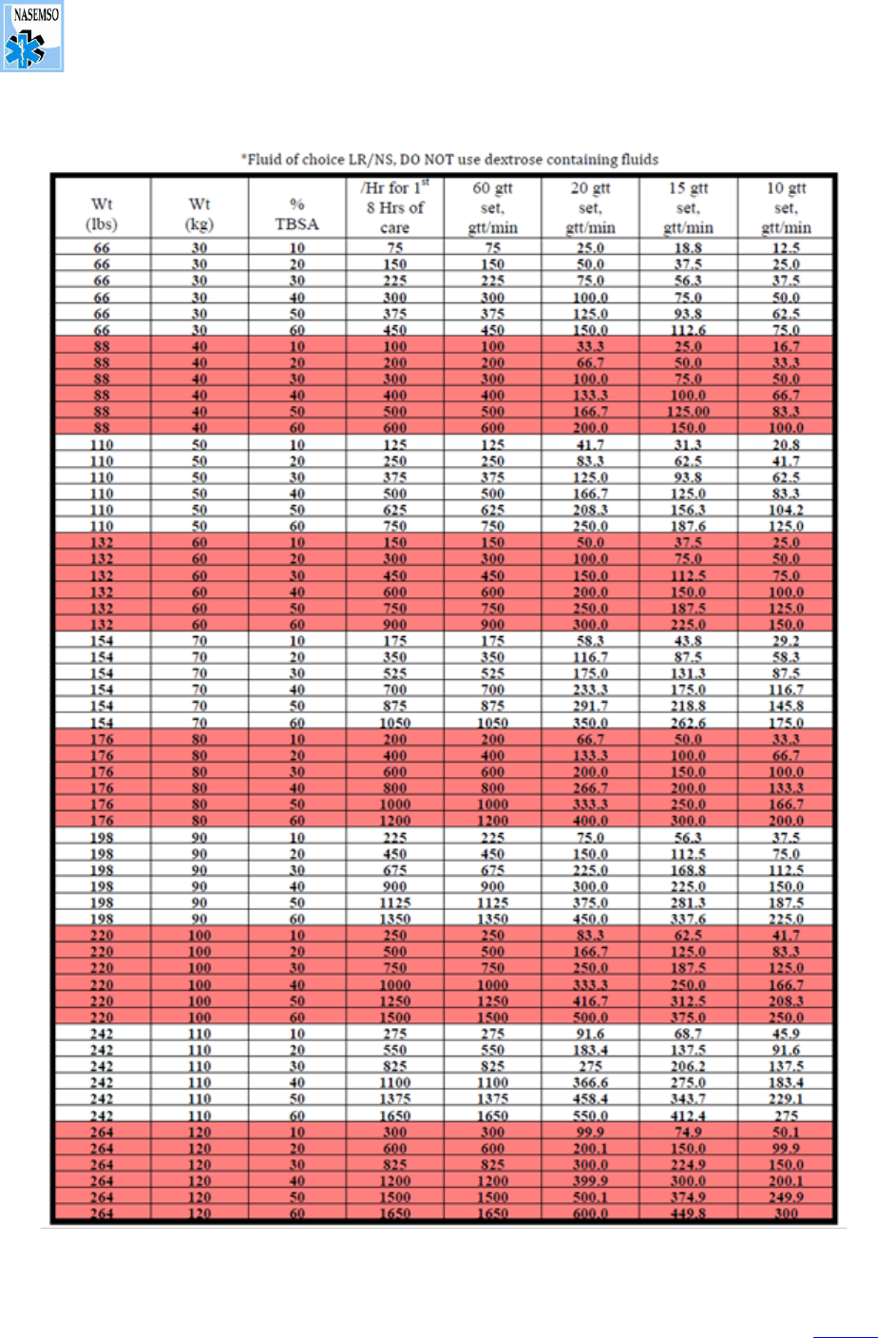

NASEMSO
National Model EMS Clinical Guidelines
________________________ Go To TOC
APPENDICES Rev. March 2022
V. Burn and Burn Fluid Charts 403
Version 3.0
Burn Injury IV Fluid Rates
Fluid Infusion Rate < 30 KG
Source: Used with permission, University of Utah Burn Center (https://crisisstandardsofcare.utah.edu).

NASEMSO
National Model EMS Clinical Guidelines
________________________ Go To TOC
APPENDICES Rev. March 2022
VI. Neurologic Status Assessment 404
Version 3.0
VI. Neurologic Status Assessment
Neurologic status assessment involves establishing a baseline and then trending any change in patient
neurologic status. Glasgow Coma Score (GCS) is frequently used, but there are often errors in applying
and calculating this score. With this in consideration, Glasgow Coma Score may not be more valid than a
simpler field approach. Either AVPU (Alert, Verbal, Painful, Unresponsive) or only the motor component
of the GCS may more effectively serve in this capacity.
Glasgow Coma Score
Table X: AVPU
A:
The patient is alert
V:
The patient responds to verbal stimulus
P:
The patient responds to painful stimulus
U:
The patient is completely unresponsive
Points
Pediatric
Adult
Eyes
1
No eye opening
2
Eye opening to pain
3
Eye opening to verbal
4
Eyes open spontaneously
Verbal
1
No vocalization
No verbal response
2
Inconsolable, agitated
Incomprehensible sounds
3
Inconsistently consolable, moaning
Inappropriate words
4
Cries but consolable, inappropriate
interactions
Confused
5
Smiles, oriented to sounds, follows
objects, interacts
Oriented
Motor
1
No motor response
2
Extension to pain
3
Flexion to pain
4
Withdraws from pain
5
Localizes pain
6
Obeys commands

NASEMSO
National Model EMS Clinical Guidelines
________________________ Go To TOC
APPENDICES Rev. March 2022
VII. Abnormal Vital Signs 405
Version 3.0
VII. Abnormal Vital Signs
Abnormal Vital Signs
Age
Heart Rate
Respiratory
Rate
Systolic BP
Temp (°C)
0 d – 1 mo
>205
>60
<60
<36 or >38
≥ 1 mo – 3 mo
>205
>60
<70
<36 or >38
≥ 3 mo – 1 yr
>190
>60
<70
<36 or >38.5
≥ 1 yr – 2 yr
>190
>40
<70 + (age in yr x 2)
<36 or >38.5
≥ 2 yr – 4 yr
>140
>40
<70 + (age in yr x 2)
<36 or >38.5
≥ 4 yr – 6 yr
>140
>34
<70 + (age in yr x 2)
<36 or >38.5
≥ 6 yr – 10 yr
>140
>30
<70 + (age in yr x 2)
<36 or >38.5
≥ 10 yr – 13 yr
>100
>30
<90
<36 or >38.5
> 13 yr
>100
>16
<90
<36 or >38.5

NASEMSO
National Model EMS Clinical Guidelines
________________________ Go To TOC
APPENDICES Rev. March 2022
VIII. Evidence-Based Guidelines: GRADE Methodology 406
Version 3.0
VIII. Evidence-Based Guidelines: GRADE Methodology
An Overview of GRADE Methodology
Although engagement in quality EMS research has increased significantly, the demand for evidence-
based quality prehospital research continues to exceed its availability. The need for evidence-based
prehospital patient care protocols was clearly recognized by the Institute of Medicine of the
National Academies and clearly stated in 2007 in The Future of Emergency Care: Emergency Medical
Services at the Crossroads.
The Grading of Recommendations, Assessment, Development, and Evaluation (GRADE)
methodology is a transparent process where the available research is reviewed and assessed by a
panel of subject matter experts. Following this thorough review process, the available research is
reviewed and graded for its validity based upon the assessment of the workgroup, and an evidence-
based guideline (EBG) is developed based upon the outcome of the workgroup.
The Federal Interagency Committee on Emergency Medical Services (FICEMS) and the National EMS
Advisory Council (NEMSAC) approved a National Prehospital Evidence-based Guideline Model
Process for the development, implementation, and evaluation of evidence-based guidelines. This
Model Process recommends the use of the GRADE methodology for the guideline development tool.
The six process steps of the GRADE EBG development tool are:
• Assemble the expert panel and provide GRADE training
• Define the EBG content area and establish the specific clinical questions to address in patient,
intervention, comparison, and outcome (PICO) format
• Prioritize outcomes to facilitate systematic literature searches
• Create GRADE tables (or evidence profiles) for each PICO question
• Vet and endorse GRADE evidence tables and draft recommendations
• Synthesize recommendations into an EMS protocol and visual algorithm
Some evidence-based guidelines cited in this document were created for and released by NHTSA;
however, the GRADE methodology is not proprietary to NHTSA or any other organization. Local,
regional, and state EMS agencies and EMS systems are encouraged to support the ongoing need for
quality prehospital care, improved patient outcome, and the growing demand for EBGs for EMS.
References:
Brown KM. The development of evidence-based prehospital guidelines using a GRADE-based
methodology, Prehospital Emergency Care, 2014, Suppl 1:3-14, 2014

NASEMSO
National Model EMS Clinical Guidelines
________________________ Go To TOC
APPENDICES Rev. March 2022
IX. 2022 National Guideline for the Field Triage of Injured Patients 407
Version 3.0
IX. 2022 National Guideline for the Field Triage of Injured Patients
Note: "Low-level" refers to less than 10 feet including ground level falls
Source: The American College of Surgeons Committee on Trauma (ACS COT), 2022
